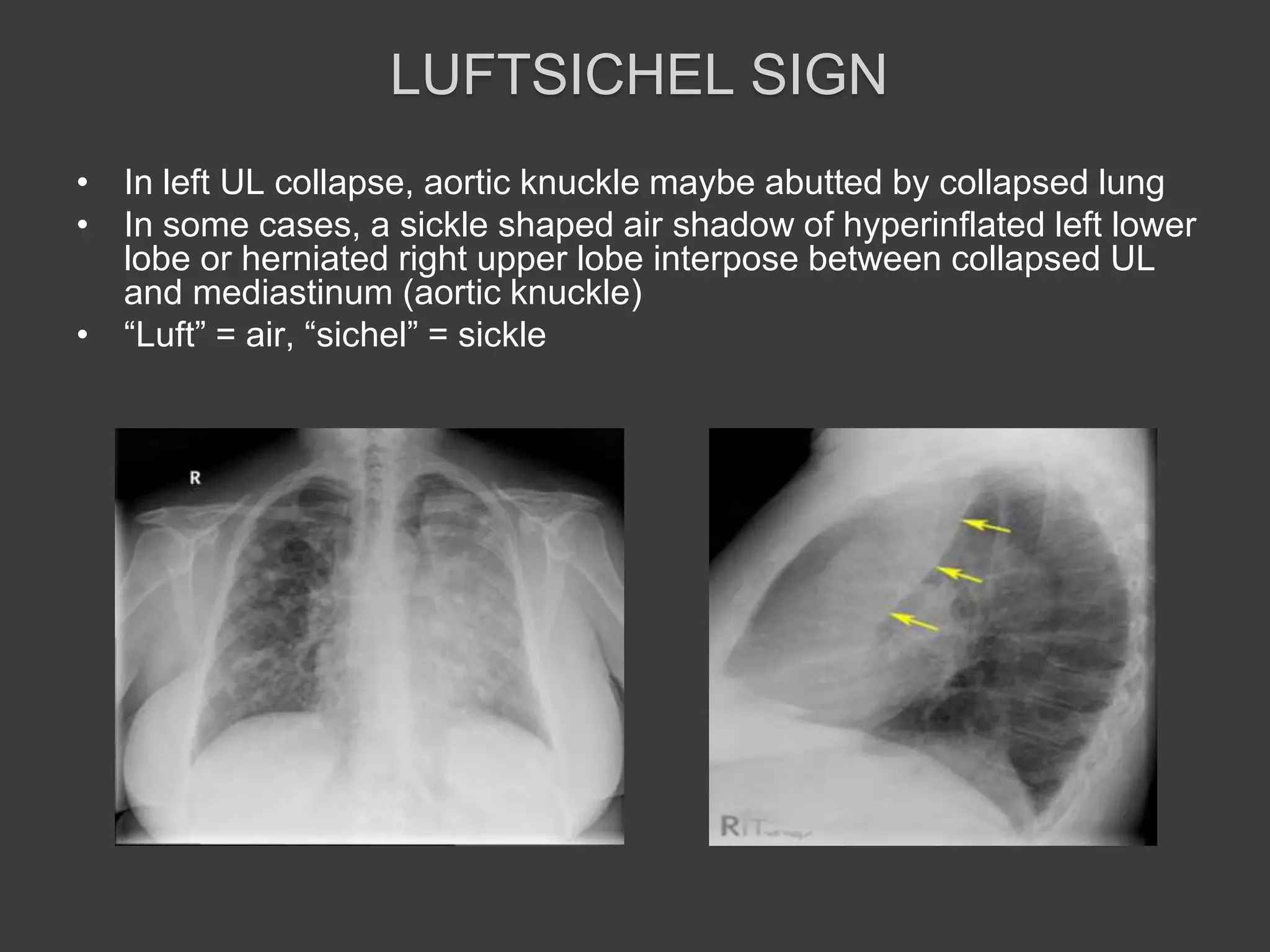
ABNORMAL CHEST X-RAYS
Dr. Tom Mishael J
PG Resident
St. John’s Medical College Bangalore
RADIOGRAPH OF CHEST
• Lung parenchyma
• Pleura
• Hilum
• Mediastinum
• Diaphragm
• Chest wall
LUNG PARENCHYMA
ALVEOLAR VS INTERSTITIAL
Alveolar disease :
Filling of alveolar air
spaces with
abnormal material
(blood, pus, water,
protein, cell debris)
Interstitial disease :
Affects the
supporting tissues of
the lung parenchyma
(connective tissue,
vessels, lymphatics,
bronchi, alveolar
wall)
ALVEOLAR VS INTERSTITIAL
CONSOLIDATION
CONSOLIDATION = PNEUMONIA ?
NO
CONSOLIDATION refers not only to
infection, but to any pathological process
that fills the alveoli with either pus, blood,
fluid, cells etc
Pneumonia - commonest cause of
consolidation
CONSOLIDATION
Pathological features :
1. Alveolar space filling with inflammatory exudate
2. Interstitium & architecture of lung remain intact
3. Airway is patent
Radiological correlation :
1. Density corresponding to a segment/ lobe
2. No significant volume loss
3. Air bronchogram
PNEUMONIA
• Bronchitis : Acute
or chronic inflammation
of the lining of a
bronchus
• Pneumonia :
Inflammation, usually
due to infection involving
the alveoli
• Lobar pneumonia :
Involving a large part of
the lobe, sometimes the
entire lobe
• Bronchopneumonia :
Pneumonia plus
bronchitis
LOBAR VS BRONCHOPNEUMONIA
BULGING FISSURE SIGN
• Refers to lobar consolidation where the affected portion of lung
expands and displaces an interlobar fissure
• Seen in infection with Klebsiella pneumoniae, Streptococcus
pneumoniae, Pseudomonas aeruginosa, Staphylococcus aureus
SILHOUETTE SIGN
Concept :
• When two structures with identical densities are in
contact with each other, the interface/ border between
them will be obscured (loss of silhouette)
Application :
• An intrathoracic lesion touching a border of the heart,
aorta or diaphragm will obliterate that border on CXR
• An intrathoracic lesion not anatomically contiguous with
a border will not obliterate that border
SILHOUETTE SIGN
SILHOUETTE SIGN
SILHOUETTE SIGN
SILHOUETTE SIGN
ABSENCE CAN TELL YOU WHERE A SHADOW IS NOT SITUATED
SILHOUETTE SIGN
SILHOUETTE SIGN
PITFALLS :
• In few normal individuals, right heart border will not be clearly seen
• A depressed sternum can produce loss of the right heart border, an appearance
which mimics the right middle lobe pneumonia
• Fat or pulmonary vessels close to the heart border may result in a false positive
silhouette sign
AIR BRONCHOGRAM SIGN
• Definition : Phenomenon of air filled bronchi (dark) being made
visible by the opacification of surrounding alveoli (grey/white) –
reverse concept of silhouette sign?
• Significance : Says that the opacity is intrapulmonary (excludes a
pleural or mediastinal lesion
AIR BRONCHOGRAM
Branching lucencies surrounded by consolidative opacity
LUNG COLLAPSE
• Concept : Diminished volume of air in lung associated with reduction in
lung volume
Direct Signs :
• Displacement of interlobar fissures
• Loss of aeration: Opacity of the affected lobe(s)
• Crowding of the vessels and bronchi within the collapsed area
Indirect Signs : Compensatory changes to volume loss
• Elevation of the ipsilateral hemi-diaphragm in case of lower lobe collapse
• Compensatory hyperinflation of the normal lung
• Displacement of the mediastinal structures towards the affected side
• Displacement of the ipsilateral hilum (elevated/depressed) which changes
shape
• Crowding of ribs on the affected side
RIGHT UPPER LOBE COLLAPSE
PA View :
• Area of whiteness in the upper
zone of right lung
• Horizontal fissure elevated
• Apparent right hilar mass
• Trachea deviated to the right
• Ribs over the area of whiteness
are closer than normal
Lateral view :
• Increased whiteness in the
upper part of the right lobe
GOLDEN S SIGN
• Due to a central mass
obstructing the upper
lobe bronchus
• Collapse of upper lobe
pulls up the lateral part of
minor fissure while the
large central mass
produces downward
convexity of the minor
fissure
RIGHT MIDDLE LOBE COLLAPSE
Lateral view:
• Triangular opacity in anterior aspect of
the chest overlying the cardiac shadow
• Apex at the hilum
• Base is running between the sternum
and the diaphragm
• Horizontal fissure is inferiorly displaced
and oblique fissure superiorly displaced
PA view:
• Right hemi diaphragm is slightly raised
with blurring of the right heart border
• Upper part of lower zone may have a
hazy appearance
RIGHT LOWER LOBE COLLAPSE
PA view:
• Complete collapse looks like a wedge
shaped shadow merging with
mediastinum
• Medial aspect of the dome of the
diaphragm is lost
• Descending right pulmonary artery is
not visualized
• Right hilum is depressed
Lateral view:
• Major (oblique) fissure moves
posteriorly but maintains its normal
slope
• Right hemi diaphragmatic outline is lost
posteriorly
• Lower thoracic vertebrae appear denser
than normally
LEFT UPPER LOBE COLLAPSE
PA view:
• Most of the left upper lobe lies in
front hence collapse causes a haze
to appear over the entire lung field
• Left hilum is drawn upwards
• Aortic knuckle appears obscured
• Trachea deviated to left
Lateral view:
• Increase in the retrosternal opacity
• Oblique fissure displaced anteriorly
• Left lower lobe is hyper expanded
LUFTSICHEL SIGN
• In left UL collapse, aortic knuckle maybe abutted by collapsed lung
• In some cases, a sickle shaped air shadow of hyperinflated left lower
lobe or herniated right upper lobe interpose between collapsed UL
and mediastinum (aortic knuckle)
• “Luft” = air, “sichel” = sickle
LEFT LOWER LOBE COLLAPSE
PA view:
• Left lung fields appear darker
• Cardiac shadow appears whiter
• White triangle behind the heart
Lateral view:
• White triangle at the bottom
posterior corner of the lung fields
• Vertebral bodies appear whiter
• Oblique fissure pulled posteriorly
COLLAPSE VS CONSOLIDATION
DIFFUSE LUNG DISEASES
Non- homogenous opacities
Various patterns :
Linear, septal lines, miliary shadows,
reticulonodular, honeycomb shadowing,
cystic, ground-glass pattern etc
Zonal distribution :
• Asbestosis – lung bases
• Sarcoidosis – spares lung bases
• Histiocytosis – reticular upper zone
opacity
• Lymphangitis carcinomatosis –
unilateral distribution
DIFFUSE INTERSTITIAL DISEASES
Reticular/ Linear shadowing :
• Fine irregular network of lines surrounding air-filled alveoli
Reticulonodular shadowing :
• Nodules are less than 1 cm in diameter
• Ill defined and irregular in outline
DIFFUSE RETICULONODULAR SHADOWING
HONEYCOMB SHADOWING
• Air containing spaces with
thick walls that are lined with
bronchiolar epithelium and
fibrous tissue
• Seen in end-stage pulmonary
fibrosis due to extensive
parenchymal destruction
• Cysts are usually 5-10 mm in
size
• Increases risk of
pneumothorax
MILIARY SHADOWING
Widespread
small discrete
opacities of
similar size (2-4
mm in diameter)
MILIARY SHADOWING
Infections :
• Tuberculosis
• Coccidioidomycosis
• Blastomycosis
• Histoplasmosis
• Chicken pox
Dust Inhalation :
• Tin, Barium
• Beryllium, Silicosis
• Coal miner’s
pneumoconiosis
Bronchiolitis obliterans
Alveolar microlithiasis
Hyaline membrane
disease
Metastases
Histiocytosis X
Hemosiderosis
Sarcoidosis
Secondary
hyperparathyroidism
Amyloidosis
LINEAR AND BAND SHADOWS
• Normal structures such as blood vessels and fissures
form linear shadows within the lung fields
• It can be pathological too
• Linear shadows – less than 5 mm wide
• Band shadows – greater than 5 mm wide
LINEAR AND BAND SHADOWS
CAUSES :
• Pulmonary infarcts
• Sentinel lines
• Thickened fissures
• Pulmonary/ pleural scars
• Bronchial wall thickening
• Curvilinear shadows (bullae, pneumatoceles)
• Anomalous vessels
• Artefacts
• Fleischner lines (plate atelectasis)
• Kerley lines
• Resolving infection
• Bronchoceles
PULMONARY INFARCTION
• Irregular thick wedge-
shaped lines with the
base adjacent to pleura
(Hampton’s Hump)
• Resolution tends to be
slow (Melting sign)
• Accompanying features :
Splinting of the diaphragm
and a pleural reaction
MELTING ICE SIGN
PLATE ATELECTASIS
• Seen post-operatively and is thought to be due to under ventilation
with obstruction of medium-sized bronchi
• Several centimetres long, 1-3 mm thick and run parallel to the
diaphragms extending to the pleural surface (Fleischner line)
• Resolution is rapid (compared to pulmonary infarct)
BRONCHOCELE
• These are bronchi distended with mucus or pus beyond an obstructing
lesion, but with aeration of the distal lung from collateral air flow
• Typical bronchocele has Gloved finger branching pattern with fingers
several millimetres wide
• Causes to consider to include malignancy, benign tumours, FB
aspiration, bronchial atresia etc
SENTINEL LINES
• Thought to be mucus-
filled bronchi and
appear as coarse lines
lying peripherally in
contact with the pleura
and curving upward
• Often left sided and
associated with left LL
collapse
KERLEY LINES
Kerley’s A lines:
• Linear opacities extending from the periphery to the hila
• Due to distention of anastomotic channels between peripheral
and central lymphatics
Kerley’s B lines:
• Short horizontal lines situated perpendicularly to the pleural
surface at the lung base
• Due to thickening of interlobular septa
Kerley’s C lines:
• Reticular opacities at the lung base representing
superimposed Kerley’s B lines
KERLEY LINES
Causes :
• Pulmonary edema
• Infections (viral, mycoplasma)
• Mitral valve disease
• Idiopathic pulmonary fibrosis
• Congenital heart disease
• Alveolar cell carcinoma
• Pneumoconiosis
• Lymphoma
• Lymphangitis carcinomatosis
• Pulmonary venous occlusive
disease
PLEURAL AND PULMONARY SCARS
• Scars are unchanged in
appearance on serial films
• Appear as a thin linear
shadows often with
associated pleural thickening
and tenting of the diaphragm
• Apical scarring is a common
finding in healed tuberculosis,
sarcoidosis and fungal
disease
BRONCHIAL WALL THICKENING
• They cast parallel tramline
shadows
• Ring shadows when seen
end-on
• Common in bronchiectasis,
recurrent asthma, pulmonary
oedema and lymphangitis
carcinomatosis
SOLITARY PULMONARY NODULE
• Discrete, well – marginated,
rounded opacity
• Less than or equal to 3 cm in
diameter
• Completely surrounded by lung
parenchyma
• Doesn’t touch the hilum or
mediastinum
• Not associated with
adenopathy, atelectasis or
pleural effusion
• Lesions larger than 4 cm are
treated as malignancies until
proven otherwise
SOLITARY PULMONARY NODULE
• Intrapulmonary mass forms an acute angle with the lung edge
• Extra-pleural and mediastinal masses form obtuse angles
SOLITARY PULMONARY NODULE
• Carcinomas often have irregular, spiculated or notched margins
• Calcification favors a benign lesion although a carcinoma may arise
coincidentally at the site of an old calcified focus
• Calcified metastasis is rare, the primary tumor being usually an
osteogenic or chondrosarcoma
• Granulomas frequently calcify and are usually well defined and
lobulated
SOLITARY PULMONARY NODULE
Hamartoma Chondrosarcoma
MULTIPLE PULMONARY NODULES
• Multiple small nodules 2-4
mm are called miliary
shadows
• Mostly metastasis or
tubercular granulomas
• Calcified nodules are
generally benign except
for metastasis from bone
or cartilaginous tumors
MULTIPLE PULMONARY NODULES
Canon ball metastasis
CAVITATING LESIONS AND CYSTS
• It’s a gas filled space
surrounded by a complete
wall which is 3mm or
greater in thickness
• Thin walled – cysts/ bullae
• Requires a patent airway to
communicate with necrotic
area
• Common cavitating
diseases are TB, staph
infections and carcinoma
CAVITATING LESIONS
Common sites :
• TB – upper zones and
apical zones of lower lobes
• Traumatic lung cysts – Sub
pleural
• Amoebic abscess – right
base
• Pulmonary infarcts –
Usually in left lower lobe
• Lung abscess – right side
and lower zone
CAVITATING LESIONS
FLUID LEVELS
• Abscesses
• Hydropneumothorax : trauma,
surgery, bronchopleural fistula
• Esophageal : pharyngeal pouch,
diverticula
• Obstruction : tumour,
achalasia
• Mediastinal : infections,
esophageal perforation
• Pneumopericardium
• Fluid levels are common in primary tumors -> irregular masses of blood clot
or necrotic tumor may be present
• Fluid levels are uncommon in cavitating metastases and tubercular cavities
HYDATID DISEASE
Signs of non-ruptured cysts :
• Honeycomb (wheel spoke, spoke wheel, rosette,
racemose) pattern: multivesicular mother cyst with
daughter cysts separated by radiating septa representing
cyst walls and hydatid sand/matrix
• Double line sign: unilocular cyst with double layered wall
representing pericyst and laminated cyst membrane
HYDATID DISEASE
Signs of partially ruptured cyst in pulmonary hydatid
disease :
• Crescent sign: when the hydatid cyst erodes the
adjacent bronchus or bronchiole, the trapped air
between the pericyst and the laminated membrane of the
endocyst give a crescent-shaped rim of air around the
cyst
• Inverted crescent sign: crescent-shaped rim of air at the
lower edge of the cyst
HYDATID DISEASE
Signs of complete rupture / cyst degeneration in pulmonary
hydatid disease :
• Cumbo (onion peel, double arch) sign: curvilinear membrane
outlined by air both inside the endocyst and a crescent of air
between the endocyst and pericyst
• Water lily (camalote) sign: folded membranes floating at the
air-fluid interface
• Empty (dry) cyst sign: air filled cyst after expectoration of
membranes and fluid
HYDATID DISEASE
Signs of ruptured endocyst in hydatid cysts :
• Serpent (snake) sign: wavy membranes within the cyst
• Spin (whirl) sign: twisting membranes within the cyst
• Ball of wool (yarn, congealed water lily, mass within a
cavity) sign: solid conglomeration of membranes settled
in the dependent portion of the cyst
HYDATID DISEASE
Cumbo sign
Water lily sign
AIR CRESCENT SIGN
• Crescent shaped
radiolucency within
parenchymal consolidation
or opacity
• Air fills the space between
the devitalized tissue and
surrounding parenchyma
• Opaque rim of haemorrhagic
tissue peripheral to the
radiolucency
• Common in aspergilloma
CALCIFICATIONS
CALCIFICATIONS
• Calcification is most easily recognized with low kVP films
• In elderly, calcification of the tracheal and bronchial cartilage
is common
• TB is the commonest calcifying pulmonary process usually
upper zone.
• Chickenpox – smaller(1-3 mm) regular in size and widely
distributed
TB CHICKEN POX
CALCIFICATIONS
Egg shell calcification :
Silicosis, Sarcoidosis
Alveolar microlithiasis:
Sand grain like calcification
APICAL SHADOWING
Common causes are :
• Pleural caps
• Pleural fluid
• Bullae
• Pancoast tumour
• Pneumothorax
• Infections –TB,
fungal infections
(histoplasmosis,
coccidioidomycosis,
aspergillosis)
UNILATERAL HYPERTRANSLUCENCY
Looks for signs of obstructive or compensatory
emphysema such as :
• Splaying of the ribs
• Separation of vascular markings
• Mediastinal displacement
• Depression of the hemidiaphragm
 Most common causes - Patient rotation and scoliosis
 With rotation to a side, that side becomes more
prominent
UNILATERAL HYPERTRANSLUCENCY
Mastectomy Pneumonectomy
OPAQUE HEMITHORAX
PLEURA
PLEURAL ABNORMALITIES
• Pleural effusion
• Pneumothorax
• Pleural plaques
• Pleural thickening
• Pleural calcification
• Pleural neoplasm
• Fibrothorax
PLEURAL EFFUSION
An area of whiteness at the
base of the lung :
• Pleural effusion
• Raised hemi diaphragm
• An area of consolidation/
collapse.
PLEURAL EFFUSION
Pleural Effusion vs Consolidation:
• Texture of whiteness ->
Consolidation usually causes
more heterogeneous shadowing
typically with the presence of an
air bronchogram
• Shape of the upper border of the
shadowing -> Fluid will have a
meniscus so the upper border of
an effusion will be concave
PLEURAL EFFUSION
Pleural effusion vs Raised hemidiaphragm :
Effusion will peak much more laterally than you would
expect the diaphragm to do
Pleural effusion vs Collapse :
Collapse usually causes shift of trachea towards the white
lung field. Hence of absence of shift excludes collapse.
Collapse can also accompany an effusion. So presence of
shift doesn’t exclude effusion.
PLEURAL EFFUSION
Look for …
• Presence of the meniscus,
which often on the lateral
view, is seen to tent up into
one of the fissures.
• Size of the heart (large =>
heart failure), enlargement of
hilum, visible parts of lung
fields for obvious masses,
bones for signs of mets, apex
pf lung for TB and tumors
PLEURAL EFFUSION
Lateral decubitus view
SMALL EFFUSION WITH BLUNTING OF
CP ANGLE
• Most dependent recess of the pleura is the CP angle.
• Small amount of effusion will tend to collect posteriorly (100-300 ml)
• 200 ml of fluid is required in Frontal film, 75 ml in lateral film.
MODERATE PLEURAL EFFUSION
• Fairly well defined upper edge, concave upwards, is higher
laterally than medially and obscures the diaphragmatic
shadow
MASSIVE EFFUSION WITH MEDIASTINAL
SHIFT
PLEURAL EFFUSION WITH COLLAPSE
No mediastinal shift in spite of the presence of
massive effusion
LAMELLAR PLEURAL EFFUSION
• Shallow collections
between the lung and
visceral pleura,
sometimes sparing the
CP angle
• Represents interstitial
pulmonary fluid
• Eg: Post cardiac
surgery
SUBPULMONIC PLEURAL EFFUSION
• Fluid is between
pleura and
diaphragm
• Lung floats above
the fluid
• Large subpulmonic
effusions mimics
elevation of hemi
diaphragm
LOCULATED PLEURAL EFFUSION
Pseudo tumor or
Phantom tumor –
effusion within the
fissures
PNEUMOTHORAX
Refers to presence of air in the pleural cavity
• Open pneumothorax : Air move freely in and out during
respiration
• Closed pneumothorax : No movement of air occurs (eg.
due to pleural adhesions)
• Valvular pneumothorax : Air enters the pleural space on
inspiration and doesn’t leave during expiration
• Tension pneumothorax : As intrapleural pressure
increases in a valvular pneumothorax
PNEUMOTHORAX
• Visible pleural edge seen as a
thin, sharp white line
• No lung markings peripheral to
this line
• Peripheral space is
radiolucent compared to
adjacent lung
• Lung may completely collapse
• No mediastinal shift unless
tension pneumothorax is
present
DEEP SULCUS SIGN
CP angle appear
abnormally deep and
lucent because of air in
the anterolateral pleural
space
DOUBLE DIAPHRAGM SIGN
Visualization of the anterior
CP angle as an edge
separate from the dome of
diaphragm but parallel to it
(especially in a supine film as
air collects and outlines the
anterior portion of
hemidiaphragm in a supine
patient with pneumothorax)
TENSION PNEUMOTHORAX
• Shift of mediastinum
away from the side of
pneumothorax
• Downward
displacement or
inversion of hemi
diaphragm
• Medical emergency
HYDROPNEUMOTHORAX
• Pneumothorax
containing a horizontal
fluid level which
separates opaque fluid
below from lucent air
above
• Classically seen as an
air-fluid level
PLEURAL PLAQUES
• Plaques are focal areas of
thickening of parietal pleura
(classically due to previous
exposure to asbestosis)
• Characteristically appear as
scattered islands of well
circumscribed pleural densities
(Holly leaf sign)
• Usually in the lower 2/3rd of the
thorax and are bilateral
• Most often affects the parietal
and diaphragmatic pleura
(virtually pathognomic)
• Do not involve the CP angles
PLEURAL CALCIFICATION
Unilateral :
• Previous empyema,
hemothorax or
pleurisy (in the
visceral pleura)
Bilateral :
• Asbestosis,
pneumoconiosis (in
the parietal pleura)
PLEURAL CALCIFICATIONS
• From previous pleurisy, calcification occurs in the
visceral pleura
• Associated pleural thickening is almost always present
and separates the calcification from the ribs
• Pleural calcification may be in a continuous sheet or in
discrete plaques usually producing dense, coarse,
irregular shadows, often sharply demarcated laterally
• In asbestosis, calcification occurs in the parietal pleura,
is more delicate and bilateral sparing the CP angles
seen
• Frequently visible over the diaphragm and adjacent to
the axillae
PLEURAL TUMOURS
• Commonest malignant disease of the
pleura is mets (most frequently from
bronchus and breast)
• Primary malignancy of pleura is
associated with asbestosis exposure
(malignant mesothelioma)
• Unilateral pleural effusion(30-95%) and
concentric or lobulated pleural
thickening
• Benign calcified or non-calcified
plaques may be present
• Frozen mediastinum sign : Because
of the pleural thickening and
mediastinal infiltration, the involved
hemithorax may be normal in volume,
despite presence of large effusion
FIBROTHORAX
• Fibrosis within the pleural
space
• Occurs secondary to the
inflammatory response
• Seen in tuberculosis,
asbestosis, hemothorax etc
HILUM
HILAR ABNORMALITIES
Superior margin of left hilum is
usually higher than the right
Whenever the left hilum appears
lower than right, check for any
evidence s/o :
• Collapse of left lower lobe or
right upper lobe
• Enlargement of right hilum
(tumour or nodes)
SMALL HILUM : CAUSES
* Mac Leod’s syndrome : U/L hemithoracic lucency as a result of post infectious obliterative
bronchiolitis
ENLARGED HILUM : CAUSES
BATWING APPEARANCE
• A pattern of b/l perihilar shadowing seen in pulmonary edema,
pneumonia (aspiration, PCP, viral, lipoid)
REVERSE BATWING APEARANCE
• Peripheral opacities in b/l lung fields with perihilar
sparing - chronic eosinophilic pneumonia,
bronchoalveolar CA, pulmonary contusion… and also
documented in COVID-19 pneumonia
GARLAND TRIAD
• Also called as 1-2-3 triad:
1. Right paratracheal nodes
2. Right hilar nodes
3. Left hilar nodes
• Typically seen in
Sarcoidosis
PULMONARY HYPERTENSION
• Prominent pulmonary outflow
tract
• Enlarged pulmonary arteries
• Pruning of peripheral
pulmonary vessels
• Elevated cardiac apex due to
right ventricular hypertrophy
• Enlarged right atrium
MEDIASTINUM
MEDIASTINAL COMPARTMENTS
MEDIASTINAL MASSES
CERVICOTHORACIC SIGN
• Any lesion with a visualized upper border above the level of
clavicle must be located posteriorly in the chest (apical
segment of upper lobes, pleura or posterior mediastinum
HILUM OVERLAY SIGN
• To distinguish between cardiac enlargement and an anterior mediastinal mass
• Hilum lateral to the lateral border of the mass – cardiac enlargement
• Hilum medial to the lateral border of the mass – mediastinal mass present
HILUM CONVERGENCE SIGN
• Allows an enlarged hilum due to pulmonary arteries to be
distinguished from enlargement due to tumor
• If the vessels arise from or converge directly onto the hilar
shadow – enlargement is vascular
• If the vessels appear to arise or converge medial to the lateral
aspect of the hilar shadow – enlargement is a mass
THORACOABDOMINAL SIGN
A sharply marginated mediastinal mass projected over the diaphragm on a CXR will be
wholly or party in the thorax because it is outlined by air in the lung
ANTERIOR MEDIASTINAL MASS
Lymphoma
MIDDLE MEDIASTINAL MASS
Thoracic aortic aneurysm
POSTERIOR MEDIASTINAL MASS
Neuroblastoma
PNEUMOMEDIASTINUM
• Presence of extra luminal gas within the mediastinum
Etiology :
Secondary to chest, neck or retroperitoneal surgery
Esophageal perforation :
• Endoscopic intervention
• Boerhaave syndrome (spontaneous perforation – increased
esophageal pressure as in vomiting, severe straining)
• CA esophagus
CONTINUOUS DIAPHRAGM SIGN
• Air in mediastinum tracks
extrapleurally between heart
and diaphragm forming a
continuous lucency outlining
base of heart representing
pneumomediastinum
• Pneumopericardium shows air
circumferentially outlining heart,
hence differentiated
SPINNAKER SAIL SIGN(ANGEL WING SIGN)
Seen in neonates
when thymic lobes are
displaced laterally by
mediastinal air
RING AROUND ARTERY SIGN
Air around pulmonary
artery and main
branches produces a
black ring
appearance
TUBULAR ARTERY SIGN
Air outlining the
major aortic
branches
PNEUMOPERICARDIUM
Air circumferentially outlining the heart
ABNORMAL HEART SHADOWS
Total anomalous pulmonary
venous connection :
Snowman / Figure of 8 sign
TOF – Boot shaped heart
ABNORMAL HEART SHADOWS
Ebstein’s anomaly (abnormal
tricuspid) :
Box shaped heart
Transposition of great
arteries :
Egg on string appearance
MITRAL STENOSIS
Double density sign – left atrial enlargement
LV ANEURYSM
PERICARDIAL EFFUSION
Retrosternal (paracardial) fat < Pericardial effusion > Epicardial fat
DIAPHRAGM
ELEVATED DIAPHRAGM
DIAPHRAGMATIC HERNIA
• Congenital or acquired
defect in the diaphragm
• More common on the
left side
Causes :
1. Hiatus hernia
2. Bochdalek hernia
3. Morgagni hernia
4. Traumatic
5. Iatrogenic
DIAPHRAGMATIC HERNIA
Bochdalek hernia Morgagni hernia
(most common, posterolateral, neonatal) (smaller, anterior, sternocostal angle,
presents later on)
*Hiatus hernia – through esophageal hiatus
EVENTRATION OF DIAPHRAGM
• A congenital condition
• Incomplete
muscularization of
diaphragm with a thin
membranous sheet
replacing the normal
muscle
• May affect only a part of
the diaphragm resulting in
a smooth hump
CHEST WALL
CLAVICULAR ABNORMALITIES
Fractures and old healed fractures
CLAVICULAR ABNORMALITIES
• Erosion of the outer ends of the clavicle is associated with RA
and hyperparathyroidism
• Hypoplastic clavicles in Holt-Oram Syndrome and Cleido
Cranial Dysostosis
Holt-Oram Syndrome Rheumatoid arthritis
STERNAL ABNORMALITIES
Pectus excavatum (funnel chest) :
• Depressed sternum
• Prominent shadowing adjacent to the right heart border
• Widening of cardiac silhouette
• Heart is displaced to the left and has a straight left heart border
STERNAL ABNORMALITIES
Pectus carinatum (pigeon
chest) : Sternum protrudes
anteriorly
ABNORMALITIES OF RIB
• Superior rib notching : RA, SLE, hyperparathyroidism, Marfan’s
syndrome, NF and in paraplegics and polio victims
• Inferior rib notching : Develops as a result of hypertrophy of the
intercostal vessels or with neurogenic tumors.
ABNORMALITIES OF RIB
CERVICAL RIB
• Cervical Rib :
Supernumerary rib
which arises from
the seventh
cervical vertebra
• Congenital rib
anomalies like bifid
ribs, hypoplasia
and bridging are
common
RIB FRACTURES
• Stress fractures – Common
in 7th rib
• 6th – 9th lines are the
common sites for cough
fractures
• Pathological fractures:
Senile osteoporosis,
myeloma, metastasis,
steroid therapy, Cushing’s
disease and other
endocrine disorders
• Cushing’s disease is
associated with abundant
callus formation
THORACIC SPINE
• Check for abnormal curvature or
alignment, bone and disc
destruction, sclerosis,
paravertebral soft-tissue masses
and congenital lesions such as
butterfly vertebrae
• Anterior erosion of vertebral
bodies sparing the disc spaces is
noted with aneurysm of
descending aorta, vascular
tumors and NF
Scoliosis
THORACIC SPINE
• A single dense vertebra, the ivory vertebra – classical
appearance of lymphoma, but also a feature of Paget’s
disease and metastasis
• Destruction of pedicle is typical of Metastasis
• Destruction of the disc with adjacent bony involvement is
characteristic of an Infective process
• Disc calcification occurs in oochronosis and ankylosing
spondylitis
SOFT TISSUE
• Skin lesions including naevi and lipomas may simulate lung tumors
• Multiple nodules occur with neurofibromatosis
• Mastectomy is one of the commonest causes of translucent
hemithorax
GINKGO LEAF SIGN
• Seen in extensive subcutaneous edema of chest wall
• Gas outlines the fibers of pectoralis major and creates a
branching pattern that resembles veins of a ginkgo leaf
If you have any questions…
THANK YOU

Abnormal Chest X rays

  • 1.
    ABNORMAL CHEST X-RAYS Dr.Tom Mishael J PG Resident St. John’s Medical College Bangalore
  • 2.
    RADIOGRAPH OF CHEST •Lung parenchyma • Pleura • Hilum • Mediastinum • Diaphragm • Chest wall
  • 3.
  • 4.
    ALVEOLAR VS INTERSTITIAL Alveolardisease : Filling of alveolar air spaces with abnormal material (blood, pus, water, protein, cell debris) Interstitial disease : Affects the supporting tissues of the lung parenchyma (connective tissue, vessels, lymphatics, bronchi, alveolar wall)
  • 5.
  • 6.
  • 7.
    CONSOLIDATION CONSOLIDATION = PNEUMONIA? NO CONSOLIDATION refers not only to infection, but to any pathological process that fills the alveoli with either pus, blood, fluid, cells etc Pneumonia - commonest cause of consolidation
  • 8.
    CONSOLIDATION Pathological features : 1.Alveolar space filling with inflammatory exudate 2. Interstitium & architecture of lung remain intact 3. Airway is patent Radiological correlation : 1. Density corresponding to a segment/ lobe 2. No significant volume loss 3. Air bronchogram
  • 9.
    PNEUMONIA • Bronchitis :Acute or chronic inflammation of the lining of a bronchus • Pneumonia : Inflammation, usually due to infection involving the alveoli • Lobar pneumonia : Involving a large part of the lobe, sometimes the entire lobe • Bronchopneumonia : Pneumonia plus bronchitis
  • 10.
  • 11.
    BULGING FISSURE SIGN •Refers to lobar consolidation where the affected portion of lung expands and displaces an interlobar fissure • Seen in infection with Klebsiella pneumoniae, Streptococcus pneumoniae, Pseudomonas aeruginosa, Staphylococcus aureus
  • 12.
    SILHOUETTE SIGN Concept : •When two structures with identical densities are in contact with each other, the interface/ border between them will be obscured (loss of silhouette) Application : • An intrathoracic lesion touching a border of the heart, aorta or diaphragm will obliterate that border on CXR • An intrathoracic lesion not anatomically contiguous with a border will not obliterate that border
  • 13.
  • 14.
  • 15.
  • 16.
    SILHOUETTE SIGN ABSENCE CANTELL YOU WHERE A SHADOW IS NOT SITUATED
  • 17.
  • 18.
    SILHOUETTE SIGN PITFALLS : •In few normal individuals, right heart border will not be clearly seen • A depressed sternum can produce loss of the right heart border, an appearance which mimics the right middle lobe pneumonia • Fat or pulmonary vessels close to the heart border may result in a false positive silhouette sign
  • 19.
    AIR BRONCHOGRAM SIGN •Definition : Phenomenon of air filled bronchi (dark) being made visible by the opacification of surrounding alveoli (grey/white) – reverse concept of silhouette sign? • Significance : Says that the opacity is intrapulmonary (excludes a pleural or mediastinal lesion
  • 20.
    AIR BRONCHOGRAM Branching lucenciessurrounded by consolidative opacity
  • 21.
    LUNG COLLAPSE • Concept: Diminished volume of air in lung associated with reduction in lung volume Direct Signs : • Displacement of interlobar fissures • Loss of aeration: Opacity of the affected lobe(s) • Crowding of the vessels and bronchi within the collapsed area Indirect Signs : Compensatory changes to volume loss • Elevation of the ipsilateral hemi-diaphragm in case of lower lobe collapse • Compensatory hyperinflation of the normal lung • Displacement of the mediastinal structures towards the affected side • Displacement of the ipsilateral hilum (elevated/depressed) which changes shape • Crowding of ribs on the affected side
  • 22.
    RIGHT UPPER LOBECOLLAPSE PA View : • Area of whiteness in the upper zone of right lung • Horizontal fissure elevated • Apparent right hilar mass • Trachea deviated to the right • Ribs over the area of whiteness are closer than normal Lateral view : • Increased whiteness in the upper part of the right lobe
  • 23.
    GOLDEN S SIGN •Due to a central mass obstructing the upper lobe bronchus • Collapse of upper lobe pulls up the lateral part of minor fissure while the large central mass produces downward convexity of the minor fissure
  • 24.
    RIGHT MIDDLE LOBECOLLAPSE Lateral view: • Triangular opacity in anterior aspect of the chest overlying the cardiac shadow • Apex at the hilum • Base is running between the sternum and the diaphragm • Horizontal fissure is inferiorly displaced and oblique fissure superiorly displaced PA view: • Right hemi diaphragm is slightly raised with blurring of the right heart border • Upper part of lower zone may have a hazy appearance
  • 25.
    RIGHT LOWER LOBECOLLAPSE PA view: • Complete collapse looks like a wedge shaped shadow merging with mediastinum • Medial aspect of the dome of the diaphragm is lost • Descending right pulmonary artery is not visualized • Right hilum is depressed Lateral view: • Major (oblique) fissure moves posteriorly but maintains its normal slope • Right hemi diaphragmatic outline is lost posteriorly • Lower thoracic vertebrae appear denser than normally
  • 26.
    LEFT UPPER LOBECOLLAPSE PA view: • Most of the left upper lobe lies in front hence collapse causes a haze to appear over the entire lung field • Left hilum is drawn upwards • Aortic knuckle appears obscured • Trachea deviated to left Lateral view: • Increase in the retrosternal opacity • Oblique fissure displaced anteriorly • Left lower lobe is hyper expanded
  • 27.
    LUFTSICHEL SIGN • Inleft UL collapse, aortic knuckle maybe abutted by collapsed lung • In some cases, a sickle shaped air shadow of hyperinflated left lower lobe or herniated right upper lobe interpose between collapsed UL and mediastinum (aortic knuckle) • “Luft” = air, “sichel” = sickle
  • 28.
    LEFT LOWER LOBECOLLAPSE PA view: • Left lung fields appear darker • Cardiac shadow appears whiter • White triangle behind the heart Lateral view: • White triangle at the bottom posterior corner of the lung fields • Vertebral bodies appear whiter • Oblique fissure pulled posteriorly
  • 29.
  • 30.
    DIFFUSE LUNG DISEASES Non-homogenous opacities Various patterns : Linear, septal lines, miliary shadows, reticulonodular, honeycomb shadowing, cystic, ground-glass pattern etc Zonal distribution : • Asbestosis – lung bases • Sarcoidosis – spares lung bases • Histiocytosis – reticular upper zone opacity • Lymphangitis carcinomatosis – unilateral distribution
  • 31.
    DIFFUSE INTERSTITIAL DISEASES Reticular/Linear shadowing : • Fine irregular network of lines surrounding air-filled alveoli Reticulonodular shadowing : • Nodules are less than 1 cm in diameter • Ill defined and irregular in outline
  • 32.
  • 33.
    HONEYCOMB SHADOWING • Aircontaining spaces with thick walls that are lined with bronchiolar epithelium and fibrous tissue • Seen in end-stage pulmonary fibrosis due to extensive parenchymal destruction • Cysts are usually 5-10 mm in size • Increases risk of pneumothorax
  • 34.
    MILIARY SHADOWING Widespread small discrete opacitiesof similar size (2-4 mm in diameter)
  • 35.
    MILIARY SHADOWING Infections : •Tuberculosis • Coccidioidomycosis • Blastomycosis • Histoplasmosis • Chicken pox Dust Inhalation : • Tin, Barium • Beryllium, Silicosis • Coal miner’s pneumoconiosis Bronchiolitis obliterans Alveolar microlithiasis Hyaline membrane disease Metastases Histiocytosis X Hemosiderosis Sarcoidosis Secondary hyperparathyroidism Amyloidosis
  • 36.
    LINEAR AND BANDSHADOWS • Normal structures such as blood vessels and fissures form linear shadows within the lung fields • It can be pathological too • Linear shadows – less than 5 mm wide • Band shadows – greater than 5 mm wide
  • 37.
    LINEAR AND BANDSHADOWS CAUSES : • Pulmonary infarcts • Sentinel lines • Thickened fissures • Pulmonary/ pleural scars • Bronchial wall thickening • Curvilinear shadows (bullae, pneumatoceles) • Anomalous vessels • Artefacts • Fleischner lines (plate atelectasis) • Kerley lines • Resolving infection • Bronchoceles
  • 38.
    PULMONARY INFARCTION • Irregularthick wedge- shaped lines with the base adjacent to pleura (Hampton’s Hump) • Resolution tends to be slow (Melting sign) • Accompanying features : Splinting of the diaphragm and a pleural reaction
  • 39.
  • 40.
    PLATE ATELECTASIS • Seenpost-operatively and is thought to be due to under ventilation with obstruction of medium-sized bronchi • Several centimetres long, 1-3 mm thick and run parallel to the diaphragms extending to the pleural surface (Fleischner line) • Resolution is rapid (compared to pulmonary infarct)
  • 41.
    BRONCHOCELE • These arebronchi distended with mucus or pus beyond an obstructing lesion, but with aeration of the distal lung from collateral air flow • Typical bronchocele has Gloved finger branching pattern with fingers several millimetres wide • Causes to consider to include malignancy, benign tumours, FB aspiration, bronchial atresia etc
  • 42.
    SENTINEL LINES • Thoughtto be mucus- filled bronchi and appear as coarse lines lying peripherally in contact with the pleura and curving upward • Often left sided and associated with left LL collapse
  • 43.
    KERLEY LINES Kerley’s Alines: • Linear opacities extending from the periphery to the hila • Due to distention of anastomotic channels between peripheral and central lymphatics Kerley’s B lines: • Short horizontal lines situated perpendicularly to the pleural surface at the lung base • Due to thickening of interlobular septa Kerley’s C lines: • Reticular opacities at the lung base representing superimposed Kerley’s B lines
  • 44.
    KERLEY LINES Causes : •Pulmonary edema • Infections (viral, mycoplasma) • Mitral valve disease • Idiopathic pulmonary fibrosis • Congenital heart disease • Alveolar cell carcinoma • Pneumoconiosis • Lymphoma • Lymphangitis carcinomatosis • Pulmonary venous occlusive disease
  • 45.
    PLEURAL AND PULMONARYSCARS • Scars are unchanged in appearance on serial films • Appear as a thin linear shadows often with associated pleural thickening and tenting of the diaphragm • Apical scarring is a common finding in healed tuberculosis, sarcoidosis and fungal disease
  • 46.
    BRONCHIAL WALL THICKENING •They cast parallel tramline shadows • Ring shadows when seen end-on • Common in bronchiectasis, recurrent asthma, pulmonary oedema and lymphangitis carcinomatosis
  • 47.
    SOLITARY PULMONARY NODULE •Discrete, well – marginated, rounded opacity • Less than or equal to 3 cm in diameter • Completely surrounded by lung parenchyma • Doesn’t touch the hilum or mediastinum • Not associated with adenopathy, atelectasis or pleural effusion • Lesions larger than 4 cm are treated as malignancies until proven otherwise
  • 48.
  • 49.
    SOLITARY PULMONARY NODULE •Intrapulmonary mass forms an acute angle with the lung edge • Extra-pleural and mediastinal masses form obtuse angles
  • 50.
    SOLITARY PULMONARY NODULE •Carcinomas often have irregular, spiculated or notched margins • Calcification favors a benign lesion although a carcinoma may arise coincidentally at the site of an old calcified focus • Calcified metastasis is rare, the primary tumor being usually an osteogenic or chondrosarcoma • Granulomas frequently calcify and are usually well defined and lobulated
  • 51.
  • 52.
    MULTIPLE PULMONARY NODULES •Multiple small nodules 2-4 mm are called miliary shadows • Mostly metastasis or tubercular granulomas • Calcified nodules are generally benign except for metastasis from bone or cartilaginous tumors
  • 53.
  • 54.
    CAVITATING LESIONS ANDCYSTS • It’s a gas filled space surrounded by a complete wall which is 3mm or greater in thickness • Thin walled – cysts/ bullae • Requires a patent airway to communicate with necrotic area • Common cavitating diseases are TB, staph infections and carcinoma
  • 55.
    CAVITATING LESIONS Common sites: • TB – upper zones and apical zones of lower lobes • Traumatic lung cysts – Sub pleural • Amoebic abscess – right base • Pulmonary infarcts – Usually in left lower lobe • Lung abscess – right side and lower zone
  • 56.
  • 57.
    FLUID LEVELS • Abscesses •Hydropneumothorax : trauma, surgery, bronchopleural fistula • Esophageal : pharyngeal pouch, diverticula • Obstruction : tumour, achalasia • Mediastinal : infections, esophageal perforation • Pneumopericardium • Fluid levels are common in primary tumors -> irregular masses of blood clot or necrotic tumor may be present • Fluid levels are uncommon in cavitating metastases and tubercular cavities
  • 58.
    HYDATID DISEASE Signs ofnon-ruptured cysts : • Honeycomb (wheel spoke, spoke wheel, rosette, racemose) pattern: multivesicular mother cyst with daughter cysts separated by radiating septa representing cyst walls and hydatid sand/matrix • Double line sign: unilocular cyst with double layered wall representing pericyst and laminated cyst membrane
  • 59.
    HYDATID DISEASE Signs ofpartially ruptured cyst in pulmonary hydatid disease : • Crescent sign: when the hydatid cyst erodes the adjacent bronchus or bronchiole, the trapped air between the pericyst and the laminated membrane of the endocyst give a crescent-shaped rim of air around the cyst • Inverted crescent sign: crescent-shaped rim of air at the lower edge of the cyst
  • 60.
    HYDATID DISEASE Signs ofcomplete rupture / cyst degeneration in pulmonary hydatid disease : • Cumbo (onion peel, double arch) sign: curvilinear membrane outlined by air both inside the endocyst and a crescent of air between the endocyst and pericyst • Water lily (camalote) sign: folded membranes floating at the air-fluid interface • Empty (dry) cyst sign: air filled cyst after expectoration of membranes and fluid
  • 61.
    HYDATID DISEASE Signs ofruptured endocyst in hydatid cysts : • Serpent (snake) sign: wavy membranes within the cyst • Spin (whirl) sign: twisting membranes within the cyst • Ball of wool (yarn, congealed water lily, mass within a cavity) sign: solid conglomeration of membranes settled in the dependent portion of the cyst
  • 62.
  • 63.
    AIR CRESCENT SIGN •Crescent shaped radiolucency within parenchymal consolidation or opacity • Air fills the space between the devitalized tissue and surrounding parenchyma • Opaque rim of haemorrhagic tissue peripheral to the radiolucency • Common in aspergilloma
  • 64.
  • 65.
    CALCIFICATIONS • Calcification ismost easily recognized with low kVP films • In elderly, calcification of the tracheal and bronchial cartilage is common • TB is the commonest calcifying pulmonary process usually upper zone. • Chickenpox – smaller(1-3 mm) regular in size and widely distributed TB CHICKEN POX
  • 66.
    CALCIFICATIONS Egg shell calcification: Silicosis, Sarcoidosis Alveolar microlithiasis: Sand grain like calcification
  • 67.
    APICAL SHADOWING Common causesare : • Pleural caps • Pleural fluid • Bullae • Pancoast tumour • Pneumothorax • Infections –TB, fungal infections (histoplasmosis, coccidioidomycosis, aspergillosis)
  • 68.
    UNILATERAL HYPERTRANSLUCENCY Looks forsigns of obstructive or compensatory emphysema such as : • Splaying of the ribs • Separation of vascular markings • Mediastinal displacement • Depression of the hemidiaphragm  Most common causes - Patient rotation and scoliosis  With rotation to a side, that side becomes more prominent
  • 69.
  • 70.
  • 71.
  • 72.
    PLEURAL ABNORMALITIES • Pleuraleffusion • Pneumothorax • Pleural plaques • Pleural thickening • Pleural calcification • Pleural neoplasm • Fibrothorax
  • 73.
    PLEURAL EFFUSION An areaof whiteness at the base of the lung : • Pleural effusion • Raised hemi diaphragm • An area of consolidation/ collapse.
  • 74.
    PLEURAL EFFUSION Pleural Effusionvs Consolidation: • Texture of whiteness -> Consolidation usually causes more heterogeneous shadowing typically with the presence of an air bronchogram • Shape of the upper border of the shadowing -> Fluid will have a meniscus so the upper border of an effusion will be concave
  • 75.
    PLEURAL EFFUSION Pleural effusionvs Raised hemidiaphragm : Effusion will peak much more laterally than you would expect the diaphragm to do Pleural effusion vs Collapse : Collapse usually causes shift of trachea towards the white lung field. Hence of absence of shift excludes collapse. Collapse can also accompany an effusion. So presence of shift doesn’t exclude effusion.
  • 76.
    PLEURAL EFFUSION Look for… • Presence of the meniscus, which often on the lateral view, is seen to tent up into one of the fissures. • Size of the heart (large => heart failure), enlargement of hilum, visible parts of lung fields for obvious masses, bones for signs of mets, apex pf lung for TB and tumors
  • 77.
  • 78.
    SMALL EFFUSION WITHBLUNTING OF CP ANGLE • Most dependent recess of the pleura is the CP angle. • Small amount of effusion will tend to collect posteriorly (100-300 ml) • 200 ml of fluid is required in Frontal film, 75 ml in lateral film.
  • 79.
    MODERATE PLEURAL EFFUSION •Fairly well defined upper edge, concave upwards, is higher laterally than medially and obscures the diaphragmatic shadow
  • 80.
    MASSIVE EFFUSION WITHMEDIASTINAL SHIFT
  • 81.
    PLEURAL EFFUSION WITHCOLLAPSE No mediastinal shift in spite of the presence of massive effusion
  • 82.
    LAMELLAR PLEURAL EFFUSION •Shallow collections between the lung and visceral pleura, sometimes sparing the CP angle • Represents interstitial pulmonary fluid • Eg: Post cardiac surgery
  • 83.
    SUBPULMONIC PLEURAL EFFUSION •Fluid is between pleura and diaphragm • Lung floats above the fluid • Large subpulmonic effusions mimics elevation of hemi diaphragm
  • 84.
    LOCULATED PLEURAL EFFUSION Pseudotumor or Phantom tumor – effusion within the fissures
  • 85.
    PNEUMOTHORAX Refers to presenceof air in the pleural cavity • Open pneumothorax : Air move freely in and out during respiration • Closed pneumothorax : No movement of air occurs (eg. due to pleural adhesions) • Valvular pneumothorax : Air enters the pleural space on inspiration and doesn’t leave during expiration • Tension pneumothorax : As intrapleural pressure increases in a valvular pneumothorax
  • 86.
    PNEUMOTHORAX • Visible pleuraledge seen as a thin, sharp white line • No lung markings peripheral to this line • Peripheral space is radiolucent compared to adjacent lung • Lung may completely collapse • No mediastinal shift unless tension pneumothorax is present
  • 87.
    DEEP SULCUS SIGN CPangle appear abnormally deep and lucent because of air in the anterolateral pleural space
  • 88.
    DOUBLE DIAPHRAGM SIGN Visualizationof the anterior CP angle as an edge separate from the dome of diaphragm but parallel to it (especially in a supine film as air collects and outlines the anterior portion of hemidiaphragm in a supine patient with pneumothorax)
  • 89.
    TENSION PNEUMOTHORAX • Shiftof mediastinum away from the side of pneumothorax • Downward displacement or inversion of hemi diaphragm • Medical emergency
  • 90.
    HYDROPNEUMOTHORAX • Pneumothorax containing ahorizontal fluid level which separates opaque fluid below from lucent air above • Classically seen as an air-fluid level
  • 91.
    PLEURAL PLAQUES • Plaquesare focal areas of thickening of parietal pleura (classically due to previous exposure to asbestosis) • Characteristically appear as scattered islands of well circumscribed pleural densities (Holly leaf sign) • Usually in the lower 2/3rd of the thorax and are bilateral • Most often affects the parietal and diaphragmatic pleura (virtually pathognomic) • Do not involve the CP angles
  • 92.
    PLEURAL CALCIFICATION Unilateral : •Previous empyema, hemothorax or pleurisy (in the visceral pleura) Bilateral : • Asbestosis, pneumoconiosis (in the parietal pleura)
  • 93.
    PLEURAL CALCIFICATIONS • Fromprevious pleurisy, calcification occurs in the visceral pleura • Associated pleural thickening is almost always present and separates the calcification from the ribs • Pleural calcification may be in a continuous sheet or in discrete plaques usually producing dense, coarse, irregular shadows, often sharply demarcated laterally • In asbestosis, calcification occurs in the parietal pleura, is more delicate and bilateral sparing the CP angles seen • Frequently visible over the diaphragm and adjacent to the axillae
  • 94.
    PLEURAL TUMOURS • Commonestmalignant disease of the pleura is mets (most frequently from bronchus and breast) • Primary malignancy of pleura is associated with asbestosis exposure (malignant mesothelioma) • Unilateral pleural effusion(30-95%) and concentric or lobulated pleural thickening • Benign calcified or non-calcified plaques may be present • Frozen mediastinum sign : Because of the pleural thickening and mediastinal infiltration, the involved hemithorax may be normal in volume, despite presence of large effusion
  • 95.
    FIBROTHORAX • Fibrosis withinthe pleural space • Occurs secondary to the inflammatory response • Seen in tuberculosis, asbestosis, hemothorax etc
  • 96.
  • 97.
    HILAR ABNORMALITIES Superior marginof left hilum is usually higher than the right Whenever the left hilum appears lower than right, check for any evidence s/o : • Collapse of left lower lobe or right upper lobe • Enlargement of right hilum (tumour or nodes)
  • 98.
    SMALL HILUM :CAUSES * Mac Leod’s syndrome : U/L hemithoracic lucency as a result of post infectious obliterative bronchiolitis
  • 99.
  • 100.
    BATWING APPEARANCE • Apattern of b/l perihilar shadowing seen in pulmonary edema, pneumonia (aspiration, PCP, viral, lipoid)
  • 101.
    REVERSE BATWING APEARANCE •Peripheral opacities in b/l lung fields with perihilar sparing - chronic eosinophilic pneumonia, bronchoalveolar CA, pulmonary contusion… and also documented in COVID-19 pneumonia
  • 102.
    GARLAND TRIAD • Alsocalled as 1-2-3 triad: 1. Right paratracheal nodes 2. Right hilar nodes 3. Left hilar nodes • Typically seen in Sarcoidosis
  • 103.
    PULMONARY HYPERTENSION • Prominentpulmonary outflow tract • Enlarged pulmonary arteries • Pruning of peripheral pulmonary vessels • Elevated cardiac apex due to right ventricular hypertrophy • Enlarged right atrium
  • 104.
  • 105.
  • 106.
  • 107.
    CERVICOTHORACIC SIGN • Anylesion with a visualized upper border above the level of clavicle must be located posteriorly in the chest (apical segment of upper lobes, pleura or posterior mediastinum
  • 108.
    HILUM OVERLAY SIGN •To distinguish between cardiac enlargement and an anterior mediastinal mass • Hilum lateral to the lateral border of the mass – cardiac enlargement • Hilum medial to the lateral border of the mass – mediastinal mass present
  • 109.
    HILUM CONVERGENCE SIGN •Allows an enlarged hilum due to pulmonary arteries to be distinguished from enlargement due to tumor • If the vessels arise from or converge directly onto the hilar shadow – enlargement is vascular • If the vessels appear to arise or converge medial to the lateral aspect of the hilar shadow – enlargement is a mass
  • 110.
    THORACOABDOMINAL SIGN A sharplymarginated mediastinal mass projected over the diaphragm on a CXR will be wholly or party in the thorax because it is outlined by air in the lung
  • 111.
  • 112.
  • 113.
  • 114.
    PNEUMOMEDIASTINUM • Presence ofextra luminal gas within the mediastinum Etiology : Secondary to chest, neck or retroperitoneal surgery Esophageal perforation : • Endoscopic intervention • Boerhaave syndrome (spontaneous perforation – increased esophageal pressure as in vomiting, severe straining) • CA esophagus
  • 115.
    CONTINUOUS DIAPHRAGM SIGN •Air in mediastinum tracks extrapleurally between heart and diaphragm forming a continuous lucency outlining base of heart representing pneumomediastinum • Pneumopericardium shows air circumferentially outlining heart, hence differentiated
  • 116.
    SPINNAKER SAIL SIGN(ANGELWING SIGN) Seen in neonates when thymic lobes are displaced laterally by mediastinal air
  • 117.
    RING AROUND ARTERYSIGN Air around pulmonary artery and main branches produces a black ring appearance
  • 118.
    TUBULAR ARTERY SIGN Airoutlining the major aortic branches
  • 119.
  • 120.
    ABNORMAL HEART SHADOWS Totalanomalous pulmonary venous connection : Snowman / Figure of 8 sign TOF – Boot shaped heart
  • 121.
    ABNORMAL HEART SHADOWS Ebstein’sanomaly (abnormal tricuspid) : Box shaped heart Transposition of great arteries : Egg on string appearance
  • 122.
    MITRAL STENOSIS Double densitysign – left atrial enlargement
  • 123.
  • 124.
    PERICARDIAL EFFUSION Retrosternal (paracardial)fat < Pericardial effusion > Epicardial fat
  • 125.
  • 126.
  • 127.
    DIAPHRAGMATIC HERNIA • Congenitalor acquired defect in the diaphragm • More common on the left side Causes : 1. Hiatus hernia 2. Bochdalek hernia 3. Morgagni hernia 4. Traumatic 5. Iatrogenic
  • 128.
    DIAPHRAGMATIC HERNIA Bochdalek herniaMorgagni hernia (most common, posterolateral, neonatal) (smaller, anterior, sternocostal angle, presents later on) *Hiatus hernia – through esophageal hiatus
  • 129.
    EVENTRATION OF DIAPHRAGM •A congenital condition • Incomplete muscularization of diaphragm with a thin membranous sheet replacing the normal muscle • May affect only a part of the diaphragm resulting in a smooth hump
  • 130.
  • 131.
  • 132.
    CLAVICULAR ABNORMALITIES • Erosionof the outer ends of the clavicle is associated with RA and hyperparathyroidism • Hypoplastic clavicles in Holt-Oram Syndrome and Cleido Cranial Dysostosis Holt-Oram Syndrome Rheumatoid arthritis
  • 133.
    STERNAL ABNORMALITIES Pectus excavatum(funnel chest) : • Depressed sternum • Prominent shadowing adjacent to the right heart border • Widening of cardiac silhouette • Heart is displaced to the left and has a straight left heart border
  • 134.
    STERNAL ABNORMALITIES Pectus carinatum(pigeon chest) : Sternum protrudes anteriorly
  • 135.
    ABNORMALITIES OF RIB •Superior rib notching : RA, SLE, hyperparathyroidism, Marfan’s syndrome, NF and in paraplegics and polio victims • Inferior rib notching : Develops as a result of hypertrophy of the intercostal vessels or with neurogenic tumors.
  • 136.
  • 137.
    CERVICAL RIB • CervicalRib : Supernumerary rib which arises from the seventh cervical vertebra • Congenital rib anomalies like bifid ribs, hypoplasia and bridging are common
  • 138.
    RIB FRACTURES • Stressfractures – Common in 7th rib • 6th – 9th lines are the common sites for cough fractures • Pathological fractures: Senile osteoporosis, myeloma, metastasis, steroid therapy, Cushing’s disease and other endocrine disorders • Cushing’s disease is associated with abundant callus formation
  • 139.
    THORACIC SPINE • Checkfor abnormal curvature or alignment, bone and disc destruction, sclerosis, paravertebral soft-tissue masses and congenital lesions such as butterfly vertebrae • Anterior erosion of vertebral bodies sparing the disc spaces is noted with aneurysm of descending aorta, vascular tumors and NF Scoliosis
  • 140.
    THORACIC SPINE • Asingle dense vertebra, the ivory vertebra – classical appearance of lymphoma, but also a feature of Paget’s disease and metastasis • Destruction of pedicle is typical of Metastasis • Destruction of the disc with adjacent bony involvement is characteristic of an Infective process • Disc calcification occurs in oochronosis and ankylosing spondylitis
  • 141.
    SOFT TISSUE • Skinlesions including naevi and lipomas may simulate lung tumors • Multiple nodules occur with neurofibromatosis • Mastectomy is one of the commonest causes of translucent hemithorax
  • 142.
    GINKGO LEAF SIGN •Seen in extensive subcutaneous edema of chest wall • Gas outlines the fibers of pectoralis major and creates a branching pattern that resembles veins of a ginkgo leaf
  • 143.
    If you haveany questions… THANK YOU