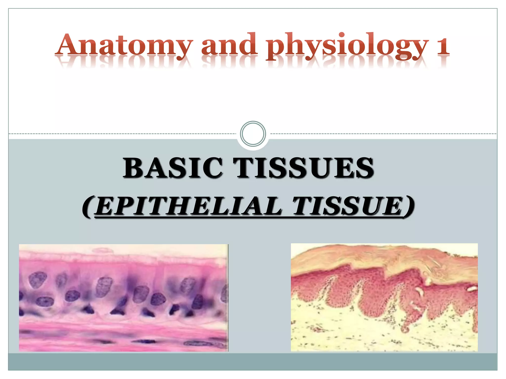
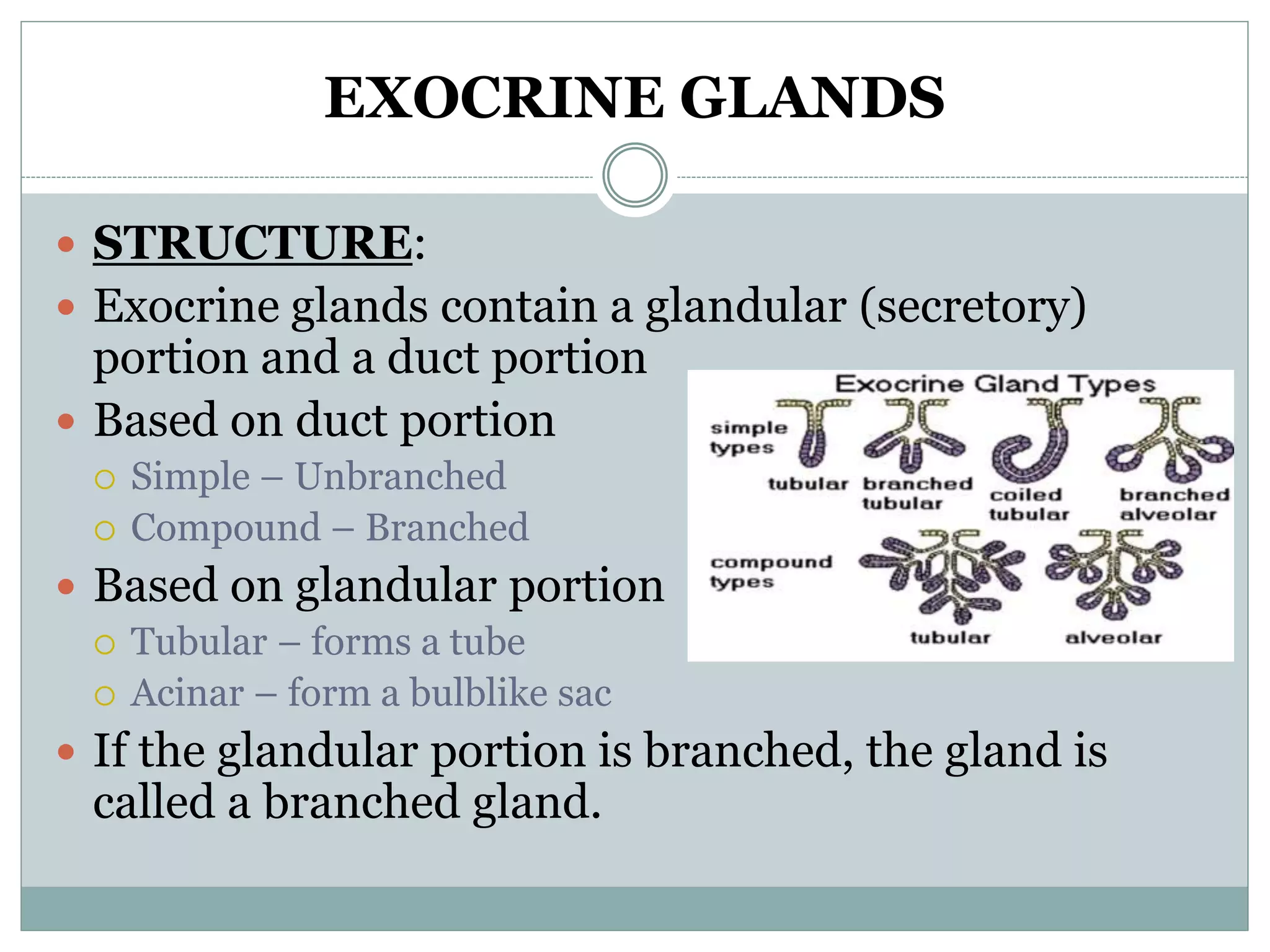
BASIC TISSUES
(EPITHELIAL TISSUE)
WHAT IS A TISSUE?
 A tissue is a group of similar cells which work
together to perform a specific function in the body.
 All cells in the body can be categorized into Four (4)
major types of tissues (Basic/Primary Tissues):
1. Epithelial Tissue
2. Connective Tissue
3. Muscular Tissue
4. Nervous Tissue
TISSUE
 The study of tissues and how they are arranged into
organs – Histology or Microscopic Anatomy
 Aside cells, tissues are also made up of extracellular
material which surrounds the cells – Matrix
 The 4 basic tissue types differ in the types and functions
of their cells and characteristics of their matrix.
 The matrix is composed of fibrous proteins and usually a
clear gel (ground substance/ tissue fluid/ extracellular
fluid/ interstitial fluid/ tissue gel)
Embryonic Tissues
 The first tissues (embryonic tissues) arise in the early
embryo when similar cells organise together into
layers (3) called Primary Germ Layers.
1. Ectoderm
2. Mesoderm
3. Endoderm
 Germ layers give rise to all the body’s mature tissues
Embryonic Tissues
 Endoderm – Inner layer which gives rise to various
membranes of the digestive and respiratory tracts.
 Ectoderm – Outer layer which forms the outer
covering of the body (epidermis).
 Mesoderm – Middle layer that forms the skeleton
and muscles of the body.
TISSUE
 Body organs usually contain all four tissue types.
 Example the stomach
Lined with epithelial tissue
Walls contain smooth muscle and connective tissue
Nerve tissue supply which control motility and
gastric secretions.
EPITHELIAL TISSUE
(EPITHELIUM)
 This group of tissues is found covering the body and
lining cavities and tubes.
 It is also found in glands.
 Functions of epithelial tissues include:
1. Protection e.g. skin
2. Absorption e.g. small intestines
3. Filtration e.g. kidneys
4. Secretion e.g. glands such as salivary
CHARACTERISTICS OF EPITHELIAL TISSUE
1. Limited intercellular spaces
 Flat sheet of closely packed cells with very little extracellular
material (matrix) between the cells.
2. Free apical surface
 Tissue always has one free surface (apical surface) which is
exposed to the body’s exterior or to the cavity of an internal
organ.
3. Basement Membrane
 Lower surface of tissue rests on a basement membrane which
anchors the tissue to the underlying connective tissue
 Basement membrane usually very thin
 Contains an aggregate of carbohydrates and proteins
CHARACTERISTICS OF EPITHELIAL TISSUE
4. Avascular
 Contain no blood vessels.
 Depend on blood vessels in underlying connective tissue for
nourishment and waste removal
5. Mitotic Capabilities
 Epithelial cells readily divide to produce new cells that replace
lost or damaged ones
CLASSIFICATION OF EPITHELIUM
 Epithelial tissues are classified according to 3 key
criteria which describe their unique traits:
1. Number of Cell Layers
2. Shape of Cells
3. Surface modifications
CLASSIFICATION OF EPITHELIUM
 Number of Cell Layers:
 Two types:
1. Simple Epithelium:
 Composed of a single layer with each cell touching the basement
membrane
2. Stratified Epithelium:
 Composed of two or more layers.
 Here some of the cells rest on top of other cells and do not touch
the basement membrane.
CLASSIFICATION OF EPITHELIUM
 Shape of Cell:
 Three categories:
1. Squamous Epithelium:
 Flattened cells.
2. Cuboidal Epithelium:
 Cube shaped cells.
3. Columnar Epithelium:
 Elongated cells.
 Cells longer than wide.
CLASSIFICATION OF EPITHELIUM
 Surface Modification:
 Usually 2 types:
1. Cilia and Stereocilia:
 Cilia and stereocilia are similar filament-like structures
projecting from the apical cell surfaces.
 Cilia beat in rhythmic motions to propel substances across the
apical surfaces of cells. E.g. Fallopian tube
 Stereocilia are longer and not uniform in length and do not beat
like cilia. E.g. Epididymis and Inner Ear
2. Microvilli
 Uniform folds of the membrane on the apical cell surfaces.
 Much shorter than cilia
CLASSIFICATION OF EPITHELIUM
 Epithelial tissues are given at least two names.
 The first name indicates the number of cell layers
 The second name describes the shape of the cell
 Where applicable, a third name is given which
denotes the type surface modification.
 Type of surface modification is a prefix to the first two names
 Naming:
(Surface modification) – No. of layers – Shape of Cell
EPITHELIAL TISSUES
 Simple Epithelial:
 Usually concerned with absorption, secretion and filtration.
 Very thin and protection is not one of their specialties
EPITHELIAL TISSUES
 Stratified Epithelial:
 Consists of two or more cell layers.
 More durable than the simple epithelial and as such function
primarily to protect.
 Locations which have to withstand mechanical or chemical wear
 Named according to cells on the outer layer (superficial layer)
 2 Specialisations occur in stratified epithelial tissues
 1. Keratinized/ (Non- Keratinized):
 Cells in apical layers are dead and lose their nucleus and
cytoplasm and instead contain keratin
 Keratin is a tough resistant protein which is waterproof and
provides strength
 2. Transitional:
SQUAMOUS EPITHELIUM
 Simple Squamous:
 Composed of a single layer of flattened cells.
 Usually found in areas where filtration or exchange of
substances by rapid diffusion occurs
 Forms tiny air sacs of lungs, where O2 and CO2 exchange occurs
 Forms walls of capillaries, where nutrients and waste exchange
occurs
 Forms serous membranes that line the ventral body cavity and
organs in that cavity
SQUAMOUS EPITHELIUM
 Stratified Squamous:
 Most common type of stratified epithelium in the body
 Has many layers and plays a protective role
 Found in sites that receive a good deal of abuse or
friction e.g. oesophagus, mouth and outer part of skin.
 In the skin, stratified squamous epithelium is keratinised
 Non-keratinised found in oesophagus, rectum, vagina,
cervix
CUBOIDAL EPITHELIUM
 Simple Cuboidal Epithelium:
 Consists of a single layer of cube-shaped cells
attached to a basement membrane
 Commonly found in glands and their ducts
 Salivary gland, thyroid gland, pancreas
 Also forms the walls of kidney tubules and surface of
ovaries
CUBOIDAL EPITHELIUM
 Stratified Cuboidal Epithelium:
 Mostly found lining larger ducts of certain glands
 Mammary gland, salivary gland
 Usually has just two layers
COLUMNAR EPITHELIUM
 Simple Columnar Epithelium:
 Columnar cells are longer than they are wide.
 Simple columnar epithelium made up of a single
layer of tall cells that fit closely together
 Ultimate cells for absorption and secretion
 Largest cytoplasmic volumes of all epithelial cells
 Line digestive organs (stomach – Rectum)
 Have microvilli (small intestines) to increase surface area
 Line small bronchioles and uterine tubes
 Have cilia which aid in movement of mucous and reproductive
cells respectively
COLUMNAR EPITHELIUM
 Goblet cells
 Specialised simple columnar cells which secrete
mucous onto the free surface of epithelium
COLUMNAR EPITHELIUM
 Stratified Columnar Epithelium:
 Very rare
 Lines ducts of large glands (e.g. salivary)
 Parts of pharynx
 Male urethra
COLUMNAR EPITHELIUM
 Pseudostratified Columnar Epithelium:
 Pseudo – false
 Appear to be layered but all cells touch the basement
membrane
 Nuclei appear at different heights above the
basement membrane giving false impression that it
is stratified.
TRANSITIONAL EPITHELIUM
 Special type of epithelium
 Change shape in response to tension
 Main locations are urinary bladder, ureters and part
of urethra
 All above sites need to stretch
 Appear to be stratified cuboidal when not stretched and as
stratified squamous when the organ is distended and tissue is
stretched
 Also known as urothelium since almost exclusively
found in the urinary system
TRANSITIONAL EPITHELIUM
GLANDULAR EPITHELIUM
 A gland consists of one or more cells that produce
and secrete a particular product
 Most glands consist primarily of epithelial tissue
 Two major types of glands develop from epithelial
tissue:
1. Endocrine Glands
2. Exocrine Glands
ENDOCRINE GLANDS
 Secrete products (hormones) directly into the
bloodstream
 Have no ducts (ductless)
 Examples: thyroid and adrenal gland
EXOCRINE GLANDS
 Have ducts
 Secretions empty through the ducts to the epithelial
surface
 Example sweat glands, mammary gland
 Glands are multicellular except goblet cells which are
unicellular
 Various ways of classifying but mainly done according to
1. Structure
2. Mode or method of secretion
3. Product or nature of secretion
EXOCRINE GLANDS
 STRUCTURE:
 Exocrine glands contain a glandular (secretory)
portion and a duct portion
 Based on duct portion
 Simple – Unbranched
 Compound – Branched
 Based on glandular portion
 Tubular – forms a tube
 Acinar – form a bulblike sac
 If the glandular portion is branched, the gland is
called a branched gland.
EXOCRINE GLAND
 STRUCTURE
EXOCRINE GLANDS
 MODE OF SECRETION:
 Secretory cells release products into ducts in 3
different ways
1. Merocrine
2. Apocrine
3. Holocrine
EXOCRINE GLAND
 MEROCRINE GLAND:
 Secretions pass through the cell membrane of the
secretory cells – through exocytosis mainly
 Most glands of this type
 Salivary, Pancreas
EXOCRINE GLAND
 APOCRINE GLAND:
 Apical portions of secretory cells pinched off and lost
during secretory process
 Secretory product contains a variety of molecular
components including those of membrane
 Mammary gland
EXOCRINE GLAND
 HOLOCRINE GLAND:
 Entire secretory cell disintegrates and released along
with the content.
 Results in most complex secretory product
 Sebaceous gland, some sweat glands located in axillae, pubic
area and around areola of breasts
MODE OF SECRETION
PRODUCT/ NATURE OF SECRETION
 Nature or product of secretion is also used to classify
exocrine glands:
1. Mucous glands
 Secrete thick mucus
 Brunner’s glands (in duodenum)
2. Serous glands
 Secrete a thinner, watery substance and usually contain enzymes
 E.g. Chief cells in stomach
3. Mixed glands
 Produce both mucous and serous secretions (have both mucous
and serous cells)
 E.g. Salivary glands (submandibular gland)

Basic Tissues 1.pptx

  • 1.
  • 2.
    WHAT IS ATISSUE?  A tissue is a group of similar cells which work together to perform a specific function in the body.  All cells in the body can be categorized into Four (4) major types of tissues (Basic/Primary Tissues): 1. Epithelial Tissue 2. Connective Tissue 3. Muscular Tissue 4. Nervous Tissue
  • 3.
    TISSUE  The studyof tissues and how they are arranged into organs – Histology or Microscopic Anatomy  Aside cells, tissues are also made up of extracellular material which surrounds the cells – Matrix  The 4 basic tissue types differ in the types and functions of their cells and characteristics of their matrix.  The matrix is composed of fibrous proteins and usually a clear gel (ground substance/ tissue fluid/ extracellular fluid/ interstitial fluid/ tissue gel)
  • 4.
    Embryonic Tissues  Thefirst tissues (embryonic tissues) arise in the early embryo when similar cells organise together into layers (3) called Primary Germ Layers. 1. Ectoderm 2. Mesoderm 3. Endoderm  Germ layers give rise to all the body’s mature tissues
  • 5.
    Embryonic Tissues  Endoderm– Inner layer which gives rise to various membranes of the digestive and respiratory tracts.  Ectoderm – Outer layer which forms the outer covering of the body (epidermis).  Mesoderm – Middle layer that forms the skeleton and muscles of the body.
  • 6.
    TISSUE  Body organsusually contain all four tissue types.  Example the stomach Lined with epithelial tissue Walls contain smooth muscle and connective tissue Nerve tissue supply which control motility and gastric secretions.
  • 7.
    EPITHELIAL TISSUE (EPITHELIUM)  Thisgroup of tissues is found covering the body and lining cavities and tubes.  It is also found in glands.  Functions of epithelial tissues include: 1. Protection e.g. skin 2. Absorption e.g. small intestines 3. Filtration e.g. kidneys 4. Secretion e.g. glands such as salivary
  • 8.
    CHARACTERISTICS OF EPITHELIALTISSUE 1. Limited intercellular spaces  Flat sheet of closely packed cells with very little extracellular material (matrix) between the cells. 2. Free apical surface  Tissue always has one free surface (apical surface) which is exposed to the body’s exterior or to the cavity of an internal organ. 3. Basement Membrane  Lower surface of tissue rests on a basement membrane which anchors the tissue to the underlying connective tissue  Basement membrane usually very thin  Contains an aggregate of carbohydrates and proteins
  • 9.
    CHARACTERISTICS OF EPITHELIALTISSUE 4. Avascular  Contain no blood vessels.  Depend on blood vessels in underlying connective tissue for nourishment and waste removal 5. Mitotic Capabilities  Epithelial cells readily divide to produce new cells that replace lost or damaged ones
  • 10.
    CLASSIFICATION OF EPITHELIUM Epithelial tissues are classified according to 3 key criteria which describe their unique traits: 1. Number of Cell Layers 2. Shape of Cells 3. Surface modifications
  • 11.
    CLASSIFICATION OF EPITHELIUM Number of Cell Layers:  Two types: 1. Simple Epithelium:  Composed of a single layer with each cell touching the basement membrane 2. Stratified Epithelium:  Composed of two or more layers.  Here some of the cells rest on top of other cells and do not touch the basement membrane.
  • 12.
    CLASSIFICATION OF EPITHELIUM Shape of Cell:  Three categories: 1. Squamous Epithelium:  Flattened cells. 2. Cuboidal Epithelium:  Cube shaped cells. 3. Columnar Epithelium:  Elongated cells.  Cells longer than wide.
  • 13.
    CLASSIFICATION OF EPITHELIUM Surface Modification:  Usually 2 types: 1. Cilia and Stereocilia:  Cilia and stereocilia are similar filament-like structures projecting from the apical cell surfaces.  Cilia beat in rhythmic motions to propel substances across the apical surfaces of cells. E.g. Fallopian tube  Stereocilia are longer and not uniform in length and do not beat like cilia. E.g. Epididymis and Inner Ear 2. Microvilli  Uniform folds of the membrane on the apical cell surfaces.  Much shorter than cilia
  • 14.
    CLASSIFICATION OF EPITHELIUM Epithelial tissues are given at least two names.  The first name indicates the number of cell layers  The second name describes the shape of the cell  Where applicable, a third name is given which denotes the type surface modification.  Type of surface modification is a prefix to the first two names  Naming: (Surface modification) – No. of layers – Shape of Cell
  • 15.
    EPITHELIAL TISSUES  SimpleEpithelial:  Usually concerned with absorption, secretion and filtration.  Very thin and protection is not one of their specialties
  • 16.
    EPITHELIAL TISSUES  StratifiedEpithelial:  Consists of two or more cell layers.  More durable than the simple epithelial and as such function primarily to protect.  Locations which have to withstand mechanical or chemical wear  Named according to cells on the outer layer (superficial layer)  2 Specialisations occur in stratified epithelial tissues  1. Keratinized/ (Non- Keratinized):  Cells in apical layers are dead and lose their nucleus and cytoplasm and instead contain keratin  Keratin is a tough resistant protein which is waterproof and provides strength  2. Transitional:
  • 17.
    SQUAMOUS EPITHELIUM  SimpleSquamous:  Composed of a single layer of flattened cells.  Usually found in areas where filtration or exchange of substances by rapid diffusion occurs  Forms tiny air sacs of lungs, where O2 and CO2 exchange occurs  Forms walls of capillaries, where nutrients and waste exchange occurs  Forms serous membranes that line the ventral body cavity and organs in that cavity
  • 18.
    SQUAMOUS EPITHELIUM  StratifiedSquamous:  Most common type of stratified epithelium in the body  Has many layers and plays a protective role  Found in sites that receive a good deal of abuse or friction e.g. oesophagus, mouth and outer part of skin.  In the skin, stratified squamous epithelium is keratinised  Non-keratinised found in oesophagus, rectum, vagina, cervix
  • 19.
    CUBOIDAL EPITHELIUM  SimpleCuboidal Epithelium:  Consists of a single layer of cube-shaped cells attached to a basement membrane  Commonly found in glands and their ducts  Salivary gland, thyroid gland, pancreas  Also forms the walls of kidney tubules and surface of ovaries
  • 20.
    CUBOIDAL EPITHELIUM  StratifiedCuboidal Epithelium:  Mostly found lining larger ducts of certain glands  Mammary gland, salivary gland  Usually has just two layers
  • 21.
    COLUMNAR EPITHELIUM  SimpleColumnar Epithelium:  Columnar cells are longer than they are wide.  Simple columnar epithelium made up of a single layer of tall cells that fit closely together  Ultimate cells for absorption and secretion  Largest cytoplasmic volumes of all epithelial cells  Line digestive organs (stomach – Rectum)  Have microvilli (small intestines) to increase surface area  Line small bronchioles and uterine tubes  Have cilia which aid in movement of mucous and reproductive cells respectively
  • 22.
    COLUMNAR EPITHELIUM  Gobletcells  Specialised simple columnar cells which secrete mucous onto the free surface of epithelium
  • 23.
    COLUMNAR EPITHELIUM  StratifiedColumnar Epithelium:  Very rare  Lines ducts of large glands (e.g. salivary)  Parts of pharynx  Male urethra
  • 24.
    COLUMNAR EPITHELIUM  PseudostratifiedColumnar Epithelium:  Pseudo – false  Appear to be layered but all cells touch the basement membrane  Nuclei appear at different heights above the basement membrane giving false impression that it is stratified.
  • 25.
    TRANSITIONAL EPITHELIUM  Specialtype of epithelium  Change shape in response to tension  Main locations are urinary bladder, ureters and part of urethra  All above sites need to stretch  Appear to be stratified cuboidal when not stretched and as stratified squamous when the organ is distended and tissue is stretched  Also known as urothelium since almost exclusively found in the urinary system
  • 26.
  • 27.
    GLANDULAR EPITHELIUM  Agland consists of one or more cells that produce and secrete a particular product  Most glands consist primarily of epithelial tissue  Two major types of glands develop from epithelial tissue: 1. Endocrine Glands 2. Exocrine Glands
  • 28.
    ENDOCRINE GLANDS  Secreteproducts (hormones) directly into the bloodstream  Have no ducts (ductless)  Examples: thyroid and adrenal gland
  • 29.
    EXOCRINE GLANDS  Haveducts  Secretions empty through the ducts to the epithelial surface  Example sweat glands, mammary gland  Glands are multicellular except goblet cells which are unicellular  Various ways of classifying but mainly done according to 1. Structure 2. Mode or method of secretion 3. Product or nature of secretion
  • 30.
    EXOCRINE GLANDS  STRUCTURE: Exocrine glands contain a glandular (secretory) portion and a duct portion  Based on duct portion  Simple – Unbranched  Compound – Branched  Based on glandular portion  Tubular – forms a tube  Acinar – form a bulblike sac  If the glandular portion is branched, the gland is called a branched gland.
  • 31.
  • 32.
    EXOCRINE GLANDS  MODEOF SECRETION:  Secretory cells release products into ducts in 3 different ways 1. Merocrine 2. Apocrine 3. Holocrine
  • 33.
    EXOCRINE GLAND  MEROCRINEGLAND:  Secretions pass through the cell membrane of the secretory cells – through exocytosis mainly  Most glands of this type  Salivary, Pancreas
  • 34.
    EXOCRINE GLAND  APOCRINEGLAND:  Apical portions of secretory cells pinched off and lost during secretory process  Secretory product contains a variety of molecular components including those of membrane  Mammary gland
  • 35.
    EXOCRINE GLAND  HOLOCRINEGLAND:  Entire secretory cell disintegrates and released along with the content.  Results in most complex secretory product  Sebaceous gland, some sweat glands located in axillae, pubic area and around areola of breasts
  • 36.
  • 37.
    PRODUCT/ NATURE OFSECRETION  Nature or product of secretion is also used to classify exocrine glands: 1. Mucous glands  Secrete thick mucus  Brunner’s glands (in duodenum) 2. Serous glands  Secrete a thinner, watery substance and usually contain enzymes  E.g. Chief cells in stomach 3. Mixed glands  Produce both mucous and serous secretions (have both mucous and serous cells)  E.g. Salivary glands (submandibular gland)