Biomarkers provide objective measures of periodontal disease that can help with early diagnosis, predicting disease progression, and assessing response to treatment. Common biomarkers found in gingival crevicular fluid, saliva, and serum include enzymes, proteins, ions, hormones, bacteria, and inflammatory mediators. Specific biomarkers like alkaline phosphatase, interleukin-1β, C-reactive protein, matrix metalloproteinases, prostaglandin E2, and proinflammatory cytokines like tumor necrosis factor-α and interleukin-6 have been associated with periodontal disease severity and activity. However, no single biomarker can currently be used alone as most provide only limited diagnostic information.
Introduction to periodontal diagnosis, emphasizing clinical parameters and the significance of biomarkers in assessing periodontal risk and disease progression.
Importance of early recognition of microbial challenges and the role of biomarkers in predicting disease activity and managing periodontal patients effectively.
Defines biomarkers as biological indicators of health or disease and categorizes them into diagnostic, prognostic, and predisposition markers.
Discusses various sources for biomarkers, including oral fluids like GCF and saliva, highlighting the non-invasive collection methods and their implications.
Examples of different types of biomarkers such as enzymes, immunoglobulins, and bacteria related to periodontal disease progression.
Introduces key proteomic, genomic, and microbial approaches to studying biomarkers, highlighting ALP and IL-1β as significant indicators of periodontal disease.
CRP as a systemic marker indicative of inflammatory responses in periodontal disease, associated with chronic conditions.
Discusses IL-17 and MMP levels as indicators of periodontal disease activity, emphasizing their roles in inflammation and tissue destruction.
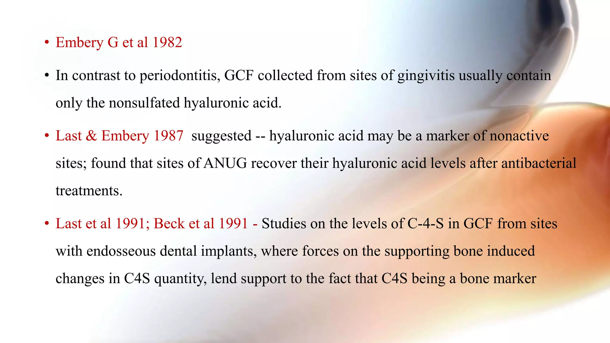
Categorizes biomarkers into host-derived enzymes and tissue breakdown products in GCF, analyzing their significance in periodontal disease.
Delineates various cytokines like IL-1, IL-6, and TNF-α, their functions in immune response, and their association with periodontal disease.
Reviews studies on cytokine profiles in GCF and their relation to periodontitis progression, highlighting variability and implications.
Details pro-inflammatory cytokines, their sources, and their association with collagen degradation markers in periodontal disease dynamics.
Discusses various biomarkers that indicate bone loss in periodontal patients, including ICTP and osteocalcin levels and their predictive values.
Review of recent studies on biomarkers for obesity and systemic diseases related to periodontal conditions, emphasizing the need for development of effective diagnostics.
Compilation of references and sources used throughout the presentation regarding biomarkers in periodontal disease.
• Periodontal Diagnosis
•Clinical diagnostic parameters
• probing depths, bleeding on probing, clinical attachment levels, plaque index, and radiographs assessing
alveolar bone level
• Require a 2- to 3-mm threshold change before a site can be identified as having experienced a significant
anatomic event. Goodson JM.
• Advances in oral and periodontal disease diagnostic research are moving toward methods whereby
periodontal risk can be identified and quantified by objective measures such as biomarkers
Chapple I. Periodontal diagnosis and treatment-where does the future lie? Periodontology 2000;
51:9-24
INTRODUCTION:
3.
• Early recognitionof the microbial challenge to the host
• To determine the presence of current disease activity, predict sites vulnerable for
future breakdown
• To assess the response to periodontal intervention
• Haffajee et al 1983
• Ultimately improve the clinical management of periodontal patients.
NEED FOR ADVANCED DIAGNOSTIC
INDICATORS
4.
• Biomarkers -“cellular,biochemical, molecular, or genetic alterations by which a normal, abnormal, or
simply biologic process can be recognized or monitored”
NIH BIOMARKER DEFINITIONS WORKING GROUP, 1998
• Also described as measures of health & disease that define the underlying biological basis to a condition
presenting as a characteristic clinical phenotype & are potentially more objective than clinical indices.
Beck et al. 2000
• A substance that is measured objectively and evaluated as an indicator of normal biologic processes,
pathogenic processes, or pharmacologic responses to a therapeutic intervention
Biomarkers Definitions Working Group.2001
DEFINITION:
SOURCES
Khiste SV, RanganathV, Nichani AS, Rajani V. Critical analysis of biomarkers in the current periodontal practice.
J Indian Soc Periodontol 2011;15:104-10
ORAL FLUIDS
Mucosal
Tramsudate
GCF Saliva
GaryC. Armitage 2004
7.
GCF SALIVA SERUM
-More than 65 GCF
constituents have
been evaluated as
potential diagnostic
markers of
periodontal disease
progression
(Armitage et al
2004 )
- Non-invasive
collection
method
- Detection of
RNA in the
salivary fluid
Zimmermann
BG
- eg: Salih E et al.
- Inflammatory
mediators are
released into
circulation
- Not the preferred
source today due to
lack of definitive
evidence
- Mainly used for
genetic risk marker
identification
- More invasive than
GCF or saliva
Biomarkers Examples
Enzymes
Alkaline phosphatise,Amino peptidase, Trypsin, â galactosidase, â
â glucoronidase, Gelatinase, Esterase, Collagenase Kininase
Immunoglobulins Ig A, Ig G, Ig M,
Protein
Cystatin, Fibronectin, Lactoferrin, Vascular endothelial growth factors, Platelet
activating factors, Epidermal growth factors
Phenotypic marker Epithelial keratin
Host cell Leukocytes (PMN’S)
Ion Calcium
Hormones Cortisol
Bacteria Aa, Pg, Pi, C. rectus, T. denticola, B. forsythus , mycoplasma
Volatile Compounds
Hydrogen sulphide, Methyl mercaptan, Picolines, Pyridines.
Possible Salivary Biomarkers
10.
• LPS-HOST DEFENSE
•MMP-1 & MMP-8,IL 1 &TNF-α
PROTEOMIC APPROACH
• IL-1 and TNFα, the anti-inflammatory cytokine IL-10
and the Fc gamma receptors
• Yoshie et al
• Reactive O2 species; 8-OHdG
GENOMIC APPROACH
• Bacteria present due to plaque, tongue, GCF, Pdl
pocket..
MICROBIAL APPROACH
DISCOVERY & AN OVERVIEW
11.
Alkaline Phosphatase (ALP):
•Mixed saliva of adult periodontitis
patients revealed the highest enzyme
activities with ALP
• Associated with alveolar bone loss, a
key feature of periodontal disease.
Interleukin (IL) 1β:
• Proinflammatory cytokine
• Functions:
- Osteoclastic activity in periodontitis
• Influences:
- Immune cell recruitment
- Cell proliferation
- Tissue destruction
- Vascular smooth muscle cell contraction
INDIVIDUAL SALIVARY BIOMARKER
Acta Odontol Scand.2015 Jul;73(5):343-7
12.
• Synthesised inLiver
• Systemic marker and indicator of acute phase of an inflammatory response.
• Circulating CRP reaches saliva via GCF or salivary glands.
• Christodoulides N et al reported that high levels CRP are associated with chronic
and aggressive periodontal diseases.
C-reactive protein (CRP) :
Journal of Indian Society of Periodontology. Jan-Feb 2013:17(1):36-41
13.
8-OHdG
• Most stableproduct of ROS
• Marker of mitochondrial DNA
damage caused by premature
oxidation in the gingival tissues of
periodontitis patients
{Canakci CF et al}
IL-17
• Proinflammatory cytokine produced by
T-helper 17 cells
• Stimulate various cell types to produce
other inflammatory cytokines and
chemokines
• Preserves immune homeostasis
• Supports immune responses (Th1) and
combines with receptor activator of
RANK and RANKL, resulting in
osteoclastic bone resorption
Yang et al
J Periodontol.2015
14.
• Key enzymein extracellular collagen matrix degradation,
• Origin: PMNs during acute stages of periodontal disease
• Significantly increased the risk of periodontal disease
• MMP-8 is not only an indicator of disease severity, but also disease activity.
• MMP-1 (interstitial collagenase) also appeared to be activated in periodontitis.
MATRIX METALLOPROTEINASES
15.
• Possible markersfor the progression of periodontitis fall into three general
categories:
1. Host-derived enzymes and their inhibitors
2. Tissue breakdown products
3. Inflammatory mediators and host response modifiers
GCF BIOMARKERS
Biomarker Type Function
Interferonalpha Cytokine Antiviral
Increased MHC Cl-I expression
Interferon gamma Cytokine Macrophage Activation,Th2
suppression
IgA Antibody Antigen Neutralization
IgG, IgG1,IgG2, G3,G4, IgM Antibody Antigen Neutralization
IL-1 ra (Receptor Antagonist) Cytokine Antagonist of IL-1
IL-1 Cytokine Regulates immune and
inflammatory reactions,
stimulates bone resorption
IL-4 Cytokine Antiinflammatory, macropge
inhibitor,Th2 differentiation
18.
Pro inflammatory Cytokines
•Monocytes, macrophages, fibroblasts and endothelial cells respond to plaque
microorganisms by secreting chemokines and inflammatory cytokines ( TNF-α,
PGE2, IL-1β and IL-6)
(Beck et al 1998)
• Can be obtained from GCF and saliva (IL1, TNF-α)
19.
TNF -α
• Origin:Mononuclear phagocyte.
• The main stimulus for release is the Lipopolysaccharide of bacterial cell walls.
• Functions:
Bone resorption
Inhibit bone collagen synthesis.
Induce collagenases
Stimulate osteoclast differentiation in the presence of M-CSF
• Act synergistically with cytokines and induces release of IL-1.
20.
TNF – β(Lymphotoxin)
• 25 KD glycoprotein
• Activated T cells
• 28% homology to TNF-
• Functions:
CTL stimulation
Osteoclast activation of PMNLs and
Antiviral activity.
• 17 KD
• Origin: Stimulated macrophages
• (+) cytoxic T lymphocytes (CTL).
• ↑ in periodontitis
Gorska et al
• ↑ bone resorption and CT
degradation by (+) PGE2 and
Collagenase
Morimoto Y et al,2008
• Shapira et al 2001, Ejeil AL et al 2003
TNF - à
21.
INTERLEUKINS
IL-1
Stimulate adhesion
molecule and
chemokine
expression
Enhance
Osteoclast
formationand
activity
Induce matrix
metalloproteinase
expression
Stimulate
production of
inflammatory
mediators(PGE
Stimulate apoptosis
of matrix producing
cells
Inflammation Bone loss
Connective
Tissue
Breakdown
Limit repair of
periodontium
Mechanism by which IL-1 could contribute to the net loss of periodontal tissues
22.
IL-6
• Produced byvarious cells such as activated monocytes or macrophages, endothelial
cells, activated T-cells, and fibroblasts.
• Earlier Names: B- cells stimulatory factor II, interferon B2 and plasmacytoma growth
factor.
• Functions :
o B cells : Promote growth and facilitate maturation of the B cells causing immunoglobulin
secretion.
o Osteoclast formation and activity
o ↑ in sites of gingival inflammation and plays a role in bone resorption.
23.
• On measuringthe level of IL-1β, IL-2, IL-4, IL-6 and TNF- α in GCF by ELISA
assays :
• Results showed high individual variability of cytokine profiles, and no association
between cytokine concentrations and clinical parameters of periodontitis
Gorska et al
• Disease activity thought to be related to Th1-Th2 cytokine profile
• Th2 cytokines like IL4, IL5, IL6 associated with progressive disease
Seymour 2002
24.
• Sakai etal 2006, Yucel et al 2008, Liu et al 1996, Homlund et al 2004,
Engebretson et al.2002, Ozmeric 1998, Toker et al 2008, Tsai et al 2007 reported
increased GCF and salivary levels of proinflammatory cytokines like IL 1, IL6, IL8
in periodontal disease.
• Th1 cytokines IFN γ, IL2 associated with progressive lesion
Berglundh 2003
• In the absence of evidence, there is no single inflammatory cytokine that can be
used as a marker as yet.
25.
Biomarker Type Function
IL-6Cytokine Regulator of t and B cell
Acute phase protein
IL-8 Cytokine Recruitment and activation of
neutrophils
IL-15 Cytokine Anti-apoptotic effect on cells
Lactoferrin Acute Phase Protein Antibacterial, creates Fe limiting
environment
TNF - alpha Cytokine Delays neutrophil apoptosis
Leukotriene B4 Proinflammatory mediator Stimultes chemotaxis, adhesion,
oxidative burst, degranulation
Prostaglandin E2 Immune Mediator Multiple pro inflammatory and
immunomodulatory effects
Pradeep AR, Manjunath SG et al. J Periodontol. 2007;78:2325-30.
Kim DM et al. J Periodontol. 2007 ;78:1620-6.
Goodson et al. in 1974
26.
PGE2
• In untreatedcases ,gcf levels not able to distinguish between progressive and non
progressive sites Offenbacher-94
• Reduction in GCF levels after nonsurgical therapy Sengupta 1990
• Attachment loss at one or more sites after 6 months had significantly higher mean GCF
PGE2 levels of 113 ng/ml at the baseline.
• Levels greater than 66 ng/ml were found to be predictive of further possible loss of
attachment and this level was used as a cut off value in a positive and negative
screening test.
27.
• Membrane-derived lipidmediator formed from arachidonic acid.
• Source : PMNLs
• Tsai et al 1998, Back et al.2006, Pradeep et al 2007,Emingil 2001 reported difference
in levels in health,disease and post treatment.
• Evidence indicates that LTB4 is elevated in inflamed deeper periodontal tissues.
• Offenbacher et al. (1991)
• AR Pradeep et al
LtB4
Beta Glucuronidase
• Degradationof the connective tissue ground substance – glycoproteins, proteoglycans
• Marker for primary granule release from PMNL
• May be obtained from both saliva and GCF
• Also positively associated with spirochetes, P. gingivalis, P.intermedia and negatively
associated with cocci.
• Lamster et al 1988 showed that sites that showed the higher β -glucuronidase activity
at baseline and again at 3 months had the highest association with loss of attachment.
• Lamster et al 1988, Harper et al 1989, Wolff et al, 1997, Lamster and Novak 2002
30.
Neutrophil elastase
• Serineendopeptidase found in primary granules
• Marker of intracrevicular PMN activity
• Degrades collagenous & non collagenous substrates, activates collagenases.
• Seen either adjacent to junctional epithelium or in granulation tissue at the
advancing front of the lesion.
• Anti bacterial activity
31.
• Palcanis etal 1992 did a 6-month longitudinal study using a test kit system, and showed
significant differences of total elastase activity at baseline in progressive and non-
progressive sites assessed 2-6 months later.
• Eley and Cox 2006 did a longitudinal study for 2 years and found levels above critical
values for total elastase activity and enzyme concentration present at all Rapid
Attachment Loss sites, both at the time of attachment loss and 3 months previously
(predictive time).
• Eley &Cox (1996) compared the GCF contents of cathepsin B to probing
depths to determine the success of cathepsin B as a marker for active disease.
• 75 patients with moderate chronic periodontitis were tested; with a high degree of specificity and
sensitivity (99.8% and 100%, respectively)
• Cathepsin B was able to better identify active pocket destruction than other markers in GCF..
• Kunimatsu et al 1990, Eley & Cox (1996), Chen et al 1998 reported its ability to
distinguish Progressive & Non progressive disease and aid in Prognosis/Therapy
• Loos et al 2005
34.
Matrix Metallo Proteinases
•Family of homologous zinc endopeptidases, that collectively cleave most if not all
constituents of ECM
• Key mediators of tissue destruction (Kinane et al 2000)
• Source: Macrophages, neutrophils, fibroblasts and keratinocyte
Gingival fibroblast-MMP8
Monocytes and macrophges:MMP9
Gingival fibroblast , epithelial cells:MMP2
35.
MMP-9
• Degrades collagenextracellular
ground substance
• Teng et al : 2x ↑ in MMP-9 with
recurrent attachment loss
• Metronidazole : ↓ MMP-9
• Most prevalent MMP found in diseased
periodontal tissue and GCF
• Mancini and co-workers: 18x ↑ MMP-8 in
patients experiencing active periodontal
tissue breakdown
• ↑ in disease and ↓ by 60% during the 2-
month protocol of LDD
Golub et al
• ↑ in PISF from periimplantitis lesion
• Collagen –I & II breakdown
MMP-8 (Collagenase 2)
Matrix Metallo Proteinases
36.
Aspartate Amino transferase(AST)
Source :Epithelial cells, gingival and PDL fibroblasts
Evidence of cell death within the periodontal tissues and, hence, possibly disease
activity.
• Chambers et al.1988, Chambers et al. (1991), Smith et al 1998, Persson et al 1992,
Oringer 2001 reported greater AST activity in periodontal disease
• Chair side Test
Pocketwatch
Perioguard
Pyridinoline cross links(ICTP) levels
• Class of collagen degradation molecules that include pyridinoline, deoxy pyridinoline,
N telopeptide and C telopeptide.
• 12 to 20 kd fragment of bone type I collagen released by digestion with trypsin or
bacterial collagenase
• Specific biomarkers for bone resorption
Eriksen et al
39.
• Palys etal. related ICTP levels to the subgingival microflora of various disease states
on GCF.
• Levels differed significantly between health, gingivitis, and periodontitis subjects
• Related modestly to several clinical disease parameters.
• Strongly correlated with whole subject levels of several periodontal pathogens including T.
forsythensis, P. gingivalis, P. intermedia, and T. denticola.
• Golub et al.
• Elevated GCF ICTP levels at baseline, especially at shallow sites, were found to be
predictive for future attachment loss as early as 1 month after sampling.
40.
Osteocalcin
• Most abundantnon-collagenous protein of mineralized tissues.
• Source: osteoblasts. It is a small calcium-binding protein of bone.
• Function:
promotes hydroxyapatite binding and accumulation of bone.
chemotactically attracts osteoclast progenitor cells and blood monocytes.
41.
• Kunimatsu etal 1993
GCF osteocalcin has positive correlation with clinical parameters in a cross-sectional
study of patients with periodontitis.
also reported that osteocalcin could not be detected in patients with gingivitis
• In contrast, Nakashima et al 1994
• GCF osteocalcin levels higher in both periodontitis and gingivitis patients.
• Contradicting results osteocalcin has a potential role as a bone specific marker of
bone turnover but not as a predictive indicator for periodontal disease.
42.
OSTEONECTIN AND BONEPHOSPHOPROTEIN
(N-PROPEPTIDE)
• Imp in initial phase of mineralization.
• Bone phosphoprotein an amino propeptide extension of alpha 1 chains of type I
collagen, appears to be involved in the attachment of connective tissue cells to the
substratum.
Bowers et al 1989
• Both detected in GCF from CP pts.
• Total amount increases with PD
Bowers et al 1989
• They therefore may be associated with periodontal disease severity.
43.
Osteopontin
• Found inbone matrix
• Highly concentrated at sites where osteoclasts are attached to the underlying
mineral surface
• Source: Both osteoblasts and osteoclasts,
• Function-
-it holds a dual function in bone maturation
44.
• GCF OPN≈ PD measures of periodontally healthy and diseased patients.
Kido et al 2001
• GCF OPN concentrations ≈ progression of disease; on nonsurgical pdl
treatment , levels were significantly reduced.
Sharma et al 2006
45.
GAGs
• Most commonGAG : nonsulfated hyaluronic acid, sulfated heparan sulfate,
chondroitin-4 sulfate and chondroitin-6 sulfate.
• chondroitin-4-sulfate : Most common GAG in periodontium but
distributions differ.
• Dermatan sulfate : rare in bone, cementum; common in pdl and gingiva.
probably reflecting a functional involvement of the molecule in the
mineralization process.
• Function: Proteoglycans bind most collagens as well as fibronectin. On
degradation of pdl tissues, GAGs are released GCF.
46.
• Embery etal 1982 : The non-sulphated GAG, hyaluronic acid was present in all
samples, and was the only major GAG found in chronic gingivitis patients.
• Sulphated GAG, c-4-sulphate, in GCF from sites with
untreated advanced periodontitis,
JP
around teeth undergoing orthodontic movement,
teeth subject to occlusal trauma
• The presence of c-4-sulphate in GCF may be a sensitive method of indicating active
phases of destructive periodontal disease.
47.
• Embery Get al 1982
• In contrast to periodontitis, GCF collected from sites of gingivitis usually contain
only the nonsulfated hyaluronic acid.
• Last & Embery 1987 suggested -- hyaluronic acid may be a marker of nonactive
sites; found that sites of ANUG recover their hyaluronic acid levels after antibacterial
treatments.
• Last et al 1991; Beck et al 1991 - Studies on the levels of C-4-S in GCF from sites
with endosseous dental implants, where forces on the supporting bone induced
changes in C4S quantity, lend support to the fact that C4S being a bone marker
48.
Rankl and OPG
•Regulation of osteoclastogenesis in bone remodeling and inflammatory osteolysis.
• Lacey et al 1998 : In vivo treatment of mice with RANKL activates osteoclasts
bone loss
• Osteoprotegerin (OPG) : a secreted glycoprotein, is a decoy receptor for RANKL
• OPG binds to RANKL the cell-to-cell signaling between marrow stromal cells and
osteoclast precursors is inhibited osteoclasts are not formed
Simonet et al 1997; Yasuda et al 1998
49.
• Thus, RANKLand decoy receptors OPG expressed by bone-associated cells play
important roles during osteoclast formation by balancing induction and inhibition
• GCF RANK-L increased in CP patients, supporting its role in the alveolar
bone loss developed in the disease.
Rolando Verna et al 2004
50.
• RANKL levels: low in health and gingivitis groups; increased in CP.
• OPG : higher in health compared to gingivitis and periodontitis.
• There were no differences in RANKL and OPG levels between CP and GAP groups
Bostanci N et al 2007
51.
• The developmentof rapid point-of-care (POC) chairside diagnostics has the
potential for the early detection of periodontal infection and progression to
identify incipient disease and reduce health care costs. However, validation of
effective diagnostics requires the identification and verification of biomarkers
correlated with disease progression (Ramseier CA et al., 2009).
Chairside kit
• OBESITY RELATED
•Chemerin - an adipokine ; CP & DM Pradeep et al,2015
• Leptin - The decreasing leptin level in GCF and gingival tissue was associated with a deteriorated
periodontal status, and smokers also showed reduced GCF leptin levels in recent studies
• SYSTEMIC DISEASE RELATED
• Progranulin – CP & type 2 DM. Pradeep et al 2013
Capsase 3 - GCF and the serum concentration of caspase-3 proportionally increases with the progression of
periodontal disease Pradeep et al 2014
• IL-29 - antiviral IL-29 level was highest in GCF of aggressive periodontitis patients while that of chronic
periodontitis lying in between. After non-surgical periodontal therapy, IL-29 levels increased both in chronic and
aggressive periodontitis patients a potential therapeutic agent in treating periodontitis.
Shivaprasad BM ,Pradeep AR 2013
RECENT BIOMARKERS
56.
• Though severalproducts show potential benefit; which gives a clue as to which tissue
components are at risk, most of the test kit, or biomarkers yield little or no additional
information, at high costing.
• It is also clear that no single marker has been able to fulfil all the criteria necessary for
assessment of the clinical state of the periodontium.
• Future research should be directed possibly at the production of "marker packages"
• As of now various efforts are on to develop an ideal test, but actual use as a chairside
diagnostic is still illusive. Therefore the development of a wide spectrum of markers is the
primary goal of periodontal research
CONCLUSION:
57.
1.Periodontology 2000 vol.39,50,51,70
2.KolokythasA et al. Salivary biomarkers associated with bone deterioration in
patients with medication-related osteonecrosis of the jaws. J Oral Maxillofac
Surg : 2015.
3.Christodoulides N et-al. Lab-on-a-chip methods for point-of-care measurements
of salivary biomarkers of periodontitis. Ann. N.Y. Acad. Sci.2007: 1098: 411–428 .
4.Carranza’s CLINICAL PERIODONTOLOGY .11th Edition
5.Pradeep A.R et-al. Correlation of human S100A12 (EN-RAGE) and high-
sensitivity C-reactive protein as gingival crevicular fluid and serum markers of
inflammation in chronic periodontitis and type 2 diabetes. Inflamm. Res. (2014)
63:317–323.
6.Gingival crevicular fluid and plasma levels of neuropeptide Substance-P in
periodontal health, disease and after nonsurgical therapy. Pradeep AR, Raj
S, Aruna G, Chowdhry S. J Periodontal Res. 2009;44:232-7.
REFERENCES: