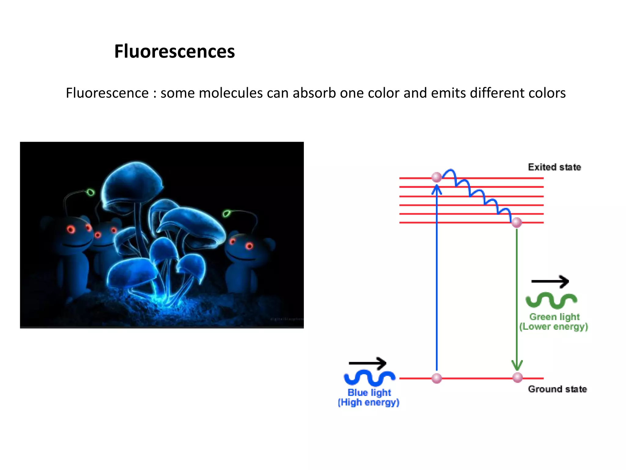
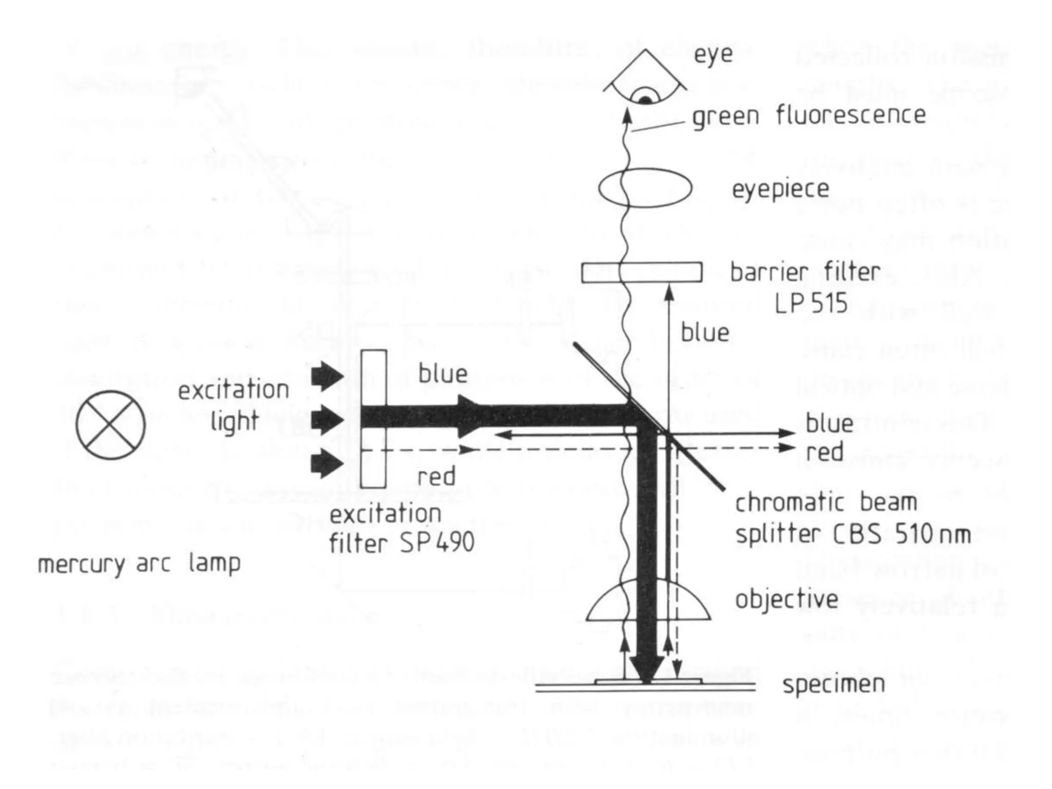
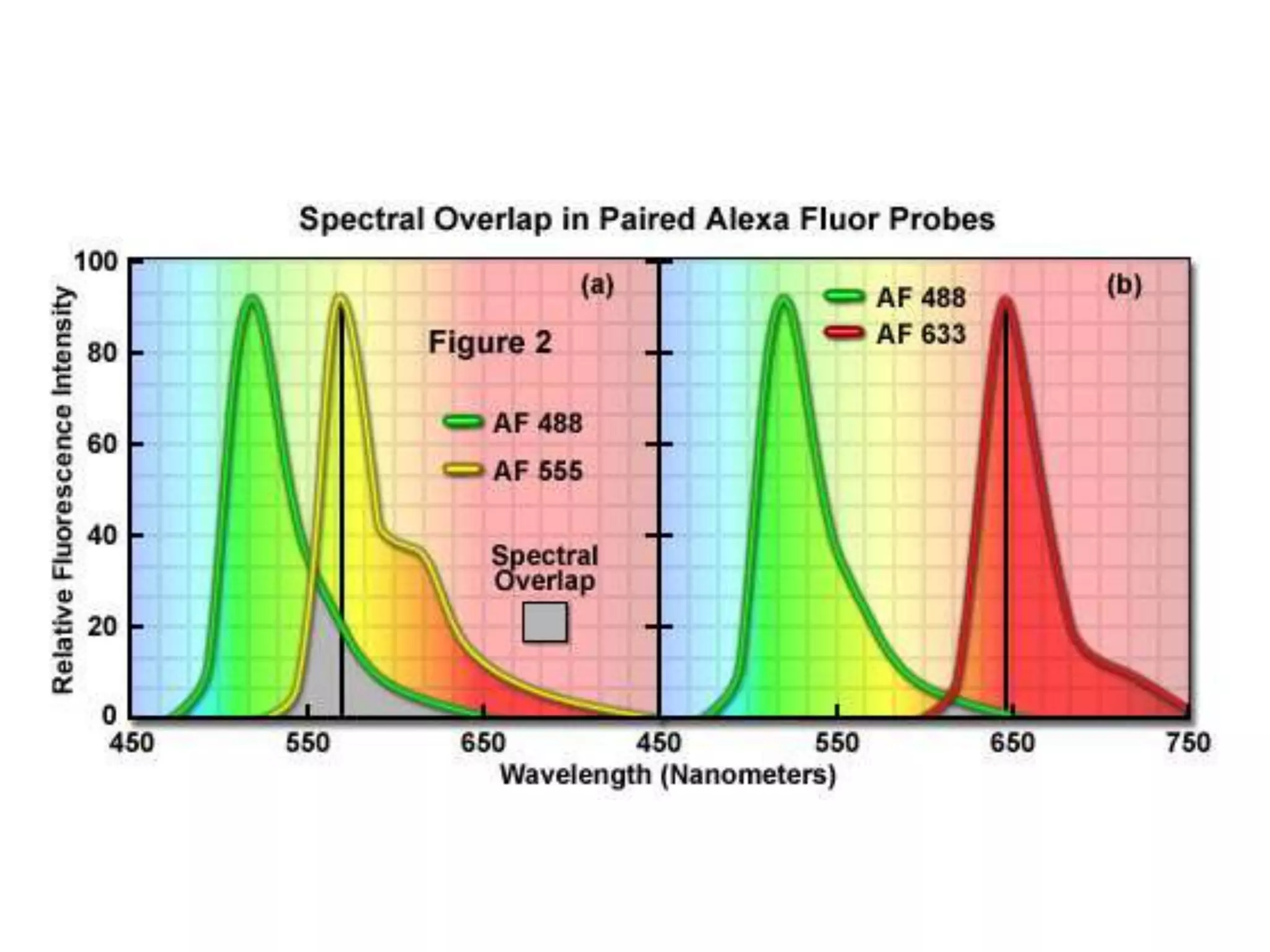
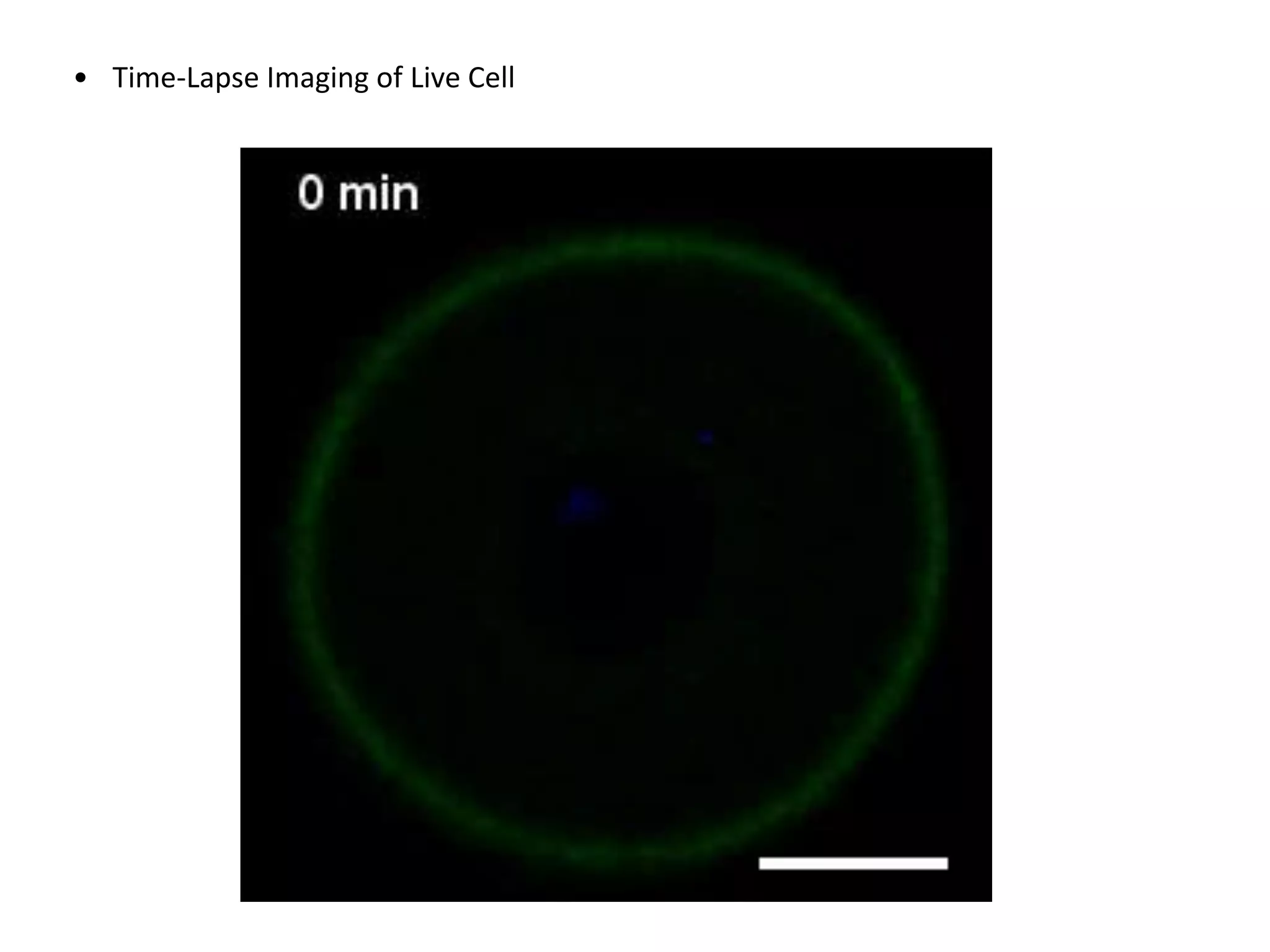
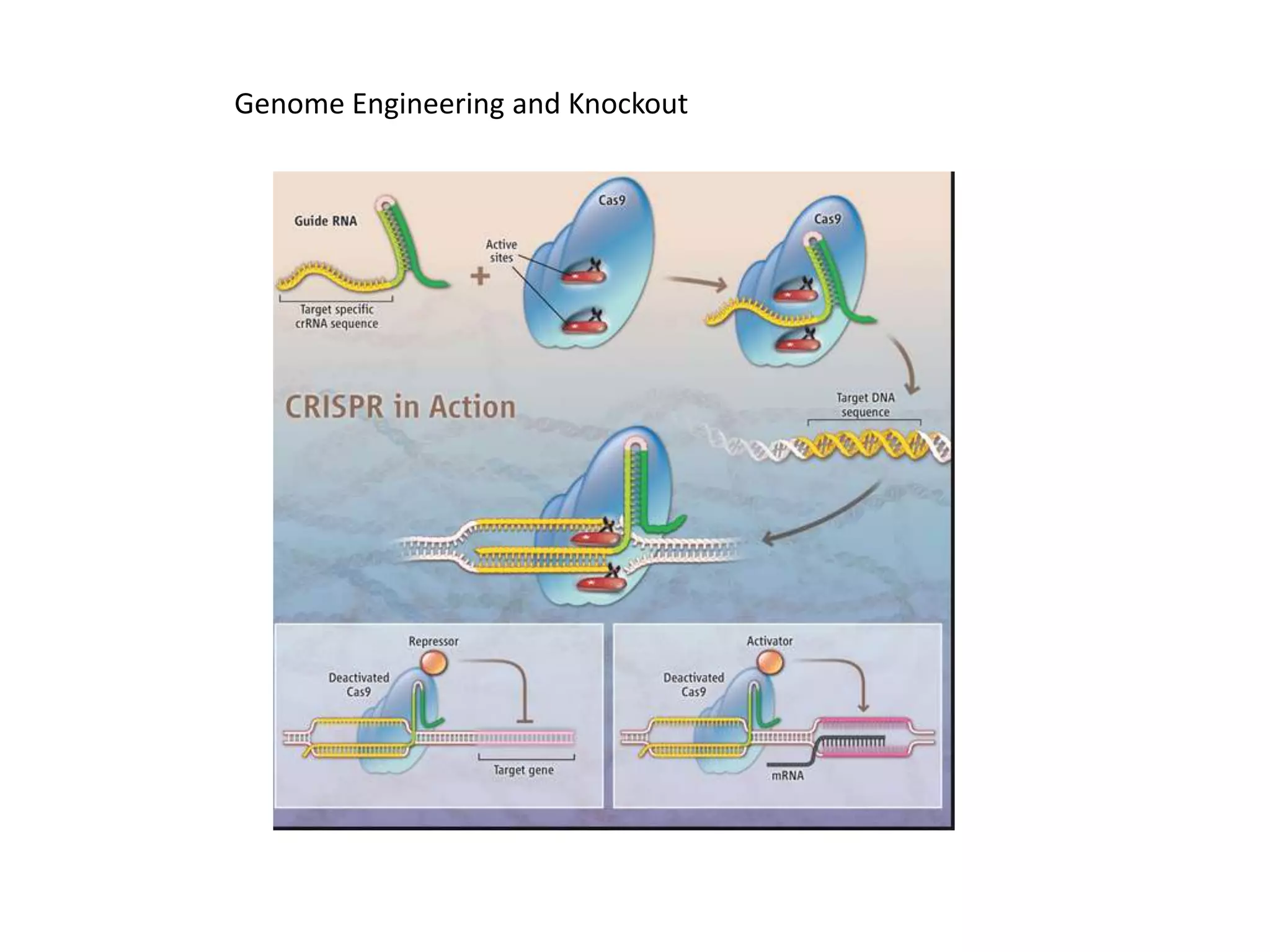
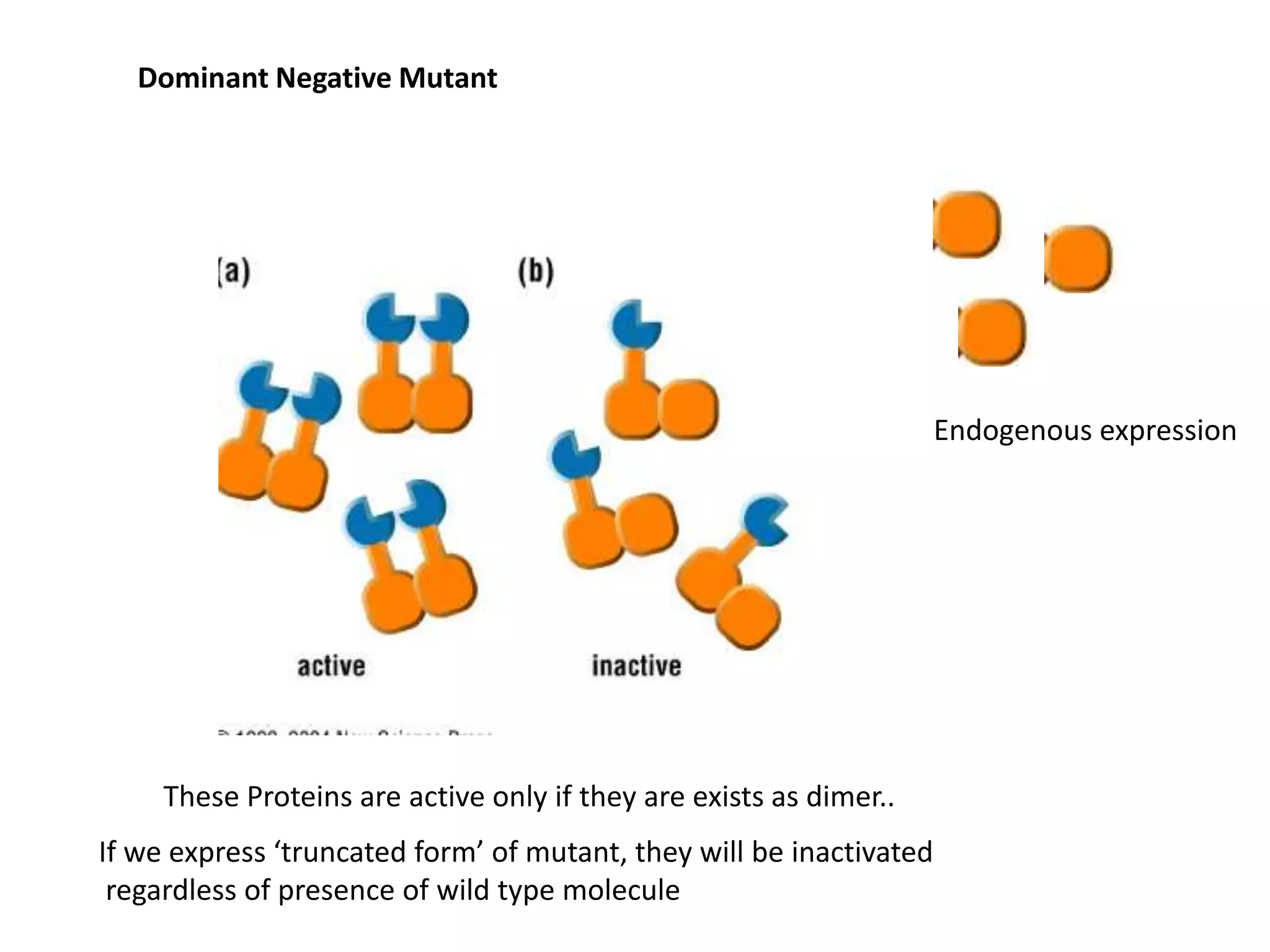
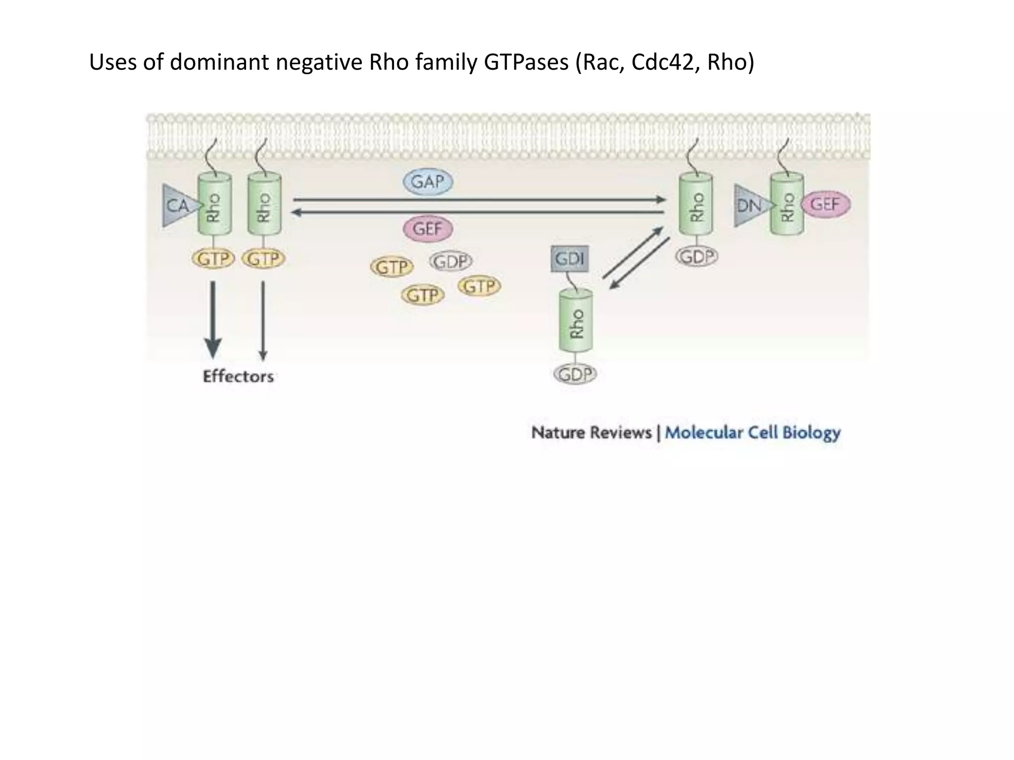
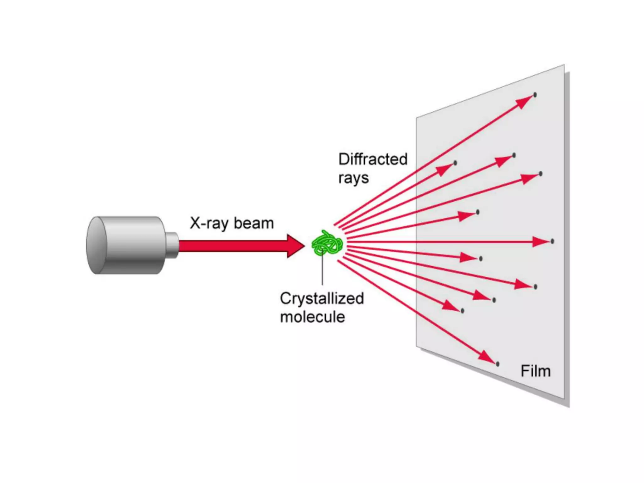
Advanced Cell Biology
2014 1nd Semester
Department of Animal Science
Chungbuk National University
2ndt Lecture
1st week : Introduction
3rd week :Research Strategies For Cell Biology
5nd week : Nucleus, Transcription and Splicing
7nd week : Membrane and Channel
9nd week : Membrane Trafficking
11nd week : Cell Signaling
13nd week : Cytoskeleton
15nd week : Cell Cycle
Cell and complicated Machine : The ways to study Cells
Component List
What kinds of component are in the machine (or cell?)
But How about the cell?
“Component” in the Cell : Protein (RNA)
Genome Sequence : Now we have most of gene list in the genome
But it does not means that we know the exact component list in a specific cells…
http://www.ncbi.nlm.nih.gov
http://genome.ucsc.edu/
Differences cells have different component (Proteins, RNA),
although they have common genome
How we can figure out the whole component list?
Expression profiling using Microarray or RNA-Seq
“What Kinds of mRNA is there?”
“How much specific RNA is there?”
RT-qPCR or Northern Blot
mRNA levels does not necessarily correlated with Protein Levels.
Nature, 2012
Proteomics
~less than 1,000 abundant Proteins
- “We want to know where the specific component is located inside in the cell”
- “We want to know whether two protein is interact each other in cell”
Immunofluorescence / Flurorescence reporter fusion
Colocalization / Immunoprecipation
- “We want to see what will happen if the specific protein / RNA was devoided in cell”
RNAi
Knockout (CRISPR/Cas9)
Ectopic Expression of Dominant Negative Mutant
Now let’s assume that we have a list of component inside the cell. Now what?
- “We want to check how the shape of the protein looks like”
X-ray Crystallography
Immunoprecipitation
Optical Microscope : Main workhorse of Cell
Research
Phase-Contrast Microscope
Contrast-enhancing techniques
Differential-Interference-Contrast Microscope
Fluorescences
Fluorescence : some molecules can absorb one color and emits different colors
Fluorescences Microscope
Filters: the key to successful
fluorescence microscopy
Staining of different components of the cell
Q : We want to localize the location of a specific protein in cell. How we can do
that?
A : Use Antibody!
By labeling antibody with fluorescence, you can locate the desired protein in
Alpha-Tubulin Actin Mitochondria
Synaptic Vesicle
Immunofluorescence
- Direct Immunofluoresence
* Antibody (or chemical) which can bind a desired protein is labeled with
fluorochrome
* Pros
- Convienient
- More Sensitive
* Cons
- You should have a primary antibody labeled
with fluorochrome
- If you don’t have it, you should do it by yourself or
use Indirect Immunofluoresence
Alexa-Phalloidin
Phalloidin : Actin binding Chemical
- Indirect Immunofluoresence
* Unlabeled Antibody is applied on the fixed tissue
* Antibody was detected by secondary antibody conjugated with fluorochrome
Primary Antibody
Recognize Antigen
Secondary Antibody recognize
Primary Antibody
It is labeled by fluorescence
Pros
• You don’t need to label primary antibody
• Based on the selection of secondary antibody, you can change wavelength of signal
Cons
* More complicated (Two step process)
Fixation and Section
We need to stop the cellular process and preserve the component inside in
cell.
Crosslinking Fixation
Commonly used for luoresence microscopy
Generate covalent cross-links between intracellular components
Most commonly used agent : aldehyde
Formation of bond between amine grouop
Glutaraldehyde
formaldehyde
- Precipitating Fixatives :
Disctrupt hydrophobic interaction Denature proteins
Methanol, Ethanol, Acetic Acid
Colocalization
Using two different fluorophore with different wavelength, we can test cellular locations of
Two protein simultaneously.
A B
Choice of fluorophore
* Choice of two closely distributed spectrum may cause bleeding
Fluorescence Protein as Reporter
GFP Gene of Interest
- Drawbacks of immunoflorescences
• You need to have (specific and high-quality) antibody against your protein of interest
• You need to fix a cell (i.e. Dead Cells), so you cannot observe live event in live cell
- You need a probe which will work in In the Living cell (and even organism)
• GFP(RFP) – Your Gene of Interest
• Transfection
• Time-Lapse Imaging of Live Cell
Confocal Microscopy
• The main problem in the florescence microscopy is that strong
illumination background from other focal planes
Biochemical Characterization of Cell
Pertubation of Component
• Loss-of-function Study
- Knockdown of functions for GOI (Gene of Interest)
- Find a Phenotype caused by the Ablation of Gene Function
- Find a function of Gene/Protein
- Can be classified as
- RNAi
- Morpholino
- Dominant Negative Mutant
• Gain-In-Function Study
- Introduction / overexpression of GOI
- Find a Phenotype caused by the (over)expression of Gene
RNAi (RNA interferences)
• Temporal knockdown of desired gene
• Loss-of function Study
Transfection
Genome Engineering and Knockout
Dominant Negative Mutant
These Proteins are active only if they are exists as dimer..
If we express ‘truncated form’ of mutant, they will be inactivated
regardless of presence of wild type molecule
Endogenous expression
Uses of dominant negative Rho family GTPases (Rac, Cdc42, Rho)
X-ray Crystallography
Protein Productions
- You need to have enough (5-10mg) pure (at least 95% purity) protein
- Overexpression (Bacteria or Insect Cell or Mammalian Cell) or Natural Source
- Purification
Crystallization
- Concentrate Proteins (at least 5mg/ml)
- Crystallization happens in the boundary of soluble and precipitation
Strong X-ray generated from synchroton is essential
Raw Data : Diffraction Images of Protein Crystal
(several hundreds)
Computer
Analysis
Final Structure and Interpretations
Do I need to know how to solve protein structure?
- Not really (In most cases..)
Do I need to know how to check my favorite protein?
- Yes for sure.
http://www.rcsb.org
OK. Let’s check some random guy’s structure 
In old days, you need very expensive workstation-level computer
To visualize Protein Structure..
Not anymore. Cheap PC or even your smartphone can do that.
Pymol (http://www.pymol.org)
Demo

Cell Biology Lecture #2

  • 1.
    Advanced Cell Biology 20141nd Semester Department of Animal Science Chungbuk National University 2ndt Lecture
  • 2.
    1st week :Introduction 3rd week :Research Strategies For Cell Biology 5nd week : Nucleus, Transcription and Splicing 7nd week : Membrane and Channel 9nd week : Membrane Trafficking 11nd week : Cell Signaling 13nd week : Cytoskeleton 15nd week : Cell Cycle
  • 3.
    Cell and complicatedMachine : The ways to study Cells
  • 4.
    Component List What kindsof component are in the machine (or cell?) But How about the cell?
  • 5.
    “Component” in theCell : Protein (RNA) Genome Sequence : Now we have most of gene list in the genome But it does not means that we know the exact component list in a specific cells… http://www.ncbi.nlm.nih.gov http://genome.ucsc.edu/
  • 6.
    Differences cells havedifferent component (Proteins, RNA), although they have common genome How we can figure out the whole component list?
  • 7.
    Expression profiling usingMicroarray or RNA-Seq “What Kinds of mRNA is there?” “How much specific RNA is there?” RT-qPCR or Northern Blot mRNA levels does not necessarily correlated with Protein Levels.
  • 8.
  • 9.
    Proteomics ~less than 1,000abundant Proteins
  • 11.
    - “We wantto know where the specific component is located inside in the cell” - “We want to know whether two protein is interact each other in cell” Immunofluorescence / Flurorescence reporter fusion Colocalization / Immunoprecipation - “We want to see what will happen if the specific protein / RNA was devoided in cell” RNAi Knockout (CRISPR/Cas9) Ectopic Expression of Dominant Negative Mutant Now let’s assume that we have a list of component inside the cell. Now what? - “We want to check how the shape of the protein looks like” X-ray Crystallography
  • 12.
  • 14.
    Optical Microscope :Main workhorse of Cell Research
  • 15.
  • 17.
  • 19.
    Fluorescences Fluorescence : somemolecules can absorb one color and emits different colors
  • 20.
  • 22.
    Filters: the keyto successful fluorescence microscopy
  • 25.
    Staining of differentcomponents of the cell Q : We want to localize the location of a specific protein in cell. How we can do that? A : Use Antibody! By labeling antibody with fluorescence, you can locate the desired protein in
  • 26.
  • 27.
    Immunofluorescence - Direct Immunofluoresence *Antibody (or chemical) which can bind a desired protein is labeled with fluorochrome * Pros - Convienient - More Sensitive * Cons - You should have a primary antibody labeled with fluorochrome - If you don’t have it, you should do it by yourself or use Indirect Immunofluoresence
  • 28.
  • 29.
    - Indirect Immunofluoresence *Unlabeled Antibody is applied on the fixed tissue * Antibody was detected by secondary antibody conjugated with fluorochrome Primary Antibody Recognize Antigen Secondary Antibody recognize Primary Antibody It is labeled by fluorescence Pros • You don’t need to label primary antibody • Based on the selection of secondary antibody, you can change wavelength of signal Cons * More complicated (Two step process)
  • 30.
    Fixation and Section Weneed to stop the cellular process and preserve the component inside in cell. Crosslinking Fixation Commonly used for luoresence microscopy Generate covalent cross-links between intracellular components Most commonly used agent : aldehyde Formation of bond between amine grouop Glutaraldehyde formaldehyde - Precipitating Fixatives : Disctrupt hydrophobic interaction Denature proteins Methanol, Ethanol, Acetic Acid
  • 31.
    Colocalization Using two differentfluorophore with different wavelength, we can test cellular locations of Two protein simultaneously. A B
  • 32.
    Choice of fluorophore *Choice of two closely distributed spectrum may cause bleeding
  • 34.
    Fluorescence Protein asReporter GFP Gene of Interest - Drawbacks of immunoflorescences • You need to have (specific and high-quality) antibody against your protein of interest • You need to fix a cell (i.e. Dead Cells), so you cannot observe live event in live cell - You need a probe which will work in In the Living cell (and even organism) • GFP(RFP) – Your Gene of Interest • Transfection
  • 35.
  • 36.
    Confocal Microscopy • Themain problem in the florescence microscopy is that strong illumination background from other focal planes
  • 38.
  • 40.
    Pertubation of Component •Loss-of-function Study - Knockdown of functions for GOI (Gene of Interest) - Find a Phenotype caused by the Ablation of Gene Function - Find a function of Gene/Protein - Can be classified as - RNAi - Morpholino - Dominant Negative Mutant • Gain-In-Function Study - Introduction / overexpression of GOI - Find a Phenotype caused by the (over)expression of Gene
  • 41.
    RNAi (RNA interferences) •Temporal knockdown of desired gene • Loss-of function Study
  • 42.
  • 44.
  • 46.
    Dominant Negative Mutant TheseProteins are active only if they are exists as dimer.. If we express ‘truncated form’ of mutant, they will be inactivated regardless of presence of wild type molecule Endogenous expression
  • 47.
    Uses of dominantnegative Rho family GTPases (Rac, Cdc42, Rho)
  • 48.
  • 49.
    Protein Productions - Youneed to have enough (5-10mg) pure (at least 95% purity) protein - Overexpression (Bacteria or Insect Cell or Mammalian Cell) or Natural Source - Purification
  • 50.
    Crystallization - Concentrate Proteins(at least 5mg/ml) - Crystallization happens in the boundary of soluble and precipitation
  • 53.
    Strong X-ray generatedfrom synchroton is essential
  • 57.
    Raw Data :Diffraction Images of Protein Crystal (several hundreds) Computer Analysis
  • 58.
    Final Structure andInterpretations
  • 59.
    Do I needto know how to solve protein structure? - Not really (In most cases..) Do I need to know how to check my favorite protein? - Yes for sure. http://www.rcsb.org
  • 60.
    OK. Let’s checksome random guy’s structure 
  • 61.
    In old days,you need very expensive workstation-level computer To visualize Protein Structure.. Not anymore. Cheap PC or even your smartphone can do that.
  • 62.