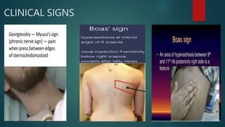
CHOLECYSTITIS
SATURDAY SURGICAL UNIT
PRESENTATION BY DR.PHANEENDRA
HOU-DR.RAM SIR
AP-DR.DMP SIR
 Inflammation of gallbladder
 Most commonly associated with gallstones
CAUSES
 Gallstones
 Tumours
 Chemical irritants
 Bile duct block due to kinking or scarring
 Infections- viral or bacterial
 Decreased blood flow to the GB due any
severe illness
TYPES
ACALCULUS
 Without evidence of gallstone
or cystic duct obstuction
 Cholesterosis (strawberry GB)
 Cholesterol
polyposis(pseudopolyps)
 Cholecystitis glandularis
proliferans
 GB diverticulosis
 Bacterial invasion
 Pancreatic reflux
 Typhoid of GB
CALCULUS
 With evidence of
gallstones 80% are of
cholesterol type.
 Acalculous cholecystitis is common in chronically icu admitted
patients
 Conditions and management leading to cholestasis
 Prolonged NBM
 Prolonged fasting
 Patients on prolonged TPN
Cholesterol laden macrophages aggregation in
mucosa (strawberry gall bladder)
Cholesterol polyps of GB
Emphysematous cholecystitis
Emphysematous cholecystitis(clostridial cholecystitis)
Acute infection caused by gas forming
Surgical emergency
Cholecystitis glandularis proliferans
 Circumscribed diffuse thickening of wall of chronically inflamed GB
 Excessive proliferation and invasion of wall by epithelium
 Formation of crypts(Rokitansky aschoff sinuses) which dilate to form cysts may
be referred as adenomatous polyp, cystadenoma, cholesterol cyst papilloma,
 Adenomyoma
GB Diverticulosis
Bacteria causing cholecystitis
 E-coli
 Klebsiella
 S.fecalis
 Salmonella
 Clostridia anaerobes (clostridium welchii) causes
emphysematous cholecystitis
ACUTE
 Sudden inflammation of GB causing
marked abdominal pain often with nausea
vomiting and fever
CHRONIC
 Low intensity inflammation of GB that lasts a
long time
 May be caused by repeated attacks of
cholecystitis
 Symptoms include only mild intermittent
abdominal pain or no symptoms at all
 Damage to the walls of GB leads to thickened and
scarred GB which ultimately shrink and looses the
ability to store and release bile
PATHOGENESIS
 Gallstone obstruct the bile flow
 Irritation and raised pressure of GB due to build
up of bile
 Inflammation and distension of GB causing
oedema of GB (bacterial and chemical)
 Blood flow compromise causing cholecystitis
CLINICAL MANIFESTATIONS
 Severe pain in right upper quadrant
 Spreading to right shoulder or back
 Nausea
 Vomiting
 Fever
 Jaundice and clay coloured stool may occur if
CBD gets blocked
CLINICAL SIGNS
CLINICAL SIGNS
Staging based on morphology
 Catarrhal- Involving only mucosa
 Phlegmonous- Inflamation penetrating deeper layers
 Gangrenous
 Gangrenous perforation
COMPLICATED CHOLECYSTITIS
 Empyema
 Mucocele
 Pancreatitis
 Icterus
 Hepatitis
 Cholangitis
 Peritonitis
COMPLICATIONS OF ACUTE CALCULOUS
CHOLECYSTITIS
 Perforation
 Usually in the fundus or in neck causing peritonitis
 Can cause cholecystoduodenal, cholecystointestinal or
 Cholecystobiliary fistula
 Pericholecystic abscess
 Fistula
 Gall stone ileus (Riggler’s triad)
 Gall stone pancreatitis
 Obstruction of cbd
 Empyema GB
 Ascending cholangitis
 Septicemia
 Emphysematous GB
 Gangrenous GB
TOKYO GUIDELINES
DIAGNOSIS AND EVALUATION
 USG-highlights any gallstones and may show the
condition of the GB
 BLOOD INVESTIGATIONS- high wbc count: raised crp
and esr indicates inflamation
 LFT- raised bilurubin
 Raised s-amylase
 CT SCAN- may reveal signs of cholecystitis
 Hepatobiliary iminodiacetic acid scan (HIDA)- creates
pictures of liver GB biliary tract and small intestine: The
HIDA scan should be done when usg is not
diagnostic and when there is clinical signs of acute
cholecystitis
 Per cutaneous Transhepatic Cholecystography (PTC)
 ERCP
 MRCP
USG
 Sensitive 85% specificity 95%
 Inexpensive and reliable
 What to look for in usg….?
 GallStone
 Pericholecystic fluid (exudative in acute cholecystitis)
 GB wall thickening
 Sonographic murphy’s sign
HIDA SCAN
 Non visualisation of GB on HIDA scan with in 1 hour along with
clinical signs is diagnostic of acute cholecystitis (without clinical
signs-cystic duct obstruction)
 Non visualisation of intestine points towards cbd obstruction
 Its importance lies in diagnosing acalculos cholecystitis
DIFFERENTIAL DIAGNOSIS
Common
 Acute pancreatitis
 Perforated duodenal/gastric
peptic ulcer
 Appendicitis
Rare
 Pyelonephritis
 Hepatitis
 MI
 Pneumonitis
Treatment (conservative approach)
 NBM: To rest the GB
 Ryle’s tube insertion and aspiration of gastric acid
decreases the secretion of bile and spasm of bladder may
come down
 Fat free diet
 Antibiotics to treat infection
 Analgesics
Indications for stopping conservative
management and early cholecystectomy
 Pain and tenderness dot decreasing and spreading
 Incresing gb size
 Increasing pulse
 Geriatric patient
 Another attack with in 48-72 hours of diagnosis
SURGICAL APPROACH
 ERCP WITH STONE REMOVAL/STENTING
 EMERGENCY CHOLECYSTECTOMY
 INTERVAL CHOLECYSTECTOMY LAPAROSCOPIC
SOS OPEN
 PERCUTANEOUS CHOLECYSTOSTOMY
 OPEN CHOLECYSTOSTOMY
 ENDOSCOPIC US GUIDED
CHOLECYSTODUODENOSTOMY
INDICATIONS OF EMERGENCY
CHOLECYSTECTOMY
 Empyema GB- High grade fever with chills, leucocytosis
 Signs of local complications- PeriGB collection, Sub hepatic local
collection
 Failure of medical management
 Emphysematous cholecystitis
 Perforation
 Peritonitis
 Acute pancreatitis
 Obstructive suppurative cholangitis
Indications for percutaneous drainage
 Seldom a first line mx
 Patients too ill to tolerate alternate procedures
 Poor surgical candidates
 Unexplained sepsis in terminally ill patients
 Access to or drainage of biliary tree following failed ercp
Percutaneous cholecystostomy
 Schematic representation
 Fluoroscopy image
 Relatively less complications and less painful
 Indicated in patients where even
percutaneous cholecystostomy is not
suitable
 Endoscopic ultrasound guided
cholecystoduodenostomy
THANK YOU

chole.pptx

  • 1.
    CHOLECYSTITIS SATURDAY SURGICAL UNIT PRESENTATIONBY DR.PHANEENDRA HOU-DR.RAM SIR AP-DR.DMP SIR
  • 2.
     Inflammation ofgallbladder  Most commonly associated with gallstones
  • 3.
    CAUSES  Gallstones  Tumours Chemical irritants  Bile duct block due to kinking or scarring  Infections- viral or bacterial  Decreased blood flow to the GB due any severe illness
  • 4.
    TYPES ACALCULUS  Without evidenceof gallstone or cystic duct obstuction  Cholesterosis (strawberry GB)  Cholesterol polyposis(pseudopolyps)  Cholecystitis glandularis proliferans  GB diverticulosis  Bacterial invasion  Pancreatic reflux  Typhoid of GB CALCULUS  With evidence of gallstones 80% are of cholesterol type.
  • 5.
     Acalculous cholecystitisis common in chronically icu admitted patients  Conditions and management leading to cholestasis  Prolonged NBM  Prolonged fasting  Patients on prolonged TPN
  • 6.
    Cholesterol laden macrophagesaggregation in mucosa (strawberry gall bladder) Cholesterol polyps of GB
  • 7.
    Emphysematous cholecystitis Emphysematous cholecystitis(clostridialcholecystitis) Acute infection caused by gas forming Surgical emergency
  • 8.
    Cholecystitis glandularis proliferans Circumscribed diffuse thickening of wall of chronically inflamed GB  Excessive proliferation and invasion of wall by epithelium  Formation of crypts(Rokitansky aschoff sinuses) which dilate to form cysts may be referred as adenomatous polyp, cystadenoma, cholesterol cyst papilloma,  Adenomyoma
  • 9.
  • 10.
    Bacteria causing cholecystitis E-coli  Klebsiella  S.fecalis  Salmonella  Clostridia anaerobes (clostridium welchii) causes emphysematous cholecystitis
  • 11.
    ACUTE  Sudden inflammationof GB causing marked abdominal pain often with nausea vomiting and fever
  • 12.
    CHRONIC  Low intensityinflammation of GB that lasts a long time  May be caused by repeated attacks of cholecystitis  Symptoms include only mild intermittent abdominal pain or no symptoms at all  Damage to the walls of GB leads to thickened and scarred GB which ultimately shrink and looses the ability to store and release bile
  • 13.
    PATHOGENESIS  Gallstone obstructthe bile flow  Irritation and raised pressure of GB due to build up of bile  Inflammation and distension of GB causing oedema of GB (bacterial and chemical)  Blood flow compromise causing cholecystitis
  • 14.
    CLINICAL MANIFESTATIONS  Severepain in right upper quadrant  Spreading to right shoulder or back  Nausea  Vomiting  Fever  Jaundice and clay coloured stool may occur if CBD gets blocked
  • 15.
  • 16.
  • 17.
    Staging based onmorphology  Catarrhal- Involving only mucosa  Phlegmonous- Inflamation penetrating deeper layers  Gangrenous  Gangrenous perforation
  • 18.
    COMPLICATED CHOLECYSTITIS  Empyema Mucocele  Pancreatitis  Icterus  Hepatitis  Cholangitis  Peritonitis
  • 19.
    COMPLICATIONS OF ACUTECALCULOUS CHOLECYSTITIS  Perforation  Usually in the fundus or in neck causing peritonitis  Can cause cholecystoduodenal, cholecystointestinal or  Cholecystobiliary fistula  Pericholecystic abscess  Fistula  Gall stone ileus (Riggler’s triad)  Gall stone pancreatitis  Obstruction of cbd  Empyema GB  Ascending cholangitis  Septicemia  Emphysematous GB  Gangrenous GB
  • 20.
  • 21.
    DIAGNOSIS AND EVALUATION USG-highlights any gallstones and may show the condition of the GB  BLOOD INVESTIGATIONS- high wbc count: raised crp and esr indicates inflamation  LFT- raised bilurubin  Raised s-amylase  CT SCAN- may reveal signs of cholecystitis  Hepatobiliary iminodiacetic acid scan (HIDA)- creates pictures of liver GB biliary tract and small intestine: The HIDA scan should be done when usg is not diagnostic and when there is clinical signs of acute cholecystitis
  • 22.
     Per cutaneousTranshepatic Cholecystography (PTC)  ERCP  MRCP
  • 23.
    USG  Sensitive 85%specificity 95%  Inexpensive and reliable  What to look for in usg….?  GallStone  Pericholecystic fluid (exudative in acute cholecystitis)  GB wall thickening  Sonographic murphy’s sign
  • 24.
  • 25.
     Non visualisationof GB on HIDA scan with in 1 hour along with clinical signs is diagnostic of acute cholecystitis (without clinical signs-cystic duct obstruction)  Non visualisation of intestine points towards cbd obstruction  Its importance lies in diagnosing acalculos cholecystitis
  • 26.
    DIFFERENTIAL DIAGNOSIS Common  Acutepancreatitis  Perforated duodenal/gastric peptic ulcer  Appendicitis Rare  Pyelonephritis  Hepatitis  MI  Pneumonitis
  • 27.
    Treatment (conservative approach) NBM: To rest the GB  Ryle’s tube insertion and aspiration of gastric acid decreases the secretion of bile and spasm of bladder may come down  Fat free diet  Antibiotics to treat infection  Analgesics
  • 28.
    Indications for stoppingconservative management and early cholecystectomy  Pain and tenderness dot decreasing and spreading  Incresing gb size  Increasing pulse  Geriatric patient  Another attack with in 48-72 hours of diagnosis
  • 29.
    SURGICAL APPROACH  ERCPWITH STONE REMOVAL/STENTING  EMERGENCY CHOLECYSTECTOMY  INTERVAL CHOLECYSTECTOMY LAPAROSCOPIC SOS OPEN  PERCUTANEOUS CHOLECYSTOSTOMY  OPEN CHOLECYSTOSTOMY  ENDOSCOPIC US GUIDED CHOLECYSTODUODENOSTOMY
  • 30.
    INDICATIONS OF EMERGENCY CHOLECYSTECTOMY Empyema GB- High grade fever with chills, leucocytosis  Signs of local complications- PeriGB collection, Sub hepatic local collection  Failure of medical management  Emphysematous cholecystitis  Perforation  Peritonitis  Acute pancreatitis  Obstructive suppurative cholangitis
  • 31.
    Indications for percutaneousdrainage  Seldom a first line mx  Patients too ill to tolerate alternate procedures  Poor surgical candidates  Unexplained sepsis in terminally ill patients  Access to or drainage of biliary tree following failed ercp
  • 32.
    Percutaneous cholecystostomy  Schematicrepresentation  Fluoroscopy image
  • 33.
     Relatively lesscomplications and less painful  Indicated in patients where even percutaneous cholecystostomy is not suitable  Endoscopic ultrasound guided cholecystoduodenostomy
  • 34.