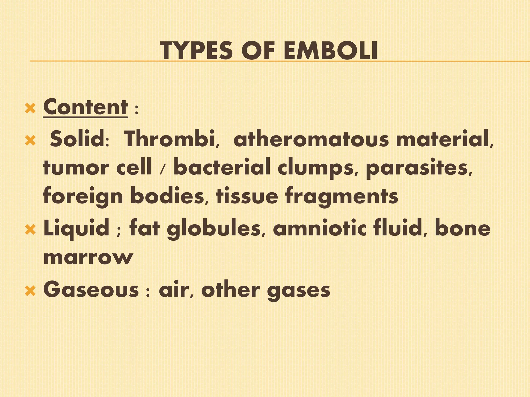
EMBOLISM
Process of obstruction of CVS by any mass carried in the circulation
Embolus:transported intravascular mass (solid liquid or gas) detached from its site of origin
Ninety percent of emboli are Thromboemboli originating from Thrombi or their parts detached from the vessel wall
TYPES OF EMBOLI
 Content :
 Solid: Thrombi, atheromatous material,
tumor cell / bacterial clumps, parasites,
foreign bodies, tissue fragments
 Liquid ; fat globules, amniotic fluid, bone
marrow
 Gaseous : air, other gases
TYPES OF EMBOLI
 Source :
 Cardiac : Left side of heart eg, from atrial
appendages, infarct in left ventricle,
vegetations of endocarditis
 Arterial : Systemic arteries in brain,
kidney spleen, small intestine
 Venous ; Pulmonary vessels
 Lymphatic emboli can also occur
 Septic ( infected) or Bland ( sterile)
 Paradoxical : from arterial to venous or
vice versa through septal defects in heart,
patent foramen ovale, AV shunts
 Retrograde : opposite to flow of blood
eg, Carcinoma metastatis to spine
THROMBOEMBOLISM
 Arterial ( systemic ) thromboembolism :
Heart – mural thrombi, vegetations ,
valves ,cardiomyopathy
 Arteries : atheromaous plaques, aortic
aneurysms, pulmonary veins
 Effects : Infarction(MI,brain,kidney,spleen)
Gangrene, Arteritis & Mycotic aneurysm (
bacterial endocarditis ), sudden death
 Venous thromboembolism
 Deep veins of lower limbs – common
 Pevic veins, upper limb veins, cavenous
sinus in brain, thrombi in right side of heart.
 Most significant effect – obstruction of
pulmonary arterial circulation leading to
sudden death, infarction of lung, Cor
Pulmonale, Pulmonary hypertension
FATAL PULMONARY EMBOLISM
PULMONARY EMBOLUS
PULMONARY THROMBOEMBOLISM
 Occlusion of pulmonary arterial tree by
thromboemboli
 95 % from large veins of lower limbs( iliac
femoral, popliteal )
 Other sources : Varicosities of superficial
veins of legs,peri prostatic,peri ovarian,
uterine, broad ligament veins
 Common in hospitalised & bedridden
 Detached thrombus from these sites go to
right side of heart
 If large – impacted at bifurcation of main
Pulmonary artery ( saddle embolus )
 Commonly multiple fragments or small
emboli to many vessels espl lower lobes
 Paradoxical – rare
PULMONARY THROMBOEMBOLISM
 Consequences :
 Sudden death
 Acute Cor Pulmonale leading to acute
right sided heart failure
 Pulmonary infarction
 Pulmonary haemorrhage
 Pulmonary hypertension, chronic cor
pulmonale
FAT EMBOLISM
 Obstruction of arterioles & capillaries by fat globules
 Traumatic : fracture long bones, post orthopaedic surgical
procedures, laceration of adipose tissue, puerperium
 Non traumatic : burns, D.M. pancreatitis, sickle cell anaemia, fatty
liver
 Mechanical ( trauma ), emulsion instability, intravascular
coagulation, toxic injury
PULMONARY FAT EMBOLISM
 Fat globules in capillaries of pts with long
bone fractures
 Lungs : hyperemia,oedema, petechial
haemorrhages & changes of ARDS
 Fat stains : Frozen sections stained with
Sudan Black, 3 & 4, Oil red O & Osmic acid
FAT EMBOLISM
FAT EMBOLISM
SYSTEMIC FAT EMBOLISM
 Fat globules pass through patent Foramen
Ovale, AV shunts in lungs & vertebral
venous plexuses.
 Capillaries of brain ,kidneys, skin
 Brain : petechiaes,microinfarcts, oedema
Delirium, convulsions, coma, death
 Kidney : < GFR, tubular damage
 Petechiae in skin,conjunctaivae, serosa
AIR EMBOLISM ( VENOUS )
 Surgeries & wounds of head/ neck Obstetric
surgeries & trauma, IV infusions
Angiography
 100 to 150 ml entry is fatal
 Entrapment of air emboli in pulmonary
arterial tree & right heart
 Autopsy : heart & pulmonary artery
opened insitu under water – escaping froth
of mixed air & blood
AIR EMBOLISM (ARTERIAL )
 Cardiothoracic surgery / trauma,
paradoxical embolism,Arteriography
 Marble skin ; blockage of skin vessels
 Pallor of tongue : occlusion of Lingual art
 Retinal vessels : air bubbles
 Death : Coronary or Cerebral arteies
AIR EMBOLISM – BRAIN
DECOMPRESSION SICKNESS
 Gas embolism : Caisson’s disease, Diver’s palsy ,
aeroembolism
 When individual decompresses suddenly from
high atmospheric pressure to normal level or from
normal to low atmospheric pressure
 Diver’s, Offshore drilling, tunnel workers:
increased Nitrogen, O2 ,CO2 is dissolved in blood
b’cos high atmospheric pressure
 When such person ascends too rapidly,
comes to normal pressure too soon –
gases bubble out which merge to form
emboli. Nitrogen – fatty tissue
 Aeroembolism ; those who ascend to high
altitudes (hot air balloon ), air flights in
unpressurised cabin – exposed to sudden
decompression
 Effects : Acute form & Chronic form
ACUTE CAISSON’S DISEASE
 Acute obstruction of small vessels of joints
& skeletal muscles
 Bends : person doubles up in bed due to
acute pain in joints, ligaments,tendons
 Chokes : accumulation in lungs causing
respiratory distress
 Cerebral effects : Vertigo, Coma, death
CHRONIC CAISSON’S DISEASE
 Ischaemic necrosis in skeletal system
 Avascular necrosis of head of femur,tibia
 Neurological : paraesthesias, paraplegia
 Lungs : haemorrhage,oedema, atelectasis
causing dysponea,cough,chest pain
 Skin ; itching,patchy erythema, cyanosis
 Lipid vacuoles in cells of Liver, pancreas
AMNIOTIC FLUID EMBOLISM
 Most serious, unpredictable and unpreventable
cause of maternal mortality
 During labour, immediate post partum period
amniotic fluid can enter uterine veins & reach right
side of heart
 Enter through tears in myometrium or endocervix
or forced into uterine sinuses by vigorous uterine
contractions
 In Pulmonary / uterine veins : squames, vernix caseosa,
lanugo hair, bile,mucus
 Lungs : haemorrhages,congestion, oedema ARDS, dilated
right heart
 Respiratory distress,dyspoena,Cyanosis, Cardiovascular
shock, coma ,death
 ? Mechanical blockage of Pulmn circln,DIC Anaphylactoid
reaction, haemorrhages due to thrombocytopenia , <
fibrinogen
AMNIOTIC FLUID EMBOLISM
ATHEROEMBOLISM
 Aortic atheromatous plaques get eroded , lodged in
medium & small arteries
 Emboli consist of cholestrol crystals, hyaline
debris,calcified material
 Ischaemia , atrophy & necrosis – distal
 Infarcts in kidneys , spleen, brain, heart
 Gangrene of lower limbs
 Hypertension if widespread renal invlvmt
SEPTIC EMBOLI IN BRAIN
TUMOR EMBOLI
INFARCTION
 Process of tissue necrosis due to some
form of circulatory insufficiency
 Infarct – the localised area of necrosis
 Interruption in arterial blood supply –
ischaemic necrosis
 Venous obstruction – stagnant hypoxia
 Sudden , complete occlusion by thrombi or
emboli
TYPES OF INFARCTS
 Colour : Pale(anaemic) –arterial occlusion
of compact organs e.g kidney,heart,
spleen Red ( haemmoragic) – soft loose
tissues lungs, intestine
 Age : Fresh ( recent ) , old ( healed )
 Infection : Bland , Septic
TYPES OF INFARCTS
PATHOGENESIS OF INFARCTION
 Localised hyperemia– haemorrhage / oedema –
cellular changes – necrosis (12 – 48 hrs) –
proteolysis of necrotic tissue – inflammatory
reaction – lysis of rbcs – deposition of blood
pigments – ingrowth of granulation tissue –
fibrous scar
 Gross – solid organs: wedge shaped, apex
pointing to occluded artery, wide base on the
surface of the organ
 Infarcts of arterial occlusion : pale
 Infarcts of venous occlusion : haemorragic
 Pulmonary infarcts never become pale
 Cerebral infarcts poorly defined with central softening
 Recent : elevated over surface old : depressed &
shrunken
 Microscopy : coagulative necrosis except brain –
liquefactive necrosis
 Some amount of haemorrhage +
 Inflammatory reaction at periphery, initially
neutrophils later macrophages
 Fibrosis, dystrophic calcification
 Cerebral infarcts : necrosis followed by
gliosis microglial cells distended with fatty
material ( glitter cells )
 Myocardial infarct : transluminal , sub
endocardial
INFARCT LUNG
 Embolism of pulmonary artery(dual blood supply of lungs saves it),
but patients with CCF, chronic lung infections – at risk
 Wedge shaped, base on pleura, vary in size ,haemorrhagic, lower
lobes, fibrinous pleuritis on surface
 C/S : dark purple,blocked vessel at apex
 Micro : coagulative necrosis, inflammation, scarring, granulation
tissue, pigments
LUNG INFARCT
LUNG INFARCT
INFARCT KIDNEY
 Thromboemboli from mural thrombi of left atrium
,vegetations, aortic aneurysm renal artery atheroma, sickle
cell anaemia
 Gross : multiple, may be bilateral, pale, wedge shaped
with apex towards medulla C/S : first 2-3 days red, centre
pale yellow by 4 th day, I week – anaemic & depressed
 Microscopy : Ghosts of glomeruli & tubules, margin shows
inflammation
RENAL INFARCT
RENAL INFARCT
RENAL INFARCT
INFARCT SPLEEN
 Occlusion of splenic artery or branches
 Thromboemboli from left side of heart or
obstruction to microcirculation e.g, SCA
Myeloproliferative diseases, Hodgkin’s disease,
bacterial infections
 Gross : multiple, anaemic, wedge shaped base at
periphery & apex towards hilum
 Microscopy : coagulative necrosis , later shrunken
fibrous scar
SPLENIC INFARCT
SPLENIC INFARCT
INFARCT LIVER
 Uncommon due to dual blood supply
 Obstruction of Portal vein – cirrhosis, carcinoma liver, ca pancreas,
pylephlebitis non ischemic infarct of Zahn
 Obstruction of Hepatic artery – arteritis, atherosclerosis, bland or
septic emboli ischaemic infarcts
 Gross : anaemic, infarcts of Zahn–red blue
 Micro : atrophy, necrosis, dilated sinusoids
MYOCARDIAL INFARCTION
 Myocardial ischaemia :coronary artery disease, shock, exercise,
hypertension,valve disease
 Atherosclerosis : thrombosis, haemorrhage
 Coronary spasm, arteritis, embolism
 Types : Transmural , Sub endocardial
 Gross : 4 – 10 cms,left venticle ,? age of infarct
 Micro : 6 hrs no change, later oedema, 12 hrs coagulative necrosis of
fibres, neutophils
MYOCARDIAL INFARCT
MYOCARDIAL INFARCTION
CEREBRAL INFARCTION
 Obstruction to major arteries e,g. middle
cerebral artery by emboli or thrombosis
 Stroke ; paralysis, hemiplegia,coma, death
 Gross: Haemorrhagic or pale
 Microscopy : Liquefactive necrosis , centre
cystic with necrotic debris, wall -
inflammatory cells , proliferating
capillaries , gliosis
CEREBRAL INFARCTION
CEREBRAL INFARCT

Embolism (Dr. MURALEEDHARA)

  • 2.
    EMBOLISM Process of obstructionof CVS by any mass carried in the circulation Embolus:transported intravascular mass (solid liquid or gas) detached from its site of origin Ninety percent of emboli are Thromboemboli originating from Thrombi or their parts detached from the vessel wall
  • 3.
    TYPES OF EMBOLI Content :  Solid: Thrombi, atheromatous material, tumor cell / bacterial clumps, parasites, foreign bodies, tissue fragments  Liquid ; fat globules, amniotic fluid, bone marrow  Gaseous : air, other gases
  • 4.
    TYPES OF EMBOLI Source :  Cardiac : Left side of heart eg, from atrial appendages, infarct in left ventricle, vegetations of endocarditis  Arterial : Systemic arteries in brain, kidney spleen, small intestine  Venous ; Pulmonary vessels  Lymphatic emboli can also occur
  • 5.
     Septic (infected) or Bland ( sterile)  Paradoxical : from arterial to venous or vice versa through septal defects in heart, patent foramen ovale, AV shunts  Retrograde : opposite to flow of blood eg, Carcinoma metastatis to spine
  • 6.
    THROMBOEMBOLISM  Arterial (systemic ) thromboembolism : Heart – mural thrombi, vegetations , valves ,cardiomyopathy  Arteries : atheromaous plaques, aortic aneurysms, pulmonary veins  Effects : Infarction(MI,brain,kidney,spleen) Gangrene, Arteritis & Mycotic aneurysm ( bacterial endocarditis ), sudden death
  • 7.
     Venous thromboembolism Deep veins of lower limbs – common  Pevic veins, upper limb veins, cavenous sinus in brain, thrombi in right side of heart.  Most significant effect – obstruction of pulmonary arterial circulation leading to sudden death, infarction of lung, Cor Pulmonale, Pulmonary hypertension
  • 8.
  • 9.
  • 10.
    PULMONARY THROMBOEMBOLISM  Occlusionof pulmonary arterial tree by thromboemboli  95 % from large veins of lower limbs( iliac femoral, popliteal )  Other sources : Varicosities of superficial veins of legs,peri prostatic,peri ovarian, uterine, broad ligament veins  Common in hospitalised & bedridden
  • 11.
     Detached thrombusfrom these sites go to right side of heart  If large – impacted at bifurcation of main Pulmonary artery ( saddle embolus )  Commonly multiple fragments or small emboli to many vessels espl lower lobes  Paradoxical – rare
  • 12.
  • 13.
     Consequences : Sudden death  Acute Cor Pulmonale leading to acute right sided heart failure  Pulmonary infarction  Pulmonary haemorrhage  Pulmonary hypertension, chronic cor pulmonale
  • 14.
    FAT EMBOLISM  Obstructionof arterioles & capillaries by fat globules  Traumatic : fracture long bones, post orthopaedic surgical procedures, laceration of adipose tissue, puerperium  Non traumatic : burns, D.M. pancreatitis, sickle cell anaemia, fatty liver  Mechanical ( trauma ), emulsion instability, intravascular coagulation, toxic injury
  • 15.
    PULMONARY FAT EMBOLISM Fat globules in capillaries of pts with long bone fractures  Lungs : hyperemia,oedema, petechial haemorrhages & changes of ARDS  Fat stains : Frozen sections stained with Sudan Black, 3 & 4, Oil red O & Osmic acid
  • 16.
  • 17.
  • 18.
    SYSTEMIC FAT EMBOLISM Fat globules pass through patent Foramen Ovale, AV shunts in lungs & vertebral venous plexuses.  Capillaries of brain ,kidneys, skin  Brain : petechiaes,microinfarcts, oedema Delirium, convulsions, coma, death  Kidney : < GFR, tubular damage  Petechiae in skin,conjunctaivae, serosa
  • 19.
    AIR EMBOLISM (VENOUS )  Surgeries & wounds of head/ neck Obstetric surgeries & trauma, IV infusions Angiography  100 to 150 ml entry is fatal  Entrapment of air emboli in pulmonary arterial tree & right heart  Autopsy : heart & pulmonary artery opened insitu under water – escaping froth of mixed air & blood
  • 20.
    AIR EMBOLISM (ARTERIAL)  Cardiothoracic surgery / trauma, paradoxical embolism,Arteriography  Marble skin ; blockage of skin vessels  Pallor of tongue : occlusion of Lingual art  Retinal vessels : air bubbles  Death : Coronary or Cerebral arteies
  • 21.
  • 22.
    DECOMPRESSION SICKNESS  Gasembolism : Caisson’s disease, Diver’s palsy , aeroembolism  When individual decompresses suddenly from high atmospheric pressure to normal level or from normal to low atmospheric pressure  Diver’s, Offshore drilling, tunnel workers: increased Nitrogen, O2 ,CO2 is dissolved in blood b’cos high atmospheric pressure
  • 23.
     When suchperson ascends too rapidly, comes to normal pressure too soon – gases bubble out which merge to form emboli. Nitrogen – fatty tissue  Aeroembolism ; those who ascend to high altitudes (hot air balloon ), air flights in unpressurised cabin – exposed to sudden decompression  Effects : Acute form & Chronic form
  • 24.
    ACUTE CAISSON’S DISEASE Acute obstruction of small vessels of joints & skeletal muscles  Bends : person doubles up in bed due to acute pain in joints, ligaments,tendons  Chokes : accumulation in lungs causing respiratory distress  Cerebral effects : Vertigo, Coma, death
  • 25.
    CHRONIC CAISSON’S DISEASE Ischaemic necrosis in skeletal system  Avascular necrosis of head of femur,tibia  Neurological : paraesthesias, paraplegia  Lungs : haemorrhage,oedema, atelectasis causing dysponea,cough,chest pain  Skin ; itching,patchy erythema, cyanosis  Lipid vacuoles in cells of Liver, pancreas
  • 26.
    AMNIOTIC FLUID EMBOLISM Most serious, unpredictable and unpreventable cause of maternal mortality  During labour, immediate post partum period amniotic fluid can enter uterine veins & reach right side of heart  Enter through tears in myometrium or endocervix or forced into uterine sinuses by vigorous uterine contractions
  • 27.
     In Pulmonary/ uterine veins : squames, vernix caseosa, lanugo hair, bile,mucus  Lungs : haemorrhages,congestion, oedema ARDS, dilated right heart  Respiratory distress,dyspoena,Cyanosis, Cardiovascular shock, coma ,death  ? Mechanical blockage of Pulmn circln,DIC Anaphylactoid reaction, haemorrhages due to thrombocytopenia , < fibrinogen
  • 28.
  • 29.
    ATHEROEMBOLISM  Aortic atheromatousplaques get eroded , lodged in medium & small arteries  Emboli consist of cholestrol crystals, hyaline debris,calcified material  Ischaemia , atrophy & necrosis – distal  Infarcts in kidneys , spleen, brain, heart  Gangrene of lower limbs  Hypertension if widespread renal invlvmt
  • 30.
  • 31.
  • 32.
    INFARCTION  Process oftissue necrosis due to some form of circulatory insufficiency  Infarct – the localised area of necrosis  Interruption in arterial blood supply – ischaemic necrosis  Venous obstruction – stagnant hypoxia  Sudden , complete occlusion by thrombi or emboli
  • 33.
    TYPES OF INFARCTS Colour : Pale(anaemic) –arterial occlusion of compact organs e.g kidney,heart, spleen Red ( haemmoragic) – soft loose tissues lungs, intestine  Age : Fresh ( recent ) , old ( healed )  Infection : Bland , Septic
  • 34.
  • 35.
    PATHOGENESIS OF INFARCTION Localised hyperemia– haemorrhage / oedema – cellular changes – necrosis (12 – 48 hrs) – proteolysis of necrotic tissue – inflammatory reaction – lysis of rbcs – deposition of blood pigments – ingrowth of granulation tissue – fibrous scar  Gross – solid organs: wedge shaped, apex pointing to occluded artery, wide base on the surface of the organ
  • 36.
     Infarcts ofarterial occlusion : pale  Infarcts of venous occlusion : haemorragic  Pulmonary infarcts never become pale  Cerebral infarcts poorly defined with central softening  Recent : elevated over surface old : depressed & shrunken  Microscopy : coagulative necrosis except brain – liquefactive necrosis
  • 37.
     Some amountof haemorrhage +  Inflammatory reaction at periphery, initially neutrophils later macrophages  Fibrosis, dystrophic calcification  Cerebral infarcts : necrosis followed by gliosis microglial cells distended with fatty material ( glitter cells )  Myocardial infarct : transluminal , sub endocardial
  • 38.
    INFARCT LUNG  Embolismof pulmonary artery(dual blood supply of lungs saves it), but patients with CCF, chronic lung infections – at risk  Wedge shaped, base on pleura, vary in size ,haemorrhagic, lower lobes, fibrinous pleuritis on surface  C/S : dark purple,blocked vessel at apex  Micro : coagulative necrosis, inflammation, scarring, granulation tissue, pigments
  • 39.
  • 40.
  • 41.
    INFARCT KIDNEY  Thromboembolifrom mural thrombi of left atrium ,vegetations, aortic aneurysm renal artery atheroma, sickle cell anaemia  Gross : multiple, may be bilateral, pale, wedge shaped with apex towards medulla C/S : first 2-3 days red, centre pale yellow by 4 th day, I week – anaemic & depressed  Microscopy : Ghosts of glomeruli & tubules, margin shows inflammation
  • 42.
  • 43.
  • 44.
  • 45.
    INFARCT SPLEEN  Occlusionof splenic artery or branches  Thromboemboli from left side of heart or obstruction to microcirculation e.g, SCA Myeloproliferative diseases, Hodgkin’s disease, bacterial infections  Gross : multiple, anaemic, wedge shaped base at periphery & apex towards hilum  Microscopy : coagulative necrosis , later shrunken fibrous scar
  • 46.
  • 47.
  • 48.
    INFARCT LIVER  Uncommondue to dual blood supply  Obstruction of Portal vein – cirrhosis, carcinoma liver, ca pancreas, pylephlebitis non ischemic infarct of Zahn  Obstruction of Hepatic artery – arteritis, atherosclerosis, bland or septic emboli ischaemic infarcts  Gross : anaemic, infarcts of Zahn–red blue  Micro : atrophy, necrosis, dilated sinusoids
  • 49.
    MYOCARDIAL INFARCTION  Myocardialischaemia :coronary artery disease, shock, exercise, hypertension,valve disease  Atherosclerosis : thrombosis, haemorrhage  Coronary spasm, arteritis, embolism  Types : Transmural , Sub endocardial  Gross : 4 – 10 cms,left venticle ,? age of infarct  Micro : 6 hrs no change, later oedema, 12 hrs coagulative necrosis of fibres, neutophils
  • 50.
  • 51.
  • 52.
    CEREBRAL INFARCTION  Obstructionto major arteries e,g. middle cerebral artery by emboli or thrombosis  Stroke ; paralysis, hemiplegia,coma, death  Gross: Haemorrhagic or pale  Microscopy : Liquefactive necrosis , centre cystic with necrotic debris, wall - inflammatory cells , proliferating capillaries , gliosis
  • 53.
  • 54.