The document details the early developmental stages of a human zygote after fertilization, highlighting processes such as cleavage, morula formation, and blastocyst development. It explains the differentiation of cells into the inner and outer cell masses, which become the embryo and trophoblast respectively, and describes the implantation process into the endometrium. Key stages, hormonal influences, and molecular signaling involved in these processes are also discussed.

![ Cleavage consists of repeated mitotic divisions of the
zygote, resulting in a rapid increase in the number of
cells
[Moore et al, 2016]
At this stage, each cell is called a blastomere
Occurs as the zygote passes along the uterine tube
towards the uterus
Zygote is still within the zona pellucida
Approximately 3 days after fertilization, cells of the
compacted embryo divide again to form a 16-cell morula
(mulberry).
CLEAVAGE](https://image.slidesharecdn.com/firstweekofdevelopment-240107120540-90e7e03b/85/First-week-of-development-after-fertilization-pptx-2-320.jpg)


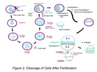
![Figure 2. Cleavage and
Blastulation [Moore et al, 2016]](https://image.slidesharecdn.com/firstweekofdevelopment-240107120540-90e7e03b/85/First-week-of-development-after-fertilization-pptx-10-320.jpg)


![ Cleavage consists of repeated mitotic divisions of the
zygote, resulting in a rapid increase in the number of
cells
[Moore et al, 2016]
At this stage, each cell is called a blastomere
Occurs as the zygote passes along the uterine tube
towards the uterus
Zygote is still within the zona pellucida
Approximately 3 days after fertilization, cells of the
compacted embryo divide again to form a 16-cell morula
(mulberry).
CLEAVAGE](https://image.slidesharecdn.com/firstweekofdevelopment-240107120540-90e7e03b/75/First-week-of-development-after-fertilization-pptx-2-2048.jpg)


![Figure 2. Cleavage and
Blastulation [Moore et al, 2016]](https://image.slidesharecdn.com/firstweekofdevelopment-240107120540-90e7e03b/75/First-week-of-development-after-fertilization-pptx-10-2048.jpg)