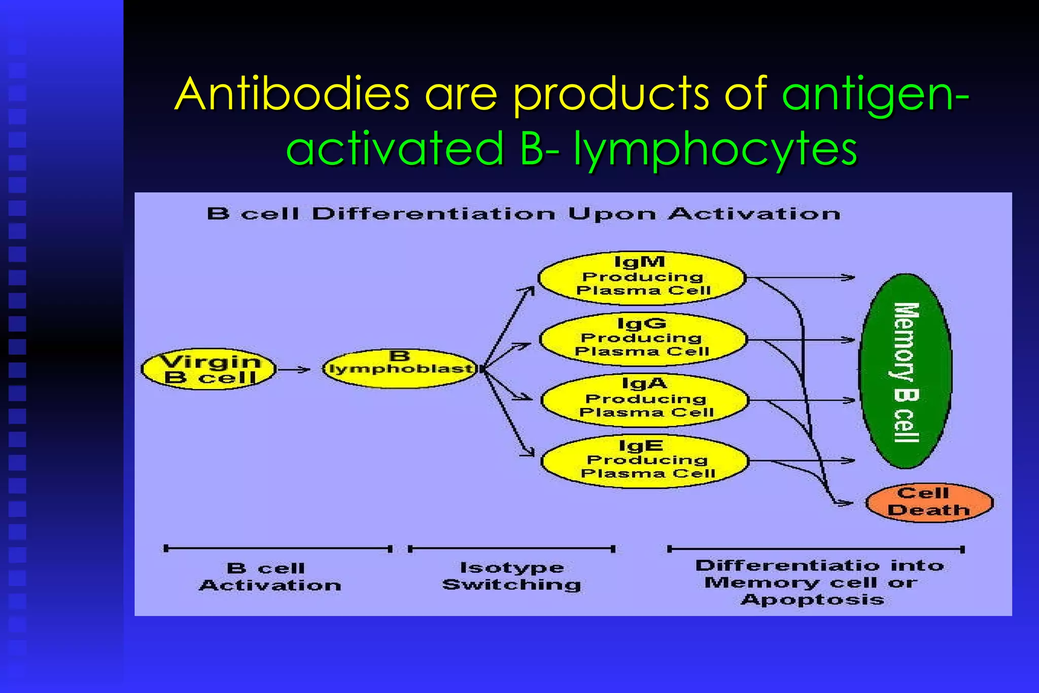
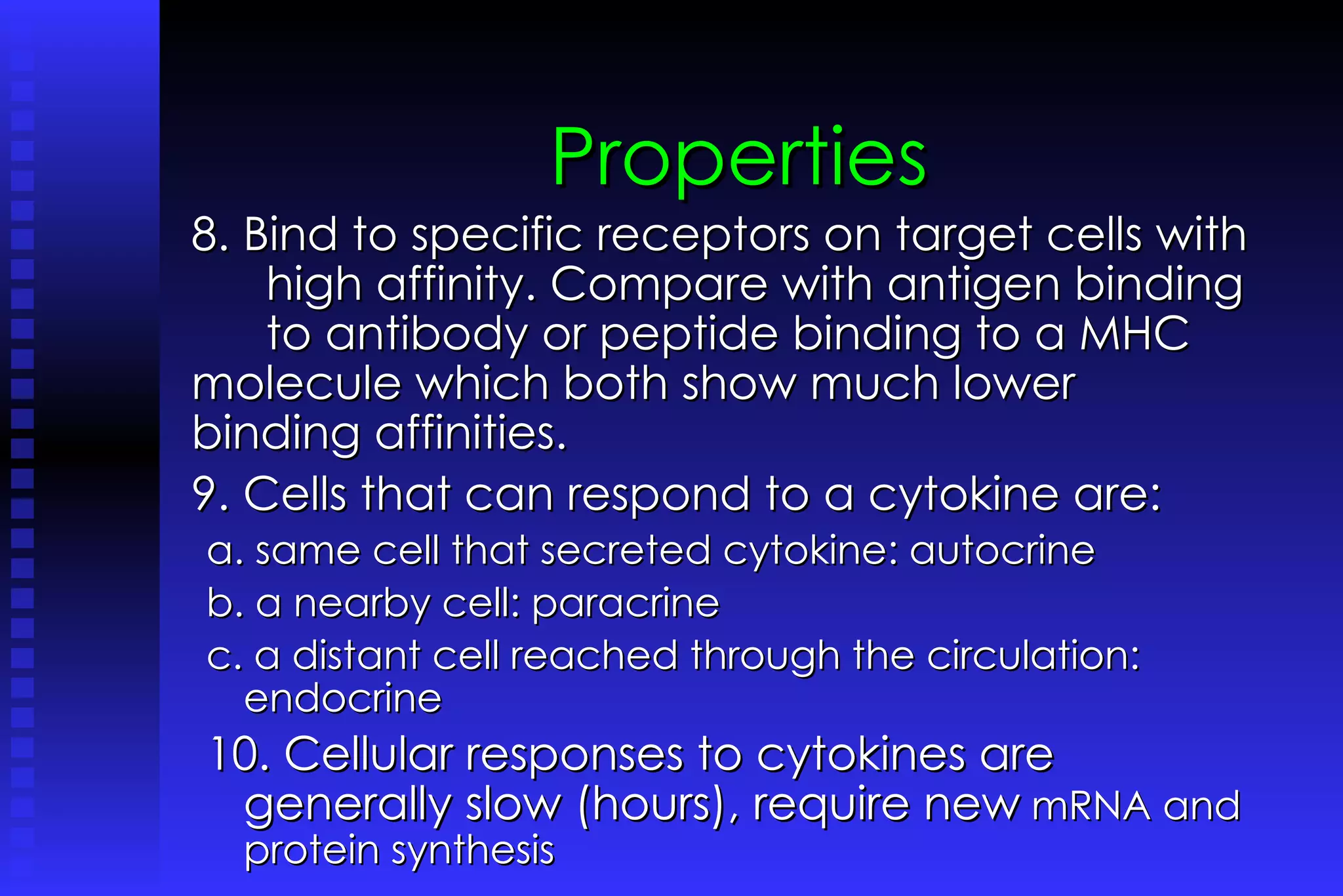
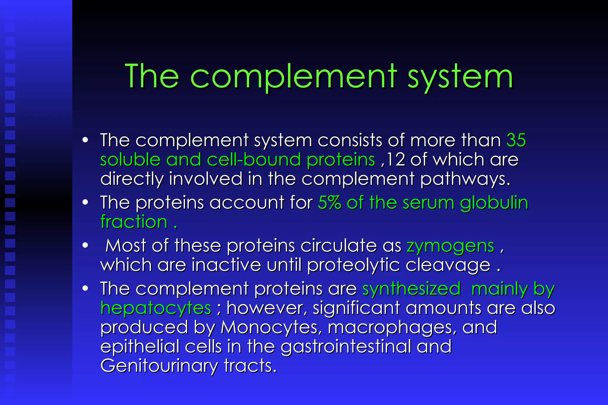
Presented by Dr. B. Victor,  St. Xavier’s College, Palayamkottai-627002,India. Functional organization  of the Immune System :  3. Immunoreactive molecules
Antibodies  (or Immunoglubulins) The chemical information  of immunoglobulin was provided by  Tiselius and Kabat  in the early 1940s. In 1950s,  Porter and Edelman  revealed the basic structure of immunoglobulin molecule. Antibodies are products of  antigen- activated B- lymphocytes. They are the  main effectors of humoral immunity . They bind antigens with  high specificity and affinity.
Antibodies are products of  antigen- activated B- lymphocytes
Subclasses of human immunoglobulins There are   nine  chemically distinct classes of human immunoglobulins,  four kinds of IgG and two kinds of IgA, plus IgM, IgE, and IgD. The classes of human immunoglobulins are  based on structure and function.  IgM -secreted during primary response  IgG -secreted during secondary response  IgD -receptors for antigens on B cells  IgA -found in external secretions  IgF -promotes histamine release Immunoglobulins G, D, and E are similar in appearance.
Kinds of Immunoglobulin
Immunoglobulin This glycoprotein is  Y-shaped molecules  and has two identical binding sites for its antigen, one on either arm of the Y.  This protein is composed of  four polypeptide chains  (two identical heavy chains and two identical and smaller light chains) held together by disulfide bonds.  -  two identical light chains  (L chains), each containing about 200 amino acids  two identical heavy chains  (H chains), each made up of 400 amino acids. Each chain is made up of several different domains.  The  antigen-binding site  is formed where a heavy chain variable domain (VH) and a light chain variable domain (VL) come close together.  These are the domains that differ most in their sequence and structure in different antibodies.
The variable (V) regions . The first 100 or so amino acids at the N-terminal of both H and L chains vary greatly from antibody to antibody.  These are the variable (V) regions .  The amino acid sequence variability in the V regions is especially pronounced in 3 hypervariable regions.  Together they construct the antigen binding site against which the epitope fits  Only a few different amino acid sequences are found in the C-terminals  of H and L chains.  These are the constant (C) regions.
The constant (C) regions.   Humans make  two different kinds of C regions for their L chains producing  kappa (κ) L chains  lambda (λ) L chains five different kinds of C regions for their H chains producing  mu (µ) chains (the H chain of IgM antibodies)  gamma (γ) chains (IgG)  alpha (α) chains (IgA)  delta (δ) chains (IgD) epsilon (ε) chains (IgE)
Fc and Fab regions  The proteolytic enzyme papain breaks each Ig molecule into 3 fragments at the hinge region. The single crystallizable fragment (Fc region) includes part of the constant domain that occupies the stem. There are 2 antigen-binding fragments (Fab region), which include the entire light chain and variable and constant portions of the heavy chain. Ig G Papain 2 Fab + Fc
Antibody Structure
Immunoglobulin
Immunoglobulin
Immunoglobulin G .  IgG,  150,000 MW, 75% of serum Ig ,   the major immunoglobulin in the blood, is also able to enter tissue spaces; Four classes; it works efficiently to coat microorganisms, speeding their destruction by other cells in the immune system. antibodies to toxins, bacteria and viruses. Crosses the placenta to give the newborn some passive immunity.  Bacteriolytic  Viricidal Precipitating ab
Immunoglobulin A IgA- -a doublet--guards the entrance to the body. 170,000 MW in serum and 400,000 MW in external secretions, 15% of Ig in serum, found in the blood as a monomer, and in tears, saliva, colustrum, nasal, vaginal, prostatic and bronchial secretions as a dimer. Blocks attachment of microbes to mucous membranes It concentrates in body fluids such as tears, saliva, and secretions of the respiratory and gastrointestinal tracts.   Secretary ab First line defense for microbes
Immunoglobulin M IgM  usually combines in star-shaped clusters. pentamer, It tends to remain in the bloodstream, 10% of blood Ig, found on the surface of B lymphocytes.  Activates the complement system.   Macroglobulin primary immune response Bacteriolytic
Immunoglobulin D 180,000 MW, activity is not well known. 0.2% of plasma Ig. 13% carbohydrate content. Also found on the surface of B lymphocytes ,  where it somehow regulates the cell's activation Membrane bound antibody Found on B-cell membrane Memory function
Immunoglobulin E !90 000 MW,  largest immunoglobulin, present in extremely low levels in a healthy individual. IgE levels rise in response to parasitic  infections and in  allergic reactions. Bind and activate mast cells. Mast cells cause acute inflammatory response (e.g. swelling, redness, pain and itchiness).  Hay fever is a condition caused by too much IgE activity. Activate mast cells Release vasoactive amines Respond to allergens Mediate hypersensitivity  reactions
Human immunoglobulin isotype monomers .
Defense mechanisms of antibodies 1. Opsonization 2. MAC Cytolysis 3. Antibody-dependent Cellular Cytotoxicity (ADCC)  by NK Cells 4. Neutralization of Exotoxins 5. Neutralization of Viruses 6. Preventing Bacterial Adherence to Host Cells 7. Agglutination of Microorganisms 8. Immobilization of Bacteria and Protozoans.
Cytokines Non- antibody proteins  produced by cells of the immune system (including T cells, B cells, monocytes, and macrophages).  Cytokines  include a  diverse group  of  interleukins ,  interferons , and  growth factors . cytokines are  chemical switches  that turn certain immune cell types on and off. One cytokine, interleukin 2 (IL-2), triggers the immune system to produce T cells.  Cytokines also are being studied for their potential  clinical benefit .
Functions of cytokines Activation of the Immune  Cells  Promotion of Cell Growth, meaning  maturation, and/or division  Attraction of Cells to the site of infection  Destruction Infected or Malignant Cells Stimulation of Phagocytic Activity
Kinds of cytokines   1.  Monokines -  produced by mononuclear  phagocytes 2.  Lymphokines -  produced by activated T  cells, primarily helper T cells 3.  Interleukins   -  name given to many  cytokines, abbreviated as IL and given a  number
Chemokines The name chemokine is a contraction of  chemo tactic cyto kines These are  a large family of substances  (more than 50) produced by many  different leukocytes and tissue cells They recruit leukocytes to sites of infection They play a role in lymphocyte trafficking
Properties Produced by cells involved in both natural and specific immunity Mediate and regulate immune and inflammatory responses Secretion is brief and limited 4. Many individual cytokines are produced by many cell types and act on many cell types (they are  pleiotropic ) 5. In many cases cytokines have similar actions (they are  redundant ).
Properties 6.  Often influence the synthesis of other cytokines  They can produce cascades, or enhance or suppress production of other cytokines They exert positive or negative regulatory mechanisms for immune and inflammatory responses 7. Often influence the action of other cytokines. Effects can be: Antagonistic Additive greater than additive (synergistic)
Properties 8. Bind to specific receptors on target cells with  high affinity. Compare with antigen binding  to antibody or peptide binding to a MHC  molecule which both show much lower  binding affinities. 9. Cells that can respond to a cytokine are: a. same cell that secreted cytokine: autocrine b. a nearby cell: paracrine c. a distant cell reached through the circulation: endocrine 10. Cellular responses to cytokines are generally slow (hours), require new  mRNA and protein synthesis
Functional types of Cytokines Cytokines can be grouped according to function 1. Mediators and regulators of Natural Immunity      Tumor Necrosis Factor (TNF)      Interleukin-1 (IL-1)      Chemokines      Interleukin-10      Interferon-gamma (IFN-gamma) 2. Mediators and regulators of specific immunity      Interleukin-2 (IL-2)      Interleukin-4 (IL-4)      Interleukin-5 (IL-5)      Interleukin-10 (IL-10)      Interferon-gamma (IFN-gamma) 3. Stimulators of hematopoeisis      Interleukin-3 (IL-3)      Colony-Stimulating Factors (CSFs)
Tumor Necrosis Factor,   TNF-gamma   produced by activated macrophages   the most important mediator of acute  inflammation  in response to Gram-negative bacteria and  other  infectious microbes mediates the recruitment of polymorphonuclear  leukocytes (PMNs) and monocytes to the  site of  infection:  acts on the hypothalamus to produce fever promotes the production of acute phase proteins  by the liver
Interleukin-2 produced mainly by helper T cells (CD4+); less by cytoxic T cells (CD8+) promote T cell division and  increase production of other cytokines has autocrine functions on T cell  proliferation.
Interleukin-4 Produced mainly by Th2 subpopulation of helper T cells (CD4+).   Stimulates immunoglobulin class switching to the IgE isotype.   Stimulates development of Th2 cells from naive CD4+ T cells promotes growth of differentiated Th2 cells
Interleukin-5 produced mainly by the Th2 subpopulation of helper T cells (CD4+) promotes growth and differentiation of eosinophils activates mature eosinophils
Interferons (IFN) There are three groups of interferons:   IFN-alpha , IFN-beta , IFN-gamma  IFN-alpha :  Twenty  variants are produced by leukocytes in response to viruses IFN-beta:  This is a single protein produced by fibroblasts and other cells in response to viruses Both IFN-alpha  and IFN-beta   inhibit viral replication and increase expression of class I MHC on cells
IFN-gamma: produced by  the Th1 subpopulation of helper T cells (CD4+), cytotoxic T cells (CD8+), and NK cells.    IFN-gamma functions in both natural and specific immunity Natural Immunity-  IFN-gamma enhances the microbicidal function of macrophages  Specific Immunity-  IFN-gamma stimulates the expression of class I and class II MHC molecules and co-stimulatory molecules on antigen presenting cells - IFN-gamma promotes the differentiation of naive helper T cells into Th1 cells  - IFN-gamma activates polymorphonuclear leukocytes (PMN) and cytotoxic T cells and increases the cytotoxicity of NK cells.
  Transforming Growth Factor  (TGF-beta) an inhibitory cytokine produced by T cells, macrophages, and many other cell types. inhibits proliferation and differentiation of T cells inhibits activation of macrophages acts on PMN and endothelial cells to block the effects of pro-inflammatory cytokines
Stimulators of Hematopoiesis : 1. Interleukin-3 - produced by helper T cells  -promotes growth and differentiation of bone marrow progenitors 2. Colony-Stimulating Factors (CSFs) - produced by T cells, macrophages, endothelial cells, fibroblasts 3.Granulocyte-macrophage colony-stimulating factor (GM-CSF)   -promotes growth and differentiation of bone marrow progenitors 4. Macrophage colony-stimulating factor (M-CSF )   -the development and function of monocytes/macrophages 5.Granulocyte colony-stimulatory factor (G-CSF )   -stimulates the production of PMN
Complement Proteins They circulate through the bloodstream in an inactive form.  When the complement cascade is triggered ( by the presence of antibody-antigen complexes), components of the cascade are successively activated, to:  Stimulate Mast Cells and Basophils to release granulocytic chemicals  Attract Neutrophils to the area  Opsonize Invading Microbes  Generate a Membrane Attack Complex,
The complement system The  complement system  helps clear  pathogens from an organism. It is derived from many small plasma  proteins that form  the biochemical  cascade of the immune system .   Activation of this system leads to  cytolysis ,  chemotaxis , opsonization ,immune  clearence, and inflamation, as well as  the  marking of pathogens  for  phagocytosis.
The complement system The complement system consists of more than  35 soluble and cell-bound proteins  ,12 of which are directly involved in the complement pathways.  The proteins account for  5% of the serum globulin fraction . Most of these proteins circulate as  zymogens  , which are inactive until proteolytic cleavage .  The complement proteins are  synthesized  mainly by hepatocytes  ; however, significant amounts are also produced by Monocytes, macrophages, and epithelial cells in the gastrointestinal and Genitourinary tracts.
Biochemical  pathways of the complement system: the classical complement pathway  the alternate complement pathway  the manon-binding –lectin pathway
Pathways of complement activation CLASSICAL PATHWAY ALTERNATIVE PATHWAY LECTIN PATHWAY activation of C5 LYTIC ATTACK PATHWAY antibody dependent antibody independent Activation of C3  and generation of C5 convertase
The Complement System
The Complement System Figure 16.10
Secretory molecules of   non-specific immunity   These include:   organic acids in skin secretions, thiocyanate in saliva,  low molecular weight fatty acids in the lower bowel;  bile acids and low molecular weight fatty acids in lower GI tract;  transferrin, lactoferrin, lysozyme, interferons, fibronectin, complement, acute phase proteins, etc. in serum;  Interferons and tumor necrosis factor (TNF) at the site of inflammation.
Secretory molecules of   non-specific immunity Transferrin and lactoferrin deprive organisms of iron.   Interferon inhibits viral replication and activates other cells which kill pathogens  Lysozyme, in serum and tears, breaks down the bacterial cell wall (peptidoglycan)  Fibronectin coats (opsonizes) bacteria and promotes their rapid phagocytosis.   Complement components and their products cause destruction of microorganism directly or with the help of phagocytic cells. Acute phase proteins (such as CRP) interact with the complement system proteins to combat infections.   TNF-alpha suppresses viral replication and activates phagocytes.
About the presenter Dr.B.Victor is a highly experienced postgraduate biology teacher, recently retired from the reputed educational institution St. Xavier’ s College, Palayamkottai, India-627001.Presently HOD of Biotech at Annai  Velankanni college, Tholayavattam. K .K Dist He was  the dean of sciences and assistant controller of examinations of St. Xavier’s college. He has more than 32 years of teaching and research experience and has guided more than 12 Ph. D scholars. He has  taught Immunology and biochemistry to graduate and  post graduate students. Send your comments to : bonfiliusvictor@gmail.com
Thank you

Functional organization of the Immune System

  • 1.
    Presented by Dr.B. Victor, St. Xavier’s College, Palayamkottai-627002,India. Functional organization of the Immune System : 3. Immunoreactive molecules
  • 2.
    Antibodies (orImmunoglubulins) The chemical information of immunoglobulin was provided by Tiselius and Kabat in the early 1940s. In 1950s, Porter and Edelman revealed the basic structure of immunoglobulin molecule. Antibodies are products of antigen- activated B- lymphocytes. They are the main effectors of humoral immunity . They bind antigens with high specificity and affinity.
  • 3.
    Antibodies are productsof antigen- activated B- lymphocytes
  • 4.
    Subclasses of humanimmunoglobulins There are nine chemically distinct classes of human immunoglobulins, four kinds of IgG and two kinds of IgA, plus IgM, IgE, and IgD. The classes of human immunoglobulins are based on structure and function. IgM -secreted during primary response IgG -secreted during secondary response IgD -receptors for antigens on B cells IgA -found in external secretions IgF -promotes histamine release Immunoglobulins G, D, and E are similar in appearance.
  • 5.
  • 6.
    Immunoglobulin This glycoproteinis Y-shaped molecules and has two identical binding sites for its antigen, one on either arm of the Y. This protein is composed of four polypeptide chains (two identical heavy chains and two identical and smaller light chains) held together by disulfide bonds. - two identical light chains (L chains), each containing about 200 amino acids two identical heavy chains (H chains), each made up of 400 amino acids. Each chain is made up of several different domains. The antigen-binding site is formed where a heavy chain variable domain (VH) and a light chain variable domain (VL) come close together. These are the domains that differ most in their sequence and structure in different antibodies.
  • 7.
    The variable (V)regions . The first 100 or so amino acids at the N-terminal of both H and L chains vary greatly from antibody to antibody. These are the variable (V) regions . The amino acid sequence variability in the V regions is especially pronounced in 3 hypervariable regions. Together they construct the antigen binding site against which the epitope fits Only a few different amino acid sequences are found in the C-terminals of H and L chains. These are the constant (C) regions.
  • 8.
    The constant (C)regions. Humans make two different kinds of C regions for their L chains producing kappa (κ) L chains lambda (λ) L chains five different kinds of C regions for their H chains producing mu (µ) chains (the H chain of IgM antibodies) gamma (γ) chains (IgG) alpha (α) chains (IgA) delta (δ) chains (IgD) epsilon (ε) chains (IgE)
  • 9.
    Fc and Fabregions The proteolytic enzyme papain breaks each Ig molecule into 3 fragments at the hinge region. The single crystallizable fragment (Fc region) includes part of the constant domain that occupies the stem. There are 2 antigen-binding fragments (Fab region), which include the entire light chain and variable and constant portions of the heavy chain. Ig G Papain 2 Fab + Fc
  • 10.
  • 11.
  • 12.
  • 13.
    Immunoglobulin G . IgG, 150,000 MW, 75% of serum Ig , the major immunoglobulin in the blood, is also able to enter tissue spaces; Four classes; it works efficiently to coat microorganisms, speeding their destruction by other cells in the immune system. antibodies to toxins, bacteria and viruses. Crosses the placenta to give the newborn some passive immunity. Bacteriolytic Viricidal Precipitating ab
  • 14.
    Immunoglobulin A IgA--a doublet--guards the entrance to the body. 170,000 MW in serum and 400,000 MW in external secretions, 15% of Ig in serum, found in the blood as a monomer, and in tears, saliva, colustrum, nasal, vaginal, prostatic and bronchial secretions as a dimer. Blocks attachment of microbes to mucous membranes It concentrates in body fluids such as tears, saliva, and secretions of the respiratory and gastrointestinal tracts. Secretary ab First line defense for microbes
  • 15.
    Immunoglobulin M IgM usually combines in star-shaped clusters. pentamer, It tends to remain in the bloodstream, 10% of blood Ig, found on the surface of B lymphocytes. Activates the complement system. Macroglobulin primary immune response Bacteriolytic
  • 16.
    Immunoglobulin D 180,000MW, activity is not well known. 0.2% of plasma Ig. 13% carbohydrate content. Also found on the surface of B lymphocytes , where it somehow regulates the cell's activation Membrane bound antibody Found on B-cell membrane Memory function
  • 17.
    Immunoglobulin E !90000 MW, largest immunoglobulin, present in extremely low levels in a healthy individual. IgE levels rise in response to parasitic  infections and in  allergic reactions. Bind and activate mast cells. Mast cells cause acute inflammatory response (e.g. swelling, redness, pain and itchiness).  Hay fever is a condition caused by too much IgE activity. Activate mast cells Release vasoactive amines Respond to allergens Mediate hypersensitivity reactions
  • 18.
  • 19.
    Defense mechanisms ofantibodies 1. Opsonization 2. MAC Cytolysis 3. Antibody-dependent Cellular Cytotoxicity (ADCC) by NK Cells 4. Neutralization of Exotoxins 5. Neutralization of Viruses 6. Preventing Bacterial Adherence to Host Cells 7. Agglutination of Microorganisms 8. Immobilization of Bacteria and Protozoans.
  • 20.
    Cytokines Non- antibodyproteins produced by cells of the immune system (including T cells, B cells, monocytes, and macrophages). Cytokines include a diverse group of interleukins , interferons , and growth factors . cytokines are chemical switches that turn certain immune cell types on and off. One cytokine, interleukin 2 (IL-2), triggers the immune system to produce T cells. Cytokines also are being studied for their potential clinical benefit .
  • 21.
    Functions of cytokinesActivation of the Immune Cells Promotion of Cell Growth, meaning maturation, and/or division Attraction of Cells to the site of infection Destruction Infected or Malignant Cells Stimulation of Phagocytic Activity
  • 22.
    Kinds of cytokines 1. Monokines - produced by mononuclear phagocytes 2. Lymphokines - produced by activated T cells, primarily helper T cells 3. Interleukins - name given to many cytokines, abbreviated as IL and given a number
  • 23.
    Chemokines The namechemokine is a contraction of chemo tactic cyto kines These are a large family of substances (more than 50) produced by many different leukocytes and tissue cells They recruit leukocytes to sites of infection They play a role in lymphocyte trafficking
  • 24.
    Properties Produced bycells involved in both natural and specific immunity Mediate and regulate immune and inflammatory responses Secretion is brief and limited 4. Many individual cytokines are produced by many cell types and act on many cell types (they are pleiotropic ) 5. In many cases cytokines have similar actions (they are redundant ).
  • 25.
    Properties 6. Often influence the synthesis of other cytokines They can produce cascades, or enhance or suppress production of other cytokines They exert positive or negative regulatory mechanisms for immune and inflammatory responses 7. Often influence the action of other cytokines. Effects can be: Antagonistic Additive greater than additive (synergistic)
  • 26.
    Properties 8. Bindto specific receptors on target cells with high affinity. Compare with antigen binding to antibody or peptide binding to a MHC molecule which both show much lower binding affinities. 9. Cells that can respond to a cytokine are: a. same cell that secreted cytokine: autocrine b. a nearby cell: paracrine c. a distant cell reached through the circulation: endocrine 10. Cellular responses to cytokines are generally slow (hours), require new mRNA and protein synthesis
  • 27.
    Functional types ofCytokines Cytokines can be grouped according to function 1. Mediators and regulators of Natural Immunity      Tumor Necrosis Factor (TNF)      Interleukin-1 (IL-1)      Chemokines      Interleukin-10      Interferon-gamma (IFN-gamma) 2. Mediators and regulators of specific immunity      Interleukin-2 (IL-2)      Interleukin-4 (IL-4)      Interleukin-5 (IL-5)      Interleukin-10 (IL-10)      Interferon-gamma (IFN-gamma) 3. Stimulators of hematopoeisis      Interleukin-3 (IL-3)      Colony-Stimulating Factors (CSFs)
  • 28.
    Tumor Necrosis Factor, TNF-gamma   produced by activated macrophages   the most important mediator of acute inflammation in response to Gram-negative bacteria and  other infectious microbes mediates the recruitment of polymorphonuclear leukocytes (PMNs) and monocytes to the  site of infection:  acts on the hypothalamus to produce fever promotes the production of acute phase proteins by the liver
  • 29.
    Interleukin-2 produced mainlyby helper T cells (CD4+); less by cytoxic T cells (CD8+) promote T cell division and increase production of other cytokines has autocrine functions on T cell proliferation.
  • 30.
    Interleukin-4 Produced mainlyby Th2 subpopulation of helper T cells (CD4+).  Stimulates immunoglobulin class switching to the IgE isotype.  Stimulates development of Th2 cells from naive CD4+ T cells promotes growth of differentiated Th2 cells
  • 31.
    Interleukin-5 produced mainlyby the Th2 subpopulation of helper T cells (CD4+) promotes growth and differentiation of eosinophils activates mature eosinophils
  • 32.
    Interferons (IFN) Thereare three groups of interferons:  IFN-alpha , IFN-beta , IFN-gamma  IFN-alpha :  Twenty  variants are produced by leukocytes in response to viruses IFN-beta: This is a single protein produced by fibroblasts and other cells in response to viruses Both IFN-alpha  and IFN-beta   inhibit viral replication and increase expression of class I MHC on cells
  • 33.
    IFN-gamma: produced by the Th1 subpopulation of helper T cells (CD4+), cytotoxic T cells (CD8+), and NK cells.   IFN-gamma functions in both natural and specific immunity Natural Immunity- IFN-gamma enhances the microbicidal function of macrophages Specific Immunity- IFN-gamma stimulates the expression of class I and class II MHC molecules and co-stimulatory molecules on antigen presenting cells - IFN-gamma promotes the differentiation of naive helper T cells into Th1 cells  - IFN-gamma activates polymorphonuclear leukocytes (PMN) and cytotoxic T cells and increases the cytotoxicity of NK cells.
  • 34.
    TransformingGrowth Factor (TGF-beta) an inhibitory cytokine produced by T cells, macrophages, and many other cell types. inhibits proliferation and differentiation of T cells inhibits activation of macrophages acts on PMN and endothelial cells to block the effects of pro-inflammatory cytokines
  • 35.
    Stimulators of Hematopoiesis: 1. Interleukin-3 - produced by helper T cells -promotes growth and differentiation of bone marrow progenitors 2. Colony-Stimulating Factors (CSFs) - produced by T cells, macrophages, endothelial cells, fibroblasts 3.Granulocyte-macrophage colony-stimulating factor (GM-CSF) -promotes growth and differentiation of bone marrow progenitors 4. Macrophage colony-stimulating factor (M-CSF ) -the development and function of monocytes/macrophages 5.Granulocyte colony-stimulatory factor (G-CSF ) -stimulates the production of PMN
  • 36.
    Complement Proteins Theycirculate through the bloodstream in an inactive form. When the complement cascade is triggered ( by the presence of antibody-antigen complexes), components of the cascade are successively activated, to: Stimulate Mast Cells and Basophils to release granulocytic chemicals Attract Neutrophils to the area Opsonize Invading Microbes Generate a Membrane Attack Complex,
  • 37.
    The complement systemThe complement system helps clear pathogens from an organism. It is derived from many small plasma proteins that form the biochemical cascade of the immune system . Activation of this system leads to cytolysis , chemotaxis , opsonization ,immune clearence, and inflamation, as well as the marking of pathogens for phagocytosis.
  • 38.
    The complement systemThe complement system consists of more than 35 soluble and cell-bound proteins ,12 of which are directly involved in the complement pathways. The proteins account for 5% of the serum globulin fraction . Most of these proteins circulate as zymogens , which are inactive until proteolytic cleavage . The complement proteins are synthesized mainly by hepatocytes ; however, significant amounts are also produced by Monocytes, macrophages, and epithelial cells in the gastrointestinal and Genitourinary tracts.
  • 39.
    Biochemical pathwaysof the complement system: the classical complement pathway the alternate complement pathway the manon-binding –lectin pathway
  • 40.
    Pathways of complementactivation CLASSICAL PATHWAY ALTERNATIVE PATHWAY LECTIN PATHWAY activation of C5 LYTIC ATTACK PATHWAY antibody dependent antibody independent Activation of C3 and generation of C5 convertase
  • 41.
  • 42.
  • 43.
    Secretory molecules of non-specific immunity These include: organic acids in skin secretions, thiocyanate in saliva, low molecular weight fatty acids in the lower bowel; bile acids and low molecular weight fatty acids in lower GI tract; transferrin, lactoferrin, lysozyme, interferons, fibronectin, complement, acute phase proteins, etc. in serum; Interferons and tumor necrosis factor (TNF) at the site of inflammation.
  • 44.
    Secretory molecules of non-specific immunity Transferrin and lactoferrin deprive organisms of iron.  Interferon inhibits viral replication and activates other cells which kill pathogens Lysozyme, in serum and tears, breaks down the bacterial cell wall (peptidoglycan) Fibronectin coats (opsonizes) bacteria and promotes their rapid phagocytosis.  Complement components and their products cause destruction of microorganism directly or with the help of phagocytic cells. Acute phase proteins (such as CRP) interact with the complement system proteins to combat infections.  TNF-alpha suppresses viral replication and activates phagocytes.
  • 45.
    About the presenterDr.B.Victor is a highly experienced postgraduate biology teacher, recently retired from the reputed educational institution St. Xavier’ s College, Palayamkottai, India-627001.Presently HOD of Biotech at Annai Velankanni college, Tholayavattam. K .K Dist He was the dean of sciences and assistant controller of examinations of St. Xavier’s college. He has more than 32 years of teaching and research experience and has guided more than 12 Ph. D scholars. He has taught Immunology and biochemistry to graduate and post graduate students. Send your comments to : [email protected]
  • 46.