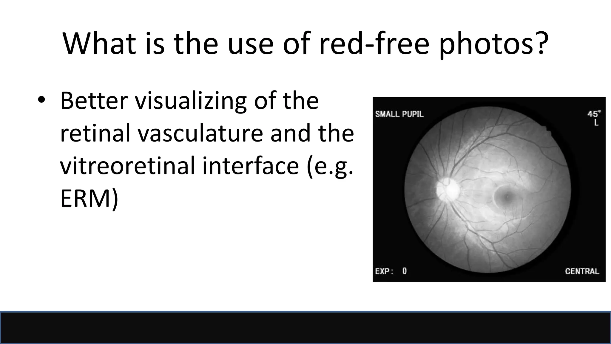
FFA/ICG
Haitham Al Mahrouqi
Oman Medical Speciality Board
Sept 2017
Acknowledgment/References
• Dr Al Yaqdhan Al Ghafri presentation
• Dr Sawsan Nawilaty (AAO 2011)
• AAO BCSC 2015, Retina and vitreous
• Ophthalmology by Yanoff&Dukar
• Retina by Ryan
• Retinal Angiography and Optical Coherence
Tomography (Arevalo [Editor])
• Google!
Definition
• Definition: Fluorescein angiography (FA) is a
diagnostic technique that uses intravenous
fluorescein dye to allow the sequential
visualization of the blood flow
simultaneously through retinal, choroidal
and iris tissue.
Concept
• Fluorescent property Sodium fluorescein
• Means: the ability of certain molecules to emit
light of longer wavelength when stimulated
by light of shorter wavelength.
Question?
• What is the excitation
wavelength and the
emitted wavelength in
FFA?
– Excited with blue light
490nm
– Emit green light 530nm
Sodium Fluorescein
• A water-soluble dye of an orange-red crystalline
hydrocarbon.
• When injected intravenously, remains largely
intravascular and circulates in the blood stream.
• 80% of fluorescein molecules bind to serum proteins
(albumin), the residue remaining unbound. Only the
unbound molecules are available for fluorescence.
• It undergoes both renal and hepatic metabolism and is
excreted in the urine over 24–48 hours.
Sodium Fluorescein
Absolute contraindication:
• Fluorescein allergy
Relative contraindications:
• History of a severe reaction to any allergen is a strong
relative
contraindication.
• Renal failure (lower the fluorescein dose if angiography
is necessary)
• Pregnancy: Although no reported birth defects.
• Moderate-severe asthma
• Significant cardiac disease.
Sodium Fluorescein
• Discolouration
of skin and urine
(invariable)
Yanoff
The procedure
1. Both pupils are dilated
2. The patient is seated at the camera and IV cannula is inserted in antecubital fossa.
3. Coloured funds photos, red-free photos +/- autofluorescence are taken.
4. Sodium fluorescein is injected 5 ml of 10% solution, or 2.5 ml of 25% solution
(15mg/kg).
5. Images are taken in the following order:
• Early rapid-sequence photographs (1 second intervals for 25–30 seconds).
• Less rapid sequences between 5 and 10 min.
• Late images at 10–20 minutes
What is the use of red-free photos?
• Better visualizing of the
retinal vasculature and the
vitreoretinal interface (e.g.
ERM)
What is the use of autofluorescence?
• Autofluorescence describe
the appearance of apparent
hyperfluorescence in the
absence of fluorescein.
• It is an imaging modality of
the RPE (lipofusin
autoflouresce).
• Described as:
– Hyperautofluoresence
– Hypoautofluorescence
Examples
• Stargardt disease
Examples
• CSR • Optic disc drusen
What is pseudofluorescence?
• Fake fluorescence
• Filters problem
Anatomical consideration
• Choroidal vasculature
– 3 layers
– Choriocappilaris are
fenestrated. They are
organised in lobules.
Supply the outer 1/3 of
retina.
– RPE makes up the outer
blood retinal barrier.
Anatomical consideration
• Retinal vessels:
– non-fenestrated (do not leak)
– Inner blood retinal barrier is
made up by the tight
junctions of the capillary
network with pericytes.
– Supply the inner 1/3
Anatomical consideration
• The choroid fills up first due to
the shorter route (1 second
before retinal arteries)
• The retinal arteries fill up in 1
second (hence the early rapid-
sequence photographs (1
second intervals for 25–30
seconds).
The procedure
1. Both pupils are dilated
2. The patient is seated at the camera and IV cannula is inserted in antecubital fossa.
3. Coloured funds photos, red-free photos +/- autofluorescence are taken.
4. Sodium fluorescein is injected 5 ml of 10% solution, or 2.5 ml of 25% solution
5. Images are taken in the following order:
• Early rapid-sequence photographs (1 second intervals for 25–30 seconds).
• Less rapid sequences between 5 and 10 min.
• Late images at 10–20 minutes
Choroidal phases
Arm-Choroid time: 8-15s
Phases:
• Early
• Patchy choroidal filling
• Choroidal flush
• Recirculation phase
Choroidal journey
Early
• Dye in the large
vessels before
leaking into the
choroidal stroma
from the CC.
Choroidal journey
Patchy choroidal
filling
• Accounted by the
mosaic lobular
pattern of the CC.
Choroidal journey
Choroidal flush (20-25s)
• Homogenous
fluorescence of the
choroid, due leaked
fluorescein in the
stroma from the CC.
Choroidal journey
Recirculation phases
Less intense fluorescence Clearance from the large
choroidal vessels makes
them silhouette darkly
Clearance from the CC,
back to “patchy choroidal
filling”!
Retinal journey
Arm-retina time ≈ 13s
Phases
• Pre-arterial
• Arterial (1s)
• Arteriovenous (8-12s)
– Capillaries phase
– Laminar flow (early venous phase)
• Venous phase (12s)
• Recirculation
– Early (30s-5mins)
– Late (10-20mins)
Retinal journey
Pre-arterial
Cilioretinal artery present in 30%
of population comes from the
posterior ciliary artery.
- Which choroidal phase is this?
Patchy choroidal filling
Retinal journey
Arterial
• Fills up in 1s
Retinal journey
Early arteriovenous (Retinal
capillaries phase)
• Complete arterial filling, no dye in
veins, 1-3 sec.
Retinal journey
Late arteriovenous (Laminar
flow).
• Dye appears in retinal venules
entering along venous walls
Retinal journey
Venous phase (12s)
• When veins are evenly filled.
Homogeneous hyperfluorescent
background with dye equally
distributed in retinal arterioles,
capillaries, venules and choroid.
• Best to visualize the foveal avascular
zone (FAZ).
FAZ
• FAZ corresponds to the
foveola (350-500um)
• Dark because:
– Avascular
– Densely packed and tall RPE
– Xanothophyls
• Ischaemic macula if FAZ >
1000um
Retinal journey
Early recirculation
phase (30s-5mins)
• Veins brighter than
arteries
• Best phase for
detecting staining.
Retinal journey
Late recirculation phase
(10-20mins)
• Fading fluorescence.
Optic disc
• Stains with FFA due
to staining of the
lamina cribrosa and
from the posterior
ciliary arteries.
Abnormal FFA
• Hyperfluorescence
– Leaking
– Window defect
– Pooling
– Staining
• Hyporfluorescence
– Blockage
– Vascular filling defect/capillary dropout.
Abnormal FFA
Hyperfluorescence: Leaking
Extravasation of dye due to
incompetent blood
retinal barriers.
• Pattern: early continuous increase in
fluorescence In intensity and/or size
• Margins: fuzzy
• Level: intravitreal, intraretinal,
subretinal
Abnormal FFA
Hyperfluorescence: Leaking
Example: Leaking microaneurysm
Abnormal FFA
Hyperfluorescence: Leaking
Example: Leaking CNVM
Abnormal FFA
Hyperfluorescence: Window defect
Increased transmission of the normal
choroidalfluorescence (Atrophic RPE
/reduced RPE pigment)
• Pattern: parallels choroidal
fluorescence
• Size: no change
• Margins: distinct
• Level: subretinal (RPE)
Abnormal FFA
Hyperfluorescence: Pooling
Pattern:
• Steady increase in the
hyperfluorescence intensity
• Stable size
• Distinct margins
PED
Abnormal FFA
• Hyperfluorescence: Staining
Leakage into tissue
Pattern: steady increase in intensity
• Size: stable
• Margins: distinct
• Level: retinal or subretinal
Staining
drusen
Abnormal FFA
• Hyporfluorescence: Blockage
Abnormal FFA
• Hyporfluorescence: Vascular
filling defect/capillary dropout.
Capillary
dropout in
BRVO
RPE is a screen
Dampens transmission of choroidal
fluorescence (melanin acts as a curtain).
• How can we then image the choroid better?
Indocyanine green angiography
• First used in cardiology to measure cardiac
output in 1957
• Large molecular weight compound
• 98% bound to plasma globulins.
• Metabolised in the liver.
Indocyanine green angiography
• Does not leak through the fenestrations of the CC.
Therefore perfect for imaging the choroidal
vasculature.
• Its maximal peak of absorption is at 790 to 805nm
and has a peak emission of 835 nm.
• The infra-red light can cross through the RPE.
• Poor efficacy in fluorescence – only 4%, thus poor
resolution.
Indocyanine green angiography
• Contraindication
- Contraindicated in iodine allergy
- Patients with liver disease
- Patients taking metformin (precipitates lactic acidosis)
- Pregnancy
• Adverse events (less than fluorescein):
- Used with caution in patients with shellfish allergy.
- Nausea and vomiting
- Anaphylaxis
- Death
Procedure
• Similar to FFA.
• Fundus camera must be equipped with infra-red
light.
• Inject 25mg in 5 mls
• Can be done sequentially with FFA
• Image acquisition: 10 images in the first 30s and
then every 15s for the first 5mins and then delayed
at 10mins, 20mins and 40mins.
Image acquisition
• Camera: Fundus
camera can be used
with filters however,
newer technologies
which block
reflections give
better resolution.
Visupac, Ziess
Indications
• AMD
• Choroidal polypoidal vasculopathy
• Retinal Angiomatous Proliferation
• Choroidal tumours
• Chorioretinal Inflammatory Diseases
Indocyanine green angiography
• Phases:
– Early : 0-3mins
– Mid (3-15mins): Fading away
– Late (15-40mins): Staining of the extrachoroidal tissue
• Interpretation: Same as FFA
– Hypocyanescence: Blockage and filling defects
– Hypercyanescence: Leakage, staining, pooling and window
defects
Indocyanine green angiography (Early phase)
Example:
AMD
Example:
AMD
Example:
PCV
Example:
Birdshot
Example:
Birdshot
Thank you

fundus fluorescein and indocyanine green angiography

  • 1.
    FFA/ICG Haitham Al Mahrouqi OmanMedical Speciality Board Sept 2017
  • 2.
    Acknowledgment/References • Dr AlYaqdhan Al Ghafri presentation • Dr Sawsan Nawilaty (AAO 2011) • AAO BCSC 2015, Retina and vitreous • Ophthalmology by Yanoff&Dukar • Retina by Ryan • Retinal Angiography and Optical Coherence Tomography (Arevalo [Editor]) • Google!
  • 3.
    Definition • Definition: Fluoresceinangiography (FA) is a diagnostic technique that uses intravenous fluorescein dye to allow the sequential visualization of the blood flow simultaneously through retinal, choroidal and iris tissue.
  • 4.
    Concept • Fluorescent propertySodium fluorescein • Means: the ability of certain molecules to emit light of longer wavelength when stimulated by light of shorter wavelength.
  • 6.
    Question? • What isthe excitation wavelength and the emitted wavelength in FFA? – Excited with blue light 490nm – Emit green light 530nm
  • 7.
    Sodium Fluorescein • Awater-soluble dye of an orange-red crystalline hydrocarbon. • When injected intravenously, remains largely intravascular and circulates in the blood stream. • 80% of fluorescein molecules bind to serum proteins (albumin), the residue remaining unbound. Only the unbound molecules are available for fluorescence. • It undergoes both renal and hepatic metabolism and is excreted in the urine over 24–48 hours.
  • 8.
    Sodium Fluorescein Absolute contraindication: •Fluorescein allergy Relative contraindications: • History of a severe reaction to any allergen is a strong relative contraindication. • Renal failure (lower the fluorescein dose if angiography is necessary) • Pregnancy: Although no reported birth defects. • Moderate-severe asthma • Significant cardiac disease.
  • 9.
    Sodium Fluorescein • Discolouration ofskin and urine (invariable) Yanoff
  • 10.
    The procedure 1. Bothpupils are dilated 2. The patient is seated at the camera and IV cannula is inserted in antecubital fossa. 3. Coloured funds photos, red-free photos +/- autofluorescence are taken. 4. Sodium fluorescein is injected 5 ml of 10% solution, or 2.5 ml of 25% solution (15mg/kg). 5. Images are taken in the following order: • Early rapid-sequence photographs (1 second intervals for 25–30 seconds). • Less rapid sequences between 5 and 10 min. • Late images at 10–20 minutes
  • 11.
    What is theuse of red-free photos? • Better visualizing of the retinal vasculature and the vitreoretinal interface (e.g. ERM)
  • 12.
    What is theuse of autofluorescence? • Autofluorescence describe the appearance of apparent hyperfluorescence in the absence of fluorescein. • It is an imaging modality of the RPE (lipofusin autoflouresce). • Described as: – Hyperautofluoresence – Hypoautofluorescence
  • 13.
  • 14.
    Examples • CSR •Optic disc drusen
  • 15.
    What is pseudofluorescence? •Fake fluorescence • Filters problem
  • 16.
    Anatomical consideration • Choroidalvasculature – 3 layers – Choriocappilaris are fenestrated. They are organised in lobules. Supply the outer 1/3 of retina. – RPE makes up the outer blood retinal barrier.
  • 17.
    Anatomical consideration • Retinalvessels: – non-fenestrated (do not leak) – Inner blood retinal barrier is made up by the tight junctions of the capillary network with pericytes. – Supply the inner 1/3
  • 18.
    Anatomical consideration • Thechoroid fills up first due to the shorter route (1 second before retinal arteries) • The retinal arteries fill up in 1 second (hence the early rapid- sequence photographs (1 second intervals for 25–30 seconds).
  • 19.
    The procedure 1. Bothpupils are dilated 2. The patient is seated at the camera and IV cannula is inserted in antecubital fossa. 3. Coloured funds photos, red-free photos +/- autofluorescence are taken. 4. Sodium fluorescein is injected 5 ml of 10% solution, or 2.5 ml of 25% solution 5. Images are taken in the following order: • Early rapid-sequence photographs (1 second intervals for 25–30 seconds). • Less rapid sequences between 5 and 10 min. • Late images at 10–20 minutes
  • 20.
    Choroidal phases Arm-Choroid time:8-15s Phases: • Early • Patchy choroidal filling • Choroidal flush • Recirculation phase
  • 21.
    Choroidal journey Early • Dyein the large vessels before leaking into the choroidal stroma from the CC.
  • 22.
    Choroidal journey Patchy choroidal filling •Accounted by the mosaic lobular pattern of the CC.
  • 23.
    Choroidal journey Choroidal flush(20-25s) • Homogenous fluorescence of the choroid, due leaked fluorescein in the stroma from the CC.
  • 24.
    Choroidal journey Recirculation phases Lessintense fluorescence Clearance from the large choroidal vessels makes them silhouette darkly Clearance from the CC, back to “patchy choroidal filling”!
  • 25.
    Retinal journey Arm-retina time≈ 13s Phases • Pre-arterial • Arterial (1s) • Arteriovenous (8-12s) – Capillaries phase – Laminar flow (early venous phase) • Venous phase (12s) • Recirculation – Early (30s-5mins) – Late (10-20mins)
  • 26.
    Retinal journey Pre-arterial Cilioretinal arterypresent in 30% of population comes from the posterior ciliary artery. - Which choroidal phase is this? Patchy choroidal filling
  • 27.
  • 28.
    Retinal journey Early arteriovenous(Retinal capillaries phase) • Complete arterial filling, no dye in veins, 1-3 sec.
  • 29.
    Retinal journey Late arteriovenous(Laminar flow). • Dye appears in retinal venules entering along venous walls
  • 30.
    Retinal journey Venous phase(12s) • When veins are evenly filled. Homogeneous hyperfluorescent background with dye equally distributed in retinal arterioles, capillaries, venules and choroid. • Best to visualize the foveal avascular zone (FAZ).
  • 31.
    FAZ • FAZ correspondsto the foveola (350-500um) • Dark because: – Avascular – Densely packed and tall RPE – Xanothophyls • Ischaemic macula if FAZ > 1000um
  • 32.
    Retinal journey Early recirculation phase(30s-5mins) • Veins brighter than arteries • Best phase for detecting staining.
  • 33.
    Retinal journey Late recirculationphase (10-20mins) • Fading fluorescence.
  • 34.
    Optic disc • Stainswith FFA due to staining of the lamina cribrosa and from the posterior ciliary arteries.
  • 35.
    Abnormal FFA • Hyperfluorescence –Leaking – Window defect – Pooling – Staining • Hyporfluorescence – Blockage – Vascular filling defect/capillary dropout.
  • 36.
    Abnormal FFA Hyperfluorescence: Leaking Extravasationof dye due to incompetent blood retinal barriers. • Pattern: early continuous increase in fluorescence In intensity and/or size • Margins: fuzzy • Level: intravitreal, intraretinal, subretinal
  • 37.
  • 38.
  • 39.
    Abnormal FFA Hyperfluorescence: Windowdefect Increased transmission of the normal choroidalfluorescence (Atrophic RPE /reduced RPE pigment) • Pattern: parallels choroidal fluorescence • Size: no change • Margins: distinct • Level: subretinal (RPE)
  • 40.
    Abnormal FFA Hyperfluorescence: Pooling Pattern: •Steady increase in the hyperfluorescence intensity • Stable size • Distinct margins PED
  • 41.
    Abnormal FFA • Hyperfluorescence:Staining Leakage into tissue Pattern: steady increase in intensity • Size: stable • Margins: distinct • Level: retinal or subretinal Staining drusen
  • 42.
  • 43.
    Abnormal FFA • Hyporfluorescence:Vascular filling defect/capillary dropout. Capillary dropout in BRVO
  • 44.
    RPE is ascreen Dampens transmission of choroidal fluorescence (melanin acts as a curtain). • How can we then image the choroid better?
  • 45.
    Indocyanine green angiography •First used in cardiology to measure cardiac output in 1957 • Large molecular weight compound • 98% bound to plasma globulins. • Metabolised in the liver.
  • 46.
    Indocyanine green angiography •Does not leak through the fenestrations of the CC. Therefore perfect for imaging the choroidal vasculature. • Its maximal peak of absorption is at 790 to 805nm and has a peak emission of 835 nm. • The infra-red light can cross through the RPE. • Poor efficacy in fluorescence – only 4%, thus poor resolution.
  • 47.
    Indocyanine green angiography •Contraindication - Contraindicated in iodine allergy - Patients with liver disease - Patients taking metformin (precipitates lactic acidosis) - Pregnancy • Adverse events (less than fluorescein): - Used with caution in patients with shellfish allergy. - Nausea and vomiting - Anaphylaxis - Death
  • 48.
    Procedure • Similar toFFA. • Fundus camera must be equipped with infra-red light. • Inject 25mg in 5 mls • Can be done sequentially with FFA • Image acquisition: 10 images in the first 30s and then every 15s for the first 5mins and then delayed at 10mins, 20mins and 40mins.
  • 49.
    Image acquisition • Camera:Fundus camera can be used with filters however, newer technologies which block reflections give better resolution. Visupac, Ziess
  • 50.
    Indications • AMD • Choroidalpolypoidal vasculopathy • Retinal Angiomatous Proliferation • Choroidal tumours • Chorioretinal Inflammatory Diseases
  • 51.
    Indocyanine green angiography •Phases: – Early : 0-3mins – Mid (3-15mins): Fading away – Late (15-40mins): Staining of the extrachoroidal tissue • Interpretation: Same as FFA – Hypocyanescence: Blockage and filling defects – Hypercyanescence: Leakage, staining, pooling and window defects
  • 52.
  • 53.
  • 54.
  • 55.
  • 56.
  • 57.
  • 58.

Editor's Notes

  • #32 The umbo (the very centre) The foveola (0.35mm) which is also the FAZ The fovea is 1.5mm (1 disc diameter) Para and then perifoveolar