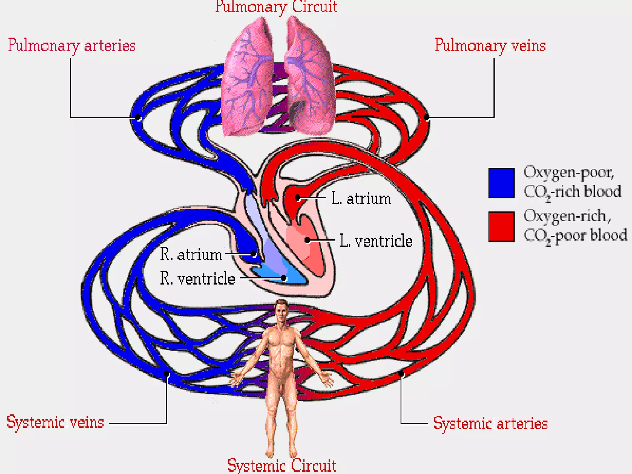
Introduction to the Cardiovascular System Chapter 13 Unit 4: Transport
Introduction Primary function of the  cardiovascular system  is to pump blood throughout the body Recall that blood is a transport medium for oxygen, nutrients, and wastes CV System is a closed circuit Blood is contained in blood vessels
Introduction There are two main components to the CV system Cardio  - refers to the presence of the heart, which is the major pump for the body Transporting 7,000 liters of blood every day. Beats 2.5 billion times in a lifetime Vascular  - refers to the vessels that contain blood Blood is forced from the heart into arteries, arterioles, capillaries, venules, and veins in that order Each blood cell circulates through the body’s CV system in about a minute
Circuits of the Cardiovascular System The cardiovascular system can be divided into two circuits: Pulmonary Circuit Sends deoxygenated blood to lungs to pick up oxygen and unload carbon dioxide Involves the right side of the heart Systemic Circuit Sends oxygenated blood and nutrients to all body cells and removes wastes Involves the left side of the heart
 
Paths of Circulation Pulmonary Circuit: Blood in the right side of the heart is O 2 -poor Right side of heart pumps blood into the  pulmonary  trunk/arteries that transport blood to the lungs Once blood becomes rich in O 2  it returns to the  left  side of the heart through the  pulmonary veins
Paths of Circulation Systemic Circuit: Blood returning to the heart from the lungs enters the  left side  of the heart Heart pumps blood into the  aorta , which branches into smaller and smaller arteries throughout the body Blood returns to the  right side  of the heart via the superior and inferior  vena cavae  after unloading its supply of O 2  in the body’s cells
Paths of Circulation Heart Animation
What would happen without circulation in the body?
Structural Characteristics of the Heart The heart is: Hollow Cone-shaped Muscular Found inside the thoracic cavity Rests on the diaphragm
 
Size and Location of the Heart Varies in size and shape, but the average adult heart is about 14 cm long and 9 cm wide. About the size of your fist. Rests in an opening called the  mediastinum Bordered laterally by the lungs, posteriorly by the vertebral column, and anteriorly by the sternum
 
Coverings of the Heart The heart is enclosed in a fibrous sack called the  Pericardium  (3 layers). Consists of an outer bag… Two layers Fibrous Pericardium  -  outermost layer Parietal Pericardium  -  deep to the fibrous p. … and an inner bag Visceral Pericardium  - deepest layer, continuous with the outermost layer of the heart.
 
 
Coverings Continued The fibrous pericardium serves as a point of attachment to: Diaphragm, sternum, vertebral column, and large blood vessels emerging from the heart The space between the parietal and visceral pericardia is called the  pericardial cavity Contains  serous fluid  secreted by the visceral pericardium Fluid serves to reduce friction between the membranes as the heart moves within them
Walls of the Heart The walls of the heart are composed of three distinct layers of tissue: Epicardium Myocardium Endocardium
 
Epicardium The  Epicardium  is the outermost layer of the heart muscle. Continuous with the visceral pericardium and protects the heart by reducing friction Composed of serous membranes (connective tissue), epithelium, and deeper adipose tissue Adipose tissue is found predominantly along the paths of coronary arteries and cardiac veins that carry blood to the heart muscle itself
Myocardium The  myocardium  is the thick middle layer of heart muscle. Composed primarily of  cardiac muscle tissue . Used to pump blood out of the heart chambers Myocardial muscle works continuously… needs a good supply of oxygen and sugar
Myocardium These muscles are supplied by the  coronary arteries , NOT THE BLOOD THAT IS IN THE CHAMBERS OF THE HEART!!!!! Coronary arteries branch off the aorta immediately after leaving the heart This is the #1 site for plaque build-up, causing  myocardial   infarction  (heart attack)
Myocardium
Endocardium The  endocardium  is the innermost layer of the heart muscle. Consists of epithelium and connective tissue that contains many elastic and collagenous fibers (helps it stretch) Contains  Purkinje Fibers  (aid in transmission of muscle impulses in heart) Continuous with the inner linings of the blood vessels attached to the heart
Heart Chambers The heart is divided into 4 hollow chambers-two on the right and two on the left: The upper chambers are called  Atria Have thin walls and receive blood returning to the heart The lower chambers are called  Ventricles Receive blood from the atria. Thick walls that contract to force blood out of the heart into the arteries The right and left chambers are separated from one another by a solid, wall-like  Septum
Heart Chambers The right side of the heart is part of the  pulmonary circuit Only deoxygenated blood passes through the right side The left side of the heart is part of the  systemic circuit Only oxygenated blood passes through the left side *O 2 -rich and O 2 -poor blood do not mix in healthy individuals
 
Valves of the Heart Valves  are flap-like tissue that prevents backflow of blood Keeps blood going in one direction only Four valves found in heart Tricuspid valve - b/w R. atrium and R. vent. Pulmonary valve - at exit of R. vent. Mitral (bicuspid) valve - b/w L. atrium and L vent. Aortic valve - at exit of L. vent.
Path of Blood Through the Heart Blood that is low in oxygen and high in carbon dioxide enters the right side of the heart and is pumped into the pulmonary circulation. After blood is oxygenated in the lungs and some carbon dioxide is removed, it returns to the left side of the heart.
Greater Detail Deoxygenated blood FROM body enters the right atrium through the vena cavae. Blood passes through the tricuspid valve and enters the right ventricle Blood moves through the pulmonary valve and into the pulmonary trunk and the pulmonary arteries Blood enters the capillaries (aveoli) of the lungs
More Detail Blood returns to the heart via the pulmonary veins into the left atrium Passes through the mitral valve into the left ventricle Blood moves through the aortic valve into the aorta and to the body
 

Introduction to the_cardiovascular_system

  • 1.
    Introduction to theCardiovascular System Chapter 13 Unit 4: Transport
  • 2.
    Introduction Primary functionof the cardiovascular system is to pump blood throughout the body Recall that blood is a transport medium for oxygen, nutrients, and wastes CV System is a closed circuit Blood is contained in blood vessels
  • 3.
    Introduction There aretwo main components to the CV system Cardio - refers to the presence of the heart, which is the major pump for the body Transporting 7,000 liters of blood every day. Beats 2.5 billion times in a lifetime Vascular - refers to the vessels that contain blood Blood is forced from the heart into arteries, arterioles, capillaries, venules, and veins in that order Each blood cell circulates through the body’s CV system in about a minute
  • 4.
    Circuits of theCardiovascular System The cardiovascular system can be divided into two circuits: Pulmonary Circuit Sends deoxygenated blood to lungs to pick up oxygen and unload carbon dioxide Involves the right side of the heart Systemic Circuit Sends oxygenated blood and nutrients to all body cells and removes wastes Involves the left side of the heart
  • 5.
  • 6.
    Paths of CirculationPulmonary Circuit: Blood in the right side of the heart is O 2 -poor Right side of heart pumps blood into the pulmonary trunk/arteries that transport blood to the lungs Once blood becomes rich in O 2 it returns to the left side of the heart through the pulmonary veins
  • 7.
    Paths of CirculationSystemic Circuit: Blood returning to the heart from the lungs enters the left side of the heart Heart pumps blood into the aorta , which branches into smaller and smaller arteries throughout the body Blood returns to the right side of the heart via the superior and inferior vena cavae after unloading its supply of O 2 in the body’s cells
  • 8.
    Paths of CirculationHeart Animation
  • 9.
    What would happenwithout circulation in the body?
  • 10.
    Structural Characteristics ofthe Heart The heart is: Hollow Cone-shaped Muscular Found inside the thoracic cavity Rests on the diaphragm
  • 11.
  • 12.
    Size and Locationof the Heart Varies in size and shape, but the average adult heart is about 14 cm long and 9 cm wide. About the size of your fist. Rests in an opening called the mediastinum Bordered laterally by the lungs, posteriorly by the vertebral column, and anteriorly by the sternum
  • 13.
  • 14.
    Coverings of theHeart The heart is enclosed in a fibrous sack called the Pericardium (3 layers). Consists of an outer bag… Two layers Fibrous Pericardium - outermost layer Parietal Pericardium - deep to the fibrous p. … and an inner bag Visceral Pericardium - deepest layer, continuous with the outermost layer of the heart.
  • 15.
  • 16.
  • 17.
    Coverings Continued Thefibrous pericardium serves as a point of attachment to: Diaphragm, sternum, vertebral column, and large blood vessels emerging from the heart The space between the parietal and visceral pericardia is called the pericardial cavity Contains serous fluid secreted by the visceral pericardium Fluid serves to reduce friction between the membranes as the heart moves within them
  • 18.
    Walls of theHeart The walls of the heart are composed of three distinct layers of tissue: Epicardium Myocardium Endocardium
  • 19.
  • 20.
    Epicardium The Epicardium is the outermost layer of the heart muscle. Continuous with the visceral pericardium and protects the heart by reducing friction Composed of serous membranes (connective tissue), epithelium, and deeper adipose tissue Adipose tissue is found predominantly along the paths of coronary arteries and cardiac veins that carry blood to the heart muscle itself
  • 21.
    Myocardium The myocardium is the thick middle layer of heart muscle. Composed primarily of cardiac muscle tissue . Used to pump blood out of the heart chambers Myocardial muscle works continuously… needs a good supply of oxygen and sugar
  • 22.
    Myocardium These musclesare supplied by the coronary arteries , NOT THE BLOOD THAT IS IN THE CHAMBERS OF THE HEART!!!!! Coronary arteries branch off the aorta immediately after leaving the heart This is the #1 site for plaque build-up, causing myocardial infarction (heart attack)
  • 23.
  • 24.
    Endocardium The endocardium is the innermost layer of the heart muscle. Consists of epithelium and connective tissue that contains many elastic and collagenous fibers (helps it stretch) Contains Purkinje Fibers (aid in transmission of muscle impulses in heart) Continuous with the inner linings of the blood vessels attached to the heart
  • 25.
    Heart Chambers Theheart is divided into 4 hollow chambers-two on the right and two on the left: The upper chambers are called Atria Have thin walls and receive blood returning to the heart The lower chambers are called Ventricles Receive blood from the atria. Thick walls that contract to force blood out of the heart into the arteries The right and left chambers are separated from one another by a solid, wall-like Septum
  • 26.
    Heart Chambers Theright side of the heart is part of the pulmonary circuit Only deoxygenated blood passes through the right side The left side of the heart is part of the systemic circuit Only oxygenated blood passes through the left side *O 2 -rich and O 2 -poor blood do not mix in healthy individuals
  • 27.
  • 28.
    Valves of theHeart Valves are flap-like tissue that prevents backflow of blood Keeps blood going in one direction only Four valves found in heart Tricuspid valve - b/w R. atrium and R. vent. Pulmonary valve - at exit of R. vent. Mitral (bicuspid) valve - b/w L. atrium and L vent. Aortic valve - at exit of L. vent.
  • 29.
    Path of BloodThrough the Heart Blood that is low in oxygen and high in carbon dioxide enters the right side of the heart and is pumped into the pulmonary circulation. After blood is oxygenated in the lungs and some carbon dioxide is removed, it returns to the left side of the heart.
  • 30.
    Greater Detail Deoxygenatedblood FROM body enters the right atrium through the vena cavae. Blood passes through the tricuspid valve and enters the right ventricle Blood moves through the pulmonary valve and into the pulmonary trunk and the pulmonary arteries Blood enters the capillaries (aveoli) of the lungs
  • 31.
    More Detail Bloodreturns to the heart via the pulmonary veins into the left atrium Passes through the mitral valve into the left ventricle Blood moves through the aortic valve into the aorta and to the body
  • 32.