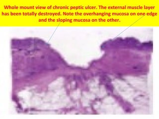
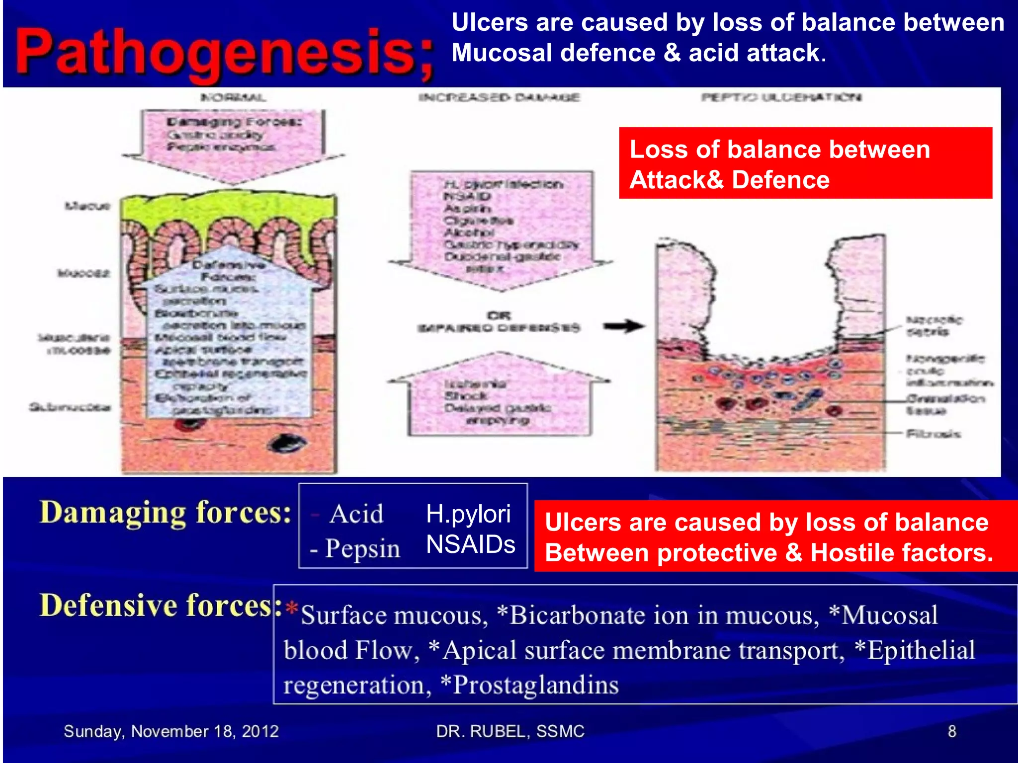
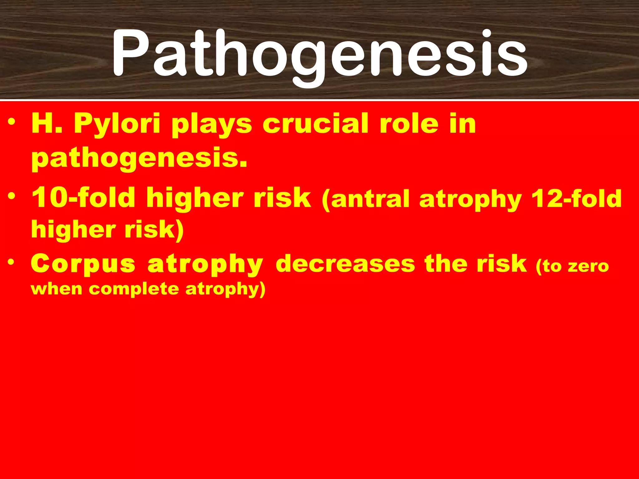
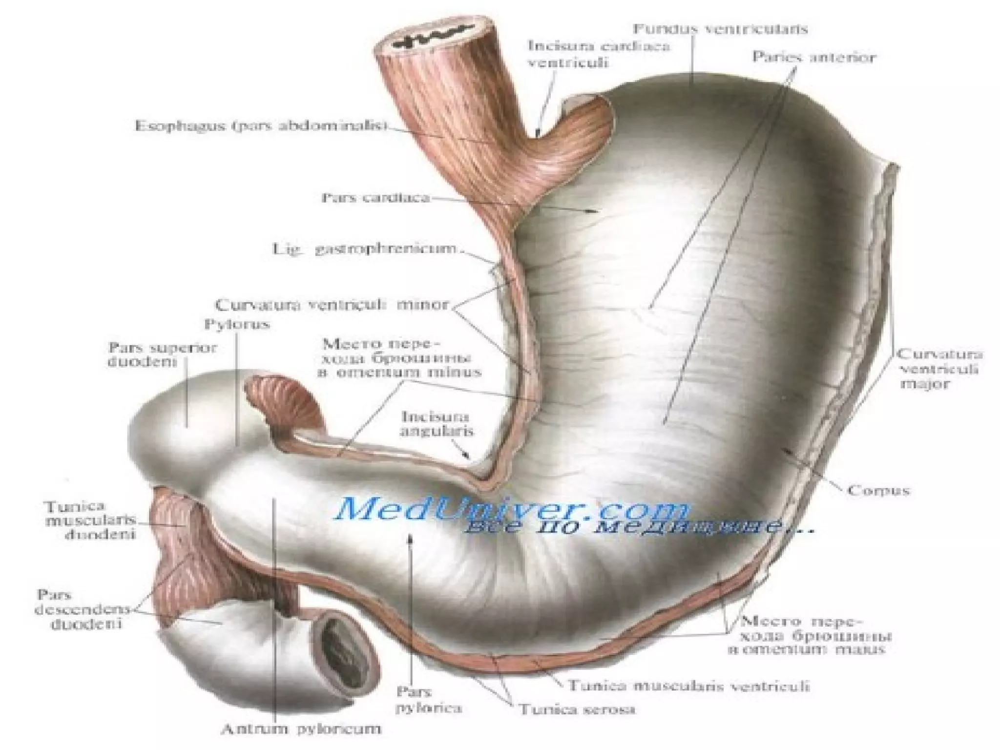
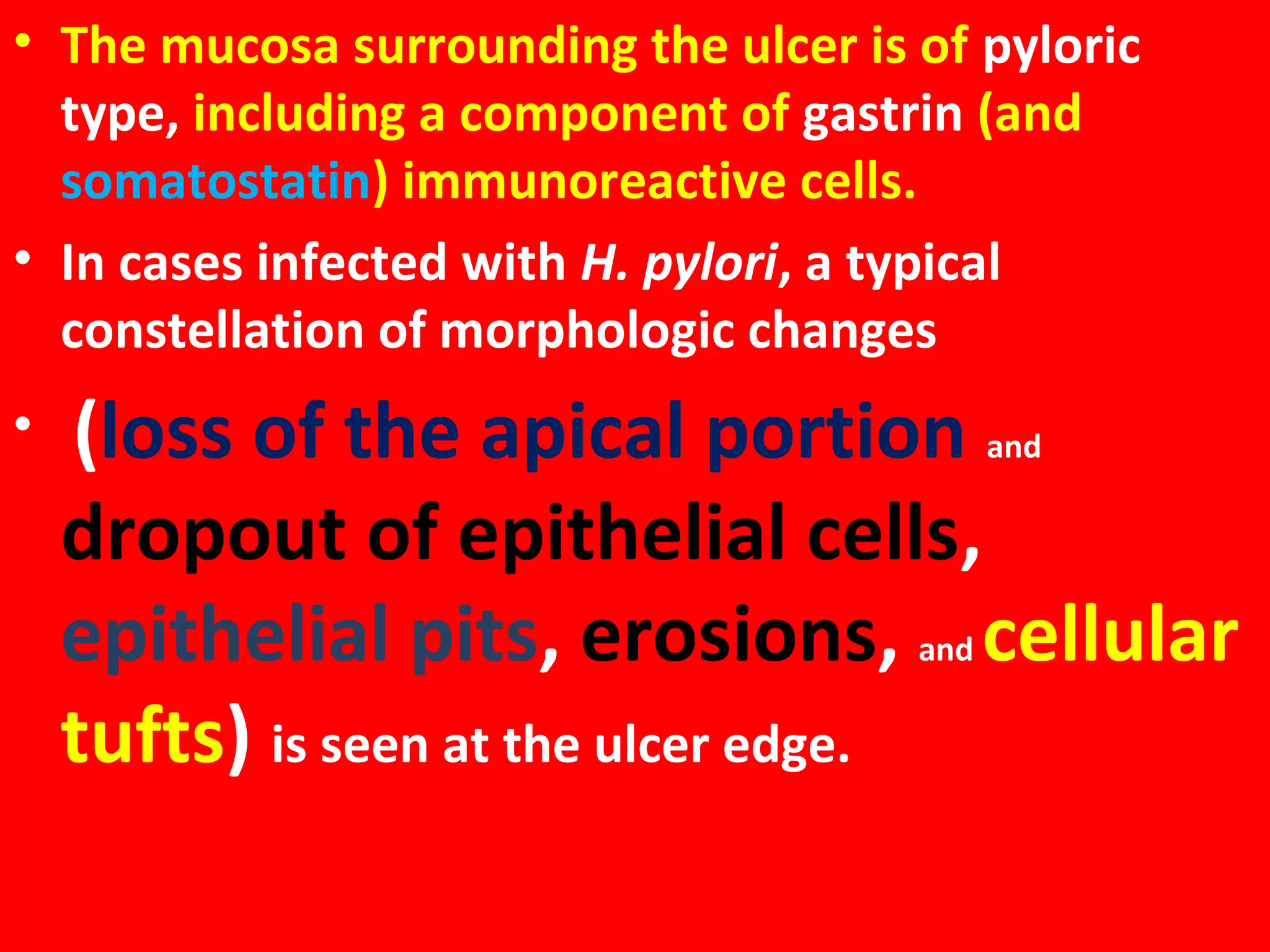
H Pylori HELICOBACTER
Campylobacter Pylori

PYLORI

H. pylori has been found in 90% of patients with
chronic gastritis, 95% with duodenal ulcer

disease, 70% with gastric ulcer, and 50% with
gastric carcinoma.

H Pylori

PePtic
ulcer
Lecture 8
Definition
• Ulcer of the GIT (Peptic ulcer) is defined
histologically as

a breach in the mucosa
that extends through the muscularis
mucosea into the submucosa or deeper.

The areas of degeneration &
necrosis of gastrointestinal mucosa
exposed to acid-peptic secretions.
Acid peptic digestion is the
ultimate cause for
Ulceration.

DU: GU= 4:1
Hurry, worry & Curry
Genetic Factors:
Blood group: O &
A
Monozygotic
twins,association
with HLA-B5 gene

Work, worry, weather

Reflux of Bile & Pancreatic Juices
Bile Reflux
L Ulcers are caused by loss of balance
Between protective & Hostile factors.

Loss of balance
between
Attack& Defence
Damagin
g
Forces

Defensive
Forces

Ulcers are caused by loss of balance
between
Mucosal defence & acid attack

Loss of balance
between
Attack& Defence

Ulcers are caused by loss of balance
Between protective & Hostile factors.
Ulcers are caused by loss of balance between
Mucosal defence & acid attack.

Loss of balance between
Attack& Defence

H.pylori
NSAIDs

Ulcers are caused by loss of balance
Between protective & Hostile factors.
Ulcers are caused by loss of balance
Between protective & Hostile factors.
Loss of balance
between
Attack& Defence
Pathogenesis
• H. Pylori plays crucial role in
pathogenesis.
• 10-fold higher risk (antral atrophy 12-fold
higher risk)
• Corpus atrophy decreases the risk
when complete atrophy)

(to zero
Pathogenesis of

DU

Duodenal Ulcer: High acid-pepsin secretions
• 1. Hypersecretion of gastric acid into the fasting stomach at night
(vagal stimulation).
• 2. Rapid emptying of the stomach exposing the duodenal mucosa to
the aggressive action of HCl.
• 3. H. pylori:
• i) Mucosal defence is broken by bacterial elaboration of urease,
protease,& phospholipase.
• ii) Host factors: H. pylori infected mucosal epithelium releases
proinflammatory cytokines such as IL-1, IL-6, IL-8 & TNF-incite
inflammatory reaction.

• iii) Bacterial factors: Epithelial injury is also induced by
cytotoxic-associated gene protein (Cag A), while vacuolating
cytotoxin (Vac A) induces elaboration of cytokines.
Pathogenesis of gastric ulcer
GU: Impaired gastric mucosal defences against

acid-pepsin secretions.
Other features in pathogenesis:
• 1. Hyperacidity due to increased serum gastrin
levels in response to ingested food in an atonic
stomach.
• 2.In case of Low to Normal HCl: Damaging influence
of gastritis, bile reflux, smoking.
• 3. Disorder of protective gastric mucus ‘barrier’ by
H. pylori.
DU & GU

• Gastric & duodenal ulcers

represent two diseases as far as
their etiology, pathogenesis &
clinical features are concerned
• but morphological findings in
both are similar.
Acute Peptic(Stress) Ulcer
• Multiple, small mucosal erosions, seen most

commonly in the stomach but may occur in the
duodenum. Mucosal erosion (loss of continuity of the
epithelial lining) is a common feature of
acute gastritis.

• Etiology:
• I. Psychological Stress

• II. Physiological stress: Shock, trauma, septicaemia, Extensive burns
•
•
•

(curling’s ulcer )
Intracranial lesions (Cushing’s ulcers developing from hyperacidity following
excessive vagal stimulation).
Drug intake (Aspirin, steroids)
Local irritants (alcohol, smocking, coffee).
ACID ?

Pathogenesis
Hyperacidity? Role of hyperacidity in acute gastritis?

• 1. Ischaemic hypoxic injury to the
mucosal cells.
• 2. Depletion of the gastric mucus
barrier rendering the mucosa
susceptible to attack by acid-peptic
secretions.
Mucosal erosion (loss of continuity of the epithelial lining) is a common feature of
acute gastritis. If the defect is severe enough to penetrate the muscularis
mucosae to involve the submucosa, this becomes—by definition—an ulcer.

Acute ulcers can be distinguished morphologically
from chronic gastritis by the lack of fibrosis in the
former.
Morphology of Acute Peptic Ulcer
• Gross: Multiple, more common in stomach, oval
or circular, small (<1cm).

• Microscopically, shallow, do not invade the

muscular layer. The margins & base may show
some inflammatory reaction. Heal without any
scar.
• Complications: Hemorrhage, Perforation.

Mucosal erosion (loss of
continuity of the epithelial
lining) is a common feature of
Chronic Peptic Ulcer
• Always occurs in an achlorhydric zone of mucosa
( an area of stomach lined by pyloric type mucosa).
Up to 95% of the ulcers are located on the lesser
curvature (Magenstrasse) near the incisura
angularis.
• Can be found anywhere in the stomach.

Pyloric antrum & lesser curvature of the stomach are the sites

most exposed for longer periods to local irritants & thus are the common
sites for occurrence of gastric ulcers.

Local Irritants: Heavily spiced
foods, alcohol, smoking &
Remitting & relapsing lesions

Once a peptic ulcer patient, always a peptic ulcer patient .
Age: DU- 5th decade, GU- 6th decade
People at risk: DU-StressExecutives, leaders
GU—Labouring groups
Vomiting, Hematemesis, melena,
Appetite, Diet, Weight loss
,deep tenderness
Periodicity: attacks—2-6
weeks,
Interval of freedom-1-6 weeks

Attacks worsened by
Indigestion

‘work,

worry,
weather
Morphology of Chronic gastritis
• Morphology of GU & Du are similar.
• Location: GU –along the Lesser curvature in the the
pyloric antrum on the posterior wall.
• Du: in the first part of the duodenum, immediately
post pyloric on the anterior wall.
• Number: Solitary (80%)
• Size: Small (1-2.5cm)
• Shape: Round to oval
• Punched out: Rounded, sharply circumscribed,
with sharply demarcated vertical margins.
>3 cm
A, Typical gross appearance of chronic peptic ulcer of stomach. B,
Sharply delimited chronic peptic ulcer with converging folds of mucosa
in the upper half. The ulcer bed is covered by fibrinopurulent exudate.

Punched Out Ulcer: rounded, sharply circumscribed, often multiple lesions with
sharply demarcated vertical margins.
Morphology of Chronic Gastritis
• Benign ulcers usually have flat margins in level with
the surrounding mucosa. The mucosal folds
converge towards the ulcer.
• Depth: The ulcers may vary in depth from being
superficial (confined to mucosa) to deep ulcers
( penetrating into the muscular layer).
• Coexistence: GU+DU in 10-20% cases
• Malignant transformation: DU never
GU 1%-carcinoma
• Malignant GU are larger, bowl-shaped with elevated &
indurated mucosa at the margins
Gross Morphology
• The proximal margin tends
to have overhanging
edges, whereas the distal
margin usually has sloping
borders.
Whole mount view of chronic peptic ulcer. The external muscle layer
has been totally destroyed. Note the overhanging mucosa on one edge
and the sloping mucosa on the other.
• On section, there is
undermining of the edges
(especially on the proximal
side) and complete
replacement of the muscle
wall by grayish white fibrous
tissue.
Morphology
On the serosal side, there
may be subserosal fibrosis
and inflammatory
enlargement of the
regional lymph nodes.
Morphology
• Prominent marginal nodularity about the ulcer
should suggest the presence of carcinoma;
however, it should be remembered that it may be
impossible to distinguish grossly a peptic ulcer from
an ulcerated carcinoma. As a matter of fact,
approximately 10–15% of gastric carcinomas
appear, grossly, to be benign ulcers.
MICROSCOPY

•

Four histological zones

Chr. Peptic ulcer have 4 histological zones. From within outside, these are as
under:

• 1. Nicrotic zone— lies in the floor of the ulcer & is
composed of fibrinous exudate containing necrotic
debris & a few leukocytes.
• 2. Superficial exudative zone –lies underneath the
necrotic zone. The tissue elements here show
coagulative necrosis giving eosinophilic, smudgy
appearance with nuclear debris.
• 3. Granulation tissue zone– is seen merging into
the necrotic zone. It is composed of nonspecific
inflammatory infiltrate & proliferative capillaries.
• 4. Zone of Cicatrization– is seen merging into

thick layer of granulation tissue. It is composed of
dense fibrocollagenic scar tissue over
which granulation tissue rests. Thrombosed or
sclerotic arteries may cross the ulcer which on
erosion may result in haemorrhage.
• Other common features in the ulcer bed include:
1)Thickening of vessels (caused by
subendothelial fibrous proliferation) and
2)Hypertrophy of nerve bundles;
both of these changes are probably secondary events.
• The necrotic surface may show superimposed
infection by Candida albicans.
• The mucosa surrounding the ulcer is of pyloric
type, including a component of gastrin (and
somatostatin) immunoreactive cells.
• In cases infected with H. pylori, a typical
constellation of morphologic changes
•

(loss of the apical portion and
dropout of epithelial cells,
epithelial pits, erosions, and cellular
tufts) is seen at the ulcer edge.
Classification of Peptic Ulcer
• DU & GU
• Acute, Subacute & Chronic
Peptic ulcers can be classified according to their
• shape and size (round-oval, giant, linear),
• activity (open ulcers or ulcer scars),
• depth of penetration (submucosa, muscularis
externa, or beyond), or
• a combination of these criteria.
Fiberoptic gastroscopy

The radiographic
diagnosis is
approximately 95%

Endoscopic gastric biopsy

multiple (about 10) biopsies
are recommended for the
standard-size ulcer.
80%

19%

Seems to be decreasing

Seems to be
decreasing

secrete either low normal or
below normal amounts of acid.

Associated with acid Hypersecretion

Mucosal Injury
Chronic, larger & deeper ulcers cause complications.

Duodenal stenosis,’hour glass’ deformity.

Cancers ulcerate but ulcers rarely cancerate
Zollinger–Ellison Syndrome (ZES)
caused by a non–beta islet cell, gastrin-secreting
tumor of the pancreas that stimulates the acidsecreting cells of the stomach to maximal activity,
with consequent gastrointestinal mucosal
ulceration.
ZES may occur sporadically or as part of an autosomal
dominant familial syndrome called multiple
endocrine neoplasia type 1 (MEN 1). The primary
tumor is usually located in the pancreas, duodenum
or abdominal lymph nodes, but ectopic locations
have also been described (e.g., heart, ovary,
gallbladder, liver, kidney).
L8 peptic ulcer

L8 peptic ulcer

  • 1.
    H Pylori HELICOBACTER CampylobacterPylori PYLORI H. pylori has been found in 90% of patients with chronic gastritis, 95% with duodenal ulcer disease, 70% with gastric ulcer, and 50% with gastric carcinoma. H Pylori PePtic ulcer Lecture 8
  • 2.
    Definition • Ulcer ofthe GIT (Peptic ulcer) is defined histologically as a breach in the mucosa that extends through the muscularis mucosea into the submucosa or deeper. The areas of degeneration & necrosis of gastrointestinal mucosa exposed to acid-peptic secretions.
  • 3.
    Acid peptic digestionis the ultimate cause for Ulceration. DU: GU= 4:1
  • 5.
    Hurry, worry &Curry Genetic Factors: Blood group: O & A Monozygotic twins,association with HLA-B5 gene Work, worry, weather Reflux of Bile & Pancreatic Juices
  • 6.
  • 10.
    L Ulcers arecaused by loss of balance Between protective & Hostile factors. Loss of balance between Attack& Defence
  • 11.
    Damagin g Forces Defensive Forces Ulcers are causedby loss of balance between Mucosal defence & acid attack Loss of balance between Attack& Defence Ulcers are caused by loss of balance Between protective & Hostile factors.
  • 12.
    Ulcers are causedby loss of balance between Mucosal defence & acid attack. Loss of balance between Attack& Defence H.pylori NSAIDs Ulcers are caused by loss of balance Between protective & Hostile factors.
  • 13.
    Ulcers are causedby loss of balance Between protective & Hostile factors. Loss of balance between Attack& Defence
  • 14.
    Pathogenesis • H. Pyloriplays crucial role in pathogenesis. • 10-fold higher risk (antral atrophy 12-fold higher risk) • Corpus atrophy decreases the risk when complete atrophy) (to zero
  • 15.
    Pathogenesis of DU Duodenal Ulcer:High acid-pepsin secretions • 1. Hypersecretion of gastric acid into the fasting stomach at night (vagal stimulation). • 2. Rapid emptying of the stomach exposing the duodenal mucosa to the aggressive action of HCl. • 3. H. pylori: • i) Mucosal defence is broken by bacterial elaboration of urease, protease,& phospholipase. • ii) Host factors: H. pylori infected mucosal epithelium releases proinflammatory cytokines such as IL-1, IL-6, IL-8 & TNF-incite inflammatory reaction. • iii) Bacterial factors: Epithelial injury is also induced by cytotoxic-associated gene protein (Cag A), while vacuolating cytotoxin (Vac A) induces elaboration of cytokines.
  • 16.
    Pathogenesis of gastriculcer GU: Impaired gastric mucosal defences against acid-pepsin secretions. Other features in pathogenesis: • 1. Hyperacidity due to increased serum gastrin levels in response to ingested food in an atonic stomach. • 2.In case of Low to Normal HCl: Damaging influence of gastritis, bile reflux, smoking. • 3. Disorder of protective gastric mucus ‘barrier’ by H. pylori.
  • 17.
    DU & GU •Gastric & duodenal ulcers represent two diseases as far as their etiology, pathogenesis & clinical features are concerned • but morphological findings in both are similar.
  • 18.
    Acute Peptic(Stress) Ulcer •Multiple, small mucosal erosions, seen most commonly in the stomach but may occur in the duodenum. Mucosal erosion (loss of continuity of the epithelial lining) is a common feature of acute gastritis. • Etiology: • I. Psychological Stress • II. Physiological stress: Shock, trauma, septicaemia, Extensive burns • • • (curling’s ulcer ) Intracranial lesions (Cushing’s ulcers developing from hyperacidity following excessive vagal stimulation). Drug intake (Aspirin, steroids) Local irritants (alcohol, smocking, coffee).
  • 19.
    ACID ? Pathogenesis Hyperacidity? Roleof hyperacidity in acute gastritis? • 1. Ischaemic hypoxic injury to the mucosal cells. • 2. Depletion of the gastric mucus barrier rendering the mucosa susceptible to attack by acid-peptic secretions. Mucosal erosion (loss of continuity of the epithelial lining) is a common feature of acute gastritis. If the defect is severe enough to penetrate the muscularis mucosae to involve the submucosa, this becomes—by definition—an ulcer. Acute ulcers can be distinguished morphologically from chronic gastritis by the lack of fibrosis in the former.
  • 20.
    Morphology of AcutePeptic Ulcer • Gross: Multiple, more common in stomach, oval or circular, small (<1cm). • Microscopically, shallow, do not invade the muscular layer. The margins & base may show some inflammatory reaction. Heal without any scar. • Complications: Hemorrhage, Perforation. Mucosal erosion (loss of continuity of the epithelial lining) is a common feature of
  • 22.
    Chronic Peptic Ulcer •Always occurs in an achlorhydric zone of mucosa ( an area of stomach lined by pyloric type mucosa). Up to 95% of the ulcers are located on the lesser curvature (Magenstrasse) near the incisura angularis. • Can be found anywhere in the stomach. Pyloric antrum & lesser curvature of the stomach are the sites most exposed for longer periods to local irritants & thus are the common sites for occurrence of gastric ulcers. Local Irritants: Heavily spiced foods, alcohol, smoking &
  • 24.
    Remitting & relapsinglesions Once a peptic ulcer patient, always a peptic ulcer patient . Age: DU- 5th decade, GU- 6th decade People at risk: DU-StressExecutives, leaders GU—Labouring groups Vomiting, Hematemesis, melena, Appetite, Diet, Weight loss ,deep tenderness Periodicity: attacks—2-6 weeks, Interval of freedom-1-6 weeks Attacks worsened by Indigestion ‘work, worry, weather
  • 25.
    Morphology of Chronicgastritis • Morphology of GU & Du are similar. • Location: GU –along the Lesser curvature in the the pyloric antrum on the posterior wall. • Du: in the first part of the duodenum, immediately post pyloric on the anterior wall. • Number: Solitary (80%) • Size: Small (1-2.5cm) • Shape: Round to oval • Punched out: Rounded, sharply circumscribed, with sharply demarcated vertical margins.
  • 26.
  • 28.
    A, Typical grossappearance of chronic peptic ulcer of stomach. B, Sharply delimited chronic peptic ulcer with converging folds of mucosa in the upper half. The ulcer bed is covered by fibrinopurulent exudate. Punched Out Ulcer: rounded, sharply circumscribed, often multiple lesions with sharply demarcated vertical margins.
  • 29.
    Morphology of ChronicGastritis • Benign ulcers usually have flat margins in level with the surrounding mucosa. The mucosal folds converge towards the ulcer. • Depth: The ulcers may vary in depth from being superficial (confined to mucosa) to deep ulcers ( penetrating into the muscular layer). • Coexistence: GU+DU in 10-20% cases • Malignant transformation: DU never GU 1%-carcinoma • Malignant GU are larger, bowl-shaped with elevated & indurated mucosa at the margins
  • 31.
    Gross Morphology • Theproximal margin tends to have overhanging edges, whereas the distal margin usually has sloping borders.
  • 32.
    Whole mount viewof chronic peptic ulcer. The external muscle layer has been totally destroyed. Note the overhanging mucosa on one edge and the sloping mucosa on the other.
  • 33.
    • On section,there is undermining of the edges (especially on the proximal side) and complete replacement of the muscle wall by grayish white fibrous tissue.
  • 34.
    Morphology On the serosalside, there may be subserosal fibrosis and inflammatory enlargement of the regional lymph nodes.
  • 35.
    Morphology • Prominent marginalnodularity about the ulcer should suggest the presence of carcinoma; however, it should be remembered that it may be impossible to distinguish grossly a peptic ulcer from an ulcerated carcinoma. As a matter of fact, approximately 10–15% of gastric carcinomas appear, grossly, to be benign ulcers.
  • 36.
    MICROSCOPY • Four histological zones Chr.Peptic ulcer have 4 histological zones. From within outside, these are as under: • 1. Nicrotic zone— lies in the floor of the ulcer & is composed of fibrinous exudate containing necrotic debris & a few leukocytes. • 2. Superficial exudative zone –lies underneath the necrotic zone. The tissue elements here show coagulative necrosis giving eosinophilic, smudgy appearance with nuclear debris. • 3. Granulation tissue zone– is seen merging into the necrotic zone. It is composed of nonspecific inflammatory infiltrate & proliferative capillaries.
  • 37.
    • 4. Zoneof Cicatrization– is seen merging into thick layer of granulation tissue. It is composed of dense fibrocollagenic scar tissue over which granulation tissue rests. Thrombosed or sclerotic arteries may cross the ulcer which on erosion may result in haemorrhage.
  • 39.
    • Other commonfeatures in the ulcer bed include: 1)Thickening of vessels (caused by subendothelial fibrous proliferation) and 2)Hypertrophy of nerve bundles; both of these changes are probably secondary events. • The necrotic surface may show superimposed infection by Candida albicans.
  • 40.
    • The mucosasurrounding the ulcer is of pyloric type, including a component of gastrin (and somatostatin) immunoreactive cells. • In cases infected with H. pylori, a typical constellation of morphologic changes • (loss of the apical portion and dropout of epithelial cells, epithelial pits, erosions, and cellular tufts) is seen at the ulcer edge.
  • 41.
    Classification of PepticUlcer • DU & GU • Acute, Subacute & Chronic Peptic ulcers can be classified according to their • shape and size (round-oval, giant, linear), • activity (open ulcers or ulcer scars), • depth of penetration (submucosa, muscularis externa, or beyond), or • a combination of these criteria.
  • 42.
    Fiberoptic gastroscopy The radiographic diagnosisis approximately 95% Endoscopic gastric biopsy multiple (about 10) biopsies are recommended for the standard-size ulcer.
  • 45.
    80% 19% Seems to bedecreasing Seems to be decreasing secrete either low normal or below normal amounts of acid. Associated with acid Hypersecretion Mucosal Injury
  • 46.
    Chronic, larger &deeper ulcers cause complications. Duodenal stenosis,’hour glass’ deformity. Cancers ulcerate but ulcers rarely cancerate
  • 48.
    Zollinger–Ellison Syndrome (ZES) causedby a non–beta islet cell, gastrin-secreting tumor of the pancreas that stimulates the acidsecreting cells of the stomach to maximal activity, with consequent gastrointestinal mucosal ulceration. ZES may occur sporadically or as part of an autosomal dominant familial syndrome called multiple endocrine neoplasia type 1 (MEN 1). The primary tumor is usually located in the pancreas, duodenum or abdominal lymph nodes, but ectopic locations have also been described (e.g., heart, ovary, gallbladder, liver, kidney).