Laparoscopy in
Gynecology
Contents
◉ Introduction
◉ History
◉ Instruments
◉ Overview procedure
◉ Use as a diagnostic tool
◉ Use as a therapeutic modality
◉ Contraindications
◉ Risk factors
◉ Complications
◉ Recent advances
◉ Summary and conclusion
Laparoscopy
A surgical procedure in which a
fibre-optic instrument is inserted
through the abdominal wall to view
the organs in the abdomen or
permit small-scale surgery with
help of pneumoperitoneum
Laparoscopic surgery
Laparoscopic surgery, also called minimally invasive
surgery (MIS), bandaid surgery, or keyhole surgery, is
a modern surgical technique in which operations are
performed far from their location through small
incisions (usually 0.5–1.5 cm)
History of laparoscopic surgery
Dr. Hans Christian
Jacobaeus, a Swedish
surgeon, was the first to
publish a description of
laparothorakoskopie in
humans in 1910.
Litynski GS. Laparoscopy--the early attempts: spotlighting Georg
Kelling and Hans Christian Jacobaeus.JSLS. 1997. 1:83-5.
He used air pneumoperitoneum and a cystoscope to evaluate
the peritoneal cavity of tuberculosis patients with ascites.
History of laparoscopic surgery
Dr. Janos Veress, a Hungarian
internist, developed a spring-loaded
needle with an inner stylet that
automatically converted the sharp
cutting edge to a rounded end.
The Veress needle is still
used today to create a
pneumoperitoneum
History of laparoscopic surgery
In 1961, Dr. Palmer described the first
laparoscopic retrieval of oocytes, and in
1974 he described the point 3 cm below the
last rib in the left mid-clavicular line.
Palmer's point is often used today for left upper quadrant
laparoscopic entry.
In addition to advocating monitoring of intra-abdominal pressure,
he pioneered intra-abdominal electrocoagulation of bleeding sites,
puncture of ovarian cysts, and lysis of pelvic adhesions.
Palmer R. Safety in laparoscopy. J Reprod Med. 1974 Jul. 13(1):1-5.
History of laparoscopic surgery
Dr. Kurt Semm, a German gynecologist
who specialized in infertility, invented the
automatic insufflator & instruments like
thermocoagulator, loop ligature, and
devices for extracorporeal and
intracorporeal endoscopic knot tying.
Mettler L, Semm K, Shive K. Endoscopic management of adnexal
masses. J Soc Laparoendosc Surg. 1997 Apr-Jun. 1(2):103-12.
History of laparoscopic surgery
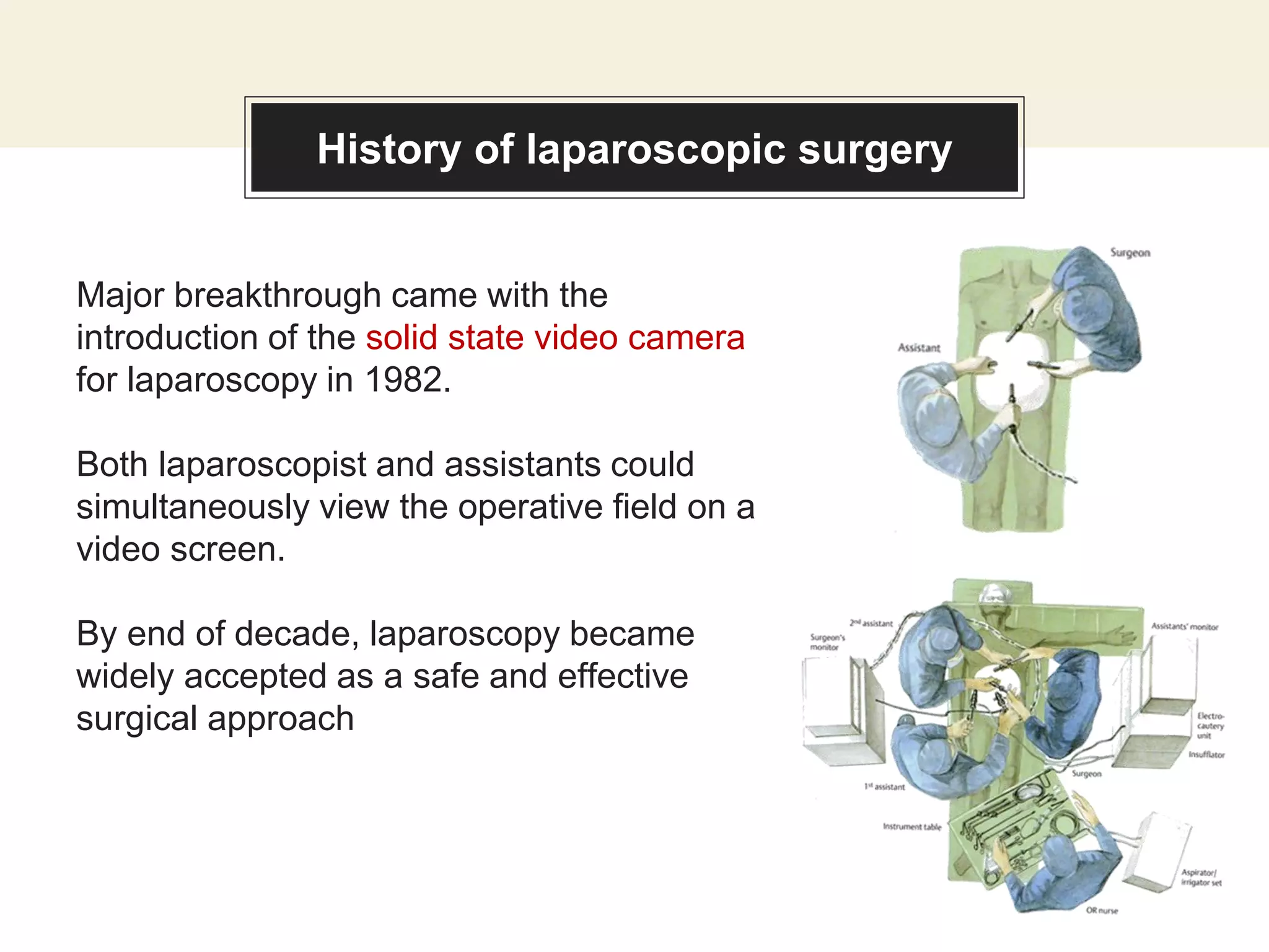
Major breakthrough came with the
introduction of the solid state video camera
for laparoscopy in 1982.
Both laparoscopist and assistants could
simultaneously view the operative field on a
video screen.
By end of decade, laparoscopy became
widely accepted as a safe and effective
surgical approach
History of laparoscopic surgery
Today, operative laparoscopy is routinely used by
gynecologists to perform a multitude of procedures,
including hysterectomies and incontinence procedures,
and for the diagnosis and treatment of gynecologic
malignancies.
Basics of laparoscopy - Instruments
Verres needle
Used to inflate air/CO2 to the peritoneal cavity
(pneumoperitoneum) through the umbilicus where there is
the thinnest abdominal wall.
Basics of laparoscopy - Instruments
Electronic laparoflator: Insufflator
Used to insufflate through the verres needle.
Maintains constant intra-abdominal pressure
without exceeding the safety limit.
Basics of laparoscopy - Instruments
Trocars
Permit access to the intra-peritoneal cavity in
which other instruments can pass.
The trocar used should be adapted to the diameter
of the telescope selected
Basics of laparoscopy - Instruments
Telescope
There are different sizes and angles, each with a
different use.
They are used to visualize the peritoneal cavity.
Basics of laparoscopy - Instruments
Camera equipment & Light source
Basics of laparoscopy - Instruments
Forceps and scissors
There are two types:
◉ Disposable
◉ Reusable
They can be either atraumatic
or grasping foreceps.
Basics of laparoscopy - Procedure
Preparation of the patient:
◉ Inform the patient about the
therapeutic benefits and potential
risks (informed consent).
◉ Intestinal preparation: Simple
intestinal emptying, for better viewing
and preventing injuries.
◉ Place the patient in the dorsolithotomy
position.
Basics of laparoscopy - Procedure
Creating a pneumoperitoneum:
◉ The abdominal wall is lifted by hand or by
grasping forceps
◉ Pnemoperitoneum is created by verres
needle introduced to the umbilical area
◉ The needle is inserted in an oblique angle
toward the uterine fundus
◉ The negative pressure will allow the
underlying structures to fall away.
◉ After making sure that the needle is in
correct position, air flow can be increased
to 2.5 L/min till a pressure of 15mmHg
Basics of laparoscopy - Procedure
Trocar introduction
Once the intra-abdominal pressure reaches
15 mmHg the main trocar is introduced after
removal of veress needle.
The position of the trocar must be verified by
inserting the laparoscope and viewing the
pelvic cavity.
Basics of laparoscopy - Procedure
Viewing the peritoneal cavity
The omentum, bowel and bifurcation of
pelvic vessels should be evaluated to avoid
injuries caused during the introduction of
Verres needle or trocar.
The site of introduction of other trocars
should be verified by finger palpation and
transillumination of abdominal wall to avoid
injury to epigastric vessels.
Identify if there is any bleeding
Basics of laparoscopy - Procedure
During the operative procedure
• 1 or 2 bottles of Ringer’s lactate are used to wash the peritoneal
cavity after laparoscopy.
• Leave 500/1000 ml of ringer’s lactate to reduce the incidence of
post operative pain.
After the procedure
• CO2 gas must be evacuated completely to reduce post-operative
pain
Applications of Laparoscopy
◉ As a diagnostic tool
◉ As a therapeutic modality
Laparoscopy - as a diagnostic tool
◉ Infertility: status of the fallopian tube (morphology and
functionality) and any pathological condition (e.g.
adhesions)
◉ Ovarian cysts or tumors.
◉ Ectopic pregnancy.
◉ PID: tubal abscess or adhesions.
◉ Endometriosis: define the sites of implants and
endometrial cysts.
Laparoscopy - as a diagnostic tool
◉ Chronic pelvic pain
◉ Ectopic pregnancy
◉ Pelvic inflammatory disease
◉ Endometriosis
◉ Adhesions
◉ Ovary: cysts, torsion
◉ Fallopian tube: torsion, salpingitis
◉ Uterus: fibroid, leiomyomata
◉ Pelvic congestion syndrome
◉ Infertility
◉ Oncologic procedures
Laparoscopy – in Chronic Pelvic Pain (CPP)
> 40% of gynecological diagnostic laparoscopies are
done for CPP
Combined results of published series of laparoscopies
for CPP shows:
◉ No visible pathology is detected in 35%
◉ Endometriosis is diagnosed in 33%
◉ Adhesive disease is found in 24%
A negative laparoscopy → not synonymous with no
disease
A meticulously performed negative laparoscopy means
that a woman does not have endometriosis-associated
or adhesion-associated pain
The role of laparoscopy as a diagnostic tool in chronic pelvic pain. Fred M Howard MS,
MD BaillieÁ re's Clinical Obstetrics and Gynaecology Vol. 14, No. 3, pp. 467-494, 2000.
Laparoscopy – in Ectopic pregnancy
Transvaginal ultrasound has replaced laparoscopy for
diagnosis of ectopic pregnancy.
Laparoscopy has diagnostic role if probable tubal
pregnancy or diagnosis in doubt.
Large uncontrolled studies have demonstrated that >
80% of ectopic pregnancies can be managed
laparoscopically
The most commonly used procedures at laparoscopy
are salpingectomy and salpingotomy.
The role of laparoscopy in the management of ectopic pregnancy. Martin Christopher Sowter, MD,
MRCOGa, Jonathan Frappell, FRCS, FRCOG Reviews in Gynaecological Practice #2 (2002) 73-82
Laparoscopy – in Pelvic Inflammatory
Disease (PID)
Clinical diagnosis of PID is often difficult especially
when symptoms are mild, as frequently when the
primary organism is C. trachomatis.
Laparoscopy is the gold standard for the diagnosis of
PID – should be used when diagnosis is uncertain,
especially in young women for whom the preservation
of fertility is important.
Sellors et al. reported that only by resorting to
diagnostic laparoscopy were they able to demonstrate
that PID was the cause of acute pelvic pain in 46% of a
group of 95 women.
Laparoscopy should be considered for patients who
have not responded to antibiotic therapy within 48 to
72 hours. The role of laparoscopy in the management of pelvic pain in women of reproductive age.
Maria Grazia Porpora, M.D. FERTILITY AND STERILITY Vol. 6M, No. 5, November 1997.
Laparoscopy – in Tubo-ovarian Abscess
Most commonly isolated pathogens from a tubo-
ovarian abscess are C. trachomatis and
peptostreptococci.
At laparoscopy the peritoneal cavity (pelvis and
abdomen) is inspected carefully.
The surgical steps include adhesiolysis, aspiration of
the abscess cavity, dissection and excision of necrotic
tissue, tubal lavage, and irrigation of the peritoneal
cavity before completion of the procedure.
Laparoscopic surgery combined with adequate broad-
spectrum antibiotic therapy has proven successful in
the treatment of more than 95% of patients.
Laparoscopy – in Endometriosis
Endometriosis is a histologically-defined disease:
“the presence of ectopic tissue which possesses
the histological structure and function of the
uterine mucosa”
- Sampson,1921
Laparoscopy has largely replaced laparotomy as
the diagnostic procedure for any patient
suspected of having endometriosis.
Based on visual appearance endometriosis are
classified as atypical (red, yellow, white or clear)
or typical (black-brown, black or puckered black
stellate) lesions.
The role of laparoscopy as a diagnostic tool in chronic pelvic pain. Fred M Howard MS,
MD BaillieÁ re's Clinical Obstetrics and Gynaecology Vol. 14, No. 3, pp. 467-494, 2000.
Laparoscopy – in Endometriosis
Endometriosis presents with a variety of
appearances that may make visual diagnosis
difficult and inaccurate. Brownish lesion on the ovary
White fibrotic lesion on the
uterosacral ligament
Peritoneal pocket of
endometriosis
Red stellate lesions in
the cul-de-sac
The role of laparoscopy as a diagnostic tool in chronic pelvic pain. Fred M Howard MS, MD
BaillieÁ re's Clinical Obstetrics and Gynaecology Vol. 14, No. 3, pp. 467-494, 2000.
Laparoscopy – in Adhesions
Presently the only definitive way to
diagnose adhesions is by surgical
visualization usually via laparoscopy
instead of laparotomy.
Laparoscopic studies reveal adhesions
on average in 24% of CPP patients
and 17% of non-CPP patients.
The role of laparoscopy as a diagnostic tool in chronic pelvic pain. Fred M Howard MS, MD
BaillieÁ re's Clinical Obstetrics and Gynaecology Vol. 14, No. 3, pp. 467-494, 2000.
Laparoscopy – in Adenomyosis
Endometrial cells penetrate the
myometrium causing either localized
(adenomyoma) or diffuse overgrowth.
Adenomyomas that penetrate the uterine
cavity become submucosal tumors.
An enlarged uterus from adenomyosis is
often misdiagnosed as being from
fibroids
Laparoscopy – in Ovarian Cysts
Most ovarian cysts are haemorrhagic
corpora lutea or follicle cysts.
They are usually asymptomatic and
when they cause pain it is almost always
acute.
Laparoscopic evaluations of patients
with CPP reveal ovarian cysts on
average in only 3% of all cases.
The role of laparoscopy as a diagnostic tool in chronic pelvic pain. Fred M Howard MS,
MD BaillieÁ re's Clinical Obstetrics and Gynaecology Vol. 14, No. 3, pp. 467-494, 2000.
Laparoscopy – in Ovarian Cysts
Even when the surgeon is ‘certain’ that
the ovary is benign, it is essential that
tissue be sent for histological evaluation.
Open the cyst and inspect the lining for
papillary structures or excrescences.
If these are noted, then a laparotomy
should be done
Laparoscopy – in Adnexal Torsion
A rare gynecologic emergency that
nearly always occurs unilaterally.
Relapse or bilateral adnexal torsion can
cause sterility interfering with fertility.
In 30% of the patients, there is torsion of
a normal adnexa, while the majority of
the cases are associated with ovarian
pathology.
Adnexal torsion in very young girls: diagnostic pitfalls. Marieke Emontsa, Heleen Doornewaardb, J.(Co’tje) F.
Admiraala, European Journal of Obstetrics & Gynecology and Reproductive Biology 116 (2004) 207-210.
Laparoscopy – in Adnexal Torsion
Conservative management by laparoscopy
is the best approach when tissues are viable
and should be carried out promptly to
preserve the adnexa
(basic principles of conservative
management are to untwist the structure
and treat the underlying cause ‘ie - ovarian
cyst’).
Once untwisted, the organ must be
observed to ensure color change to normal,
confirming viability and blood supply.
Adnexal torsion in very young girls: diagnostic pitfalls. Marieke Emontsa, Heleen Doornewaardb, J.(Co’tje) F.
Admiraala, European Journal of Obstetrics & Gynecology and Reproductive Biology 116 (2004) 207-210.
Laparoscopy – in Endosalpingiosis
Endosalpingiosis is the presence of
fallopian tubal glandular epithelium in an
ectopic location.
Visually it appears as white to yellow,
opaque or translucent, punctate, cystic
lesions.
Endosalpingiosis is generally not
recognized at the time of laparoscopic
evaluation or is misdiagnosed as
endometriosis.
The role of laparoscopy as a diagnostic tool in chronic pelvic pain. Fred M Howard MS,
MD BaillieÁ re's Clinical Obstetrics and Gynaecology Vol. 14, No. 3, pp. 467-494, 2000
Laparoscopy – in Leiomyomata (fibroids)
The role of laparoscopy as a diagnostic tool in chronic pelvic pain. Fred M Howard MS,
MD BaillieÁ re's Clinical Obstetrics and Gynaecology Vol. 14, No. 3, pp. 467-494, 2000
Laparoscopy – in Oncologic procedures
Ghezzi F, Cromi A, Uccella S, Siesto G, Zefiro F, Bolis P. Incorporating Laparoscopy in the Practice of a
Gynecologic Oncology Service: Actual Impact Beyond Clinical Trials Data. Ann Surg Oncol. 2009 May 21.
Mori KM, Neubauer NL. Minimally invasive surgery in gynecologic oncology. ISRN Obstet Gynecol. 2013
Aug 12. 2013:312982.
For second-look procedures following surgical and chemo
treatment of malignancy.
Laparoscopy has also been used for staging, including
peritoneal washes with biopsy, partial omentectomy, and
pelvic and periaortic lymphadenectomy
Laparoscopically assisted radical vaginal hysterectomy have
also been used by some gynecologic oncologists.
Laparoscopy - as a therapeutic tool
1. Management of ovarian cyst by
• Drainage
• Ovarian cystectomy
• Ovarian drilling of the cortex and stroma to decrease androgens in
the ovaries
• Correcting ovarian torsion.
• As a treatment of endometriosis By removal of the endometrial cyst,
cauterization of endometrial spots and adhesiolysis
Laparoscopy - as a therapeutic tool
2. Management of infertility:
• Adhesiolysis
• Treat the cause (endometriosis, PCOS)
3. Myomectomy for fibroids:
used for subserosal and intramural fibroids only, not used for
submucosal fibroids.
4. Management of PID
by draining tubal abscess and adhesiolysis.
Laparoscopy – in Adhesiolysis
Adhesions may form due to prior
infection, such as a ruptured appendix
or pelvic inflammatory disease (PID),
endometriosis, or previous surgery
Adhesions may be lysed by blunt or
sharp dissection
Aquadissection may aid in the
development of planes prior to lysing.
Any of the power instruments may be
used for cutting and coagulation
Laparoscopy – in Myomectomy
The fibroid may be removed by
morcellation or colpotomy.
Power morcellators are available to
expedite the process.
If the patient has a pedunculated fibroid,
the stalk may be easily incised. However,
for intramural fibroids, the risk of bleeding
increases.
Laparoscopy – in Management of Ectopic
Pregnancy
Salpingotomy
Used to preserve the tubes for desired
reproductivity.
Done if the patient is hemodynamicaly stable
If size < 5 cm
Location must be ampullary, infundibular or
isthmic.
Contralateral tube either normal or absent.
Laparoscopy – in Management of Ectopic
Pregnancy
Salpingectomy
(it is the standard for ectopic pregnancy) if:
• Ruptured tube
• Multiple recurrence of ectopic pregnancy
• Size of ectopic > 5 cm
Laparoscopy – Tubal sterilization
Tubal sterilization can be done using:
◉ Bipolar coagulation
◉ Clips (filshie clips) and rings
Before the procedure, inform patient
about:
• Chance of irreversibility
• Failure rate 1/200
• Bleeding may occur and we may shift to laparatomy.
Laparoscopic hysterectomy
Laparoscopic hysterectomy:
The 3 basic laparoscopic approaches for
hysterectomy are
• laparoscopic-assisted vaginal hysterectomy (LAVH)
• laparoscopic hysterectomy (LH)
• laparoscopic supracervical hysterectomy (LSH)
Laparoscopy – contraindications
1. Generalized peritonitis
2. Hypovolemic shock
3. Severe cardiac disease
4. Hemoglobin less than 7 g/dL
5. Uterine size > 12 wks.
6. Multiple previous abdominal procedures
7. Extreme body weight
Laparoscopy – risk factors
Patient related risk factors
◉ Obesity
◉ Age
◉ Previous abdominal surgery
Anesthetic risk factors
◉ Time since last oral intake
◉ Heart disease
◉ Pulmonary disease
Laparoscopy – Patient related risk
factors
Obesity
◉ Laparoscopy becomes more difficult and potentially
more risky.
◉ Placement of laparoscopic instruments becomes much
more difficult
◉ Bleeding from abdominal wall vessels may be more
common because these vessels become difficult to
locate.
◉ Restricted operative field secondary to retroperitoneal fat
deposits in the pelvic sidewalls and increased bowel
excursion into the operative field
Laparoscopy – Patient related risk
factors
Age
◉ Older patients are at increased risk of having
concomitant disease processes that affect their
perioperative morbidity and mortality
Laparoscopy – Patient related risk
factors
Previous abdominal surgery
◉ Risk of adhesions of omentum and/or bowel to the
anterior abdominal wall after previous abdominal surgery
is greater than 20%.
◉ As laparoscopy requires the insertion of sharp
instruments into the abdominal cavity, a reasonable
assumption is that previous surgery would increase the
risk of bowel injury
Laparoscopy – pre-op work-up
1. Complete blood cell count
2. Pregnancy test
3. Urinalysis
4. ECG
5. Other:
• In patients with known health problems, other laboratory tests, such as liver
function tests or electrolyte evaluations, may be indicated.
• A thorough preoperative medical evaluation, including appropriate
laboratory studies
6. Imaging studies:
• Chest radiography
• Intravenous pyelograph or kidney ultrasound
• Barium enema
Laparoscopy – possible
complications
Laparoscopic procedures have unique risks, related to:
• methods used for the placement of abdominal wall ports
• pneumoperitoneum required for laparoscopy
1. Pneumoperitoneum related complications
2. Injury to abdominal organs
3. Blood vessel injury
Laparoscopy complications –
Pneumoperitoneum
Pneumoperitoneum related complications
• Extra-peritoneal emphysema due to failure of introducing verres
needle correctly into the peritoneal cavity and not checking the
negative pressure on the machine.
• Gas may extend to the mediastinum and compromise cardiac
function
• Pneumo-omentum
• Increased intra-abdominal pressures may increase anesthesia-
related risks such as aspiration and increased difficulty ventilating
the patient
Laparoscopy complications – abdominal
organs
Injury to abdominal organs
• GI: if the intestine is distended or adherent to the abdominal wall
(prevented by good intestinal preparation) and putting the patient on
the telendelenburg position.
• Bladder injury: prevented by emptying the bladder.
Laparoscopy complications – blood vessel
injury
Blood vessel injury:
◉ Pelvic, omental and mesentric blood vessel injury
◉ Prevented by introducing the verres needle in an angle.
◉ Although the risk of blood loss is relatively low,
potentially massive blood loss may occur and is
complicated as control may be delayed because of the
time taken to perform an emergency laparotomy.
Laparoscopy – recent advances
3 innovations that have been introduced in the field of
laparoscopy:
◉ Robotic surgery
◉ Natural orifice transluminal surgery (NOTES)
◉ Single incision laparoscopic surgery (SILS).
Of these 3 developing technologies, robotic surgery is
having the largest impact on clinical care.
Advincula AP, Wang K. Evolving role and current state of robotics in minimally invasive
gynecologic surgery. J Minim Invasive Gynecol. 2009 May-Jun. 16(3):291-301
Swanström LL. Natural orifice transluminal endoscopic surgery. Endoscopy. 2009 Jan.
41(1):82-5.
Laparoscopy – robotic surgery
Robotic system advantages:
• 3-dimensional, high-definition imaging and magnification.
• Fully articulated instruments emulate the full range of motion of a
surgeon’s wrists and hands.
• Enhances the surgeon’s ability to remotely perform fine motor skills
such as intricate dissections and intracorporeal suturing that remain
difficult during traditional laparoscopy.
Laparoscopy – robotic surgery
Robotic system advantages:
• Robotic tools attach to traditional laparoscopic ports and the robotic
system is placed between the patient’s legs for hysterectomy. The
surgeon controls the instruments from a console located in the
same room.
• Direct correlation between hand movements and instrument
movements.
Advincula AP, Wang K. Evolving role and current state of robotics in minimally invasive
gynecologic surgery. J Minim Invasive Gynecol. 2009 May-Jun. 16(3):291-301.
Natural orifice transluminal surgery
Using an endoscope to access the abdominal cavity
through existing body openings (Ex: mouth, rectum,
and vagina)
Modern NOTES uses a flexible endoscope to access
the peritoneal cavity by creating an incision in the
stomach or colon.
ABARBANEL AR. Transvaginal pelvioscopy (peritoneoscopy); a simplified and safe
technic as an office procedure. Am J Surg. 1955 Jul. 90(1):122-8.
Single incision laparoscopic surgery
(SILS) refers to performing laparoscopy
through a single incision.
Bradford LS, Boruta DM. Laparoendoscopic single-site surgery in gynecology: a review of
the literature, tools, and techniques. Obstet Gynecol Surv. 2013 Apr. 68(4):295-304.
Advantages vs disadvantages
◉ Minimizes the number
of incisions
◉ In turn results in
decreased pain,
improved cosmetics
◉ Reduces the risks
associated with a
secondary port
placement.
Bradford LS, Boruta DM. Laparoendoscopic single-site surgery in gynecology: a review of the literature, tools, and
techniques. Obstet Gynecol Surv. 2013 Apr. 68(4):295-304.
Eom JM, Choi JS, Choi WJ, Kim YH, Lee JH. Does Single-Port Laparoscopic Surgery Reduce Postoperative Pain in Women
with Benign Gynecologic Disease?. J Laparoendosc Adv Surg Tech A. 2013 Oct 1.
visibility, depth
perception,
maneuverability,
reach, and the
ability to create
counter-traction
are all limited.
Advantages Disadvantages
Laparoscopy – risk vs benefit
Laparoscopy is a hybrid surgical approach that shares
characteristics of both minor and major surgery.
To patients, laparoscopic procedures often seem
to be minor surgery because of the small incisions,
relatively small amount of postoperative pain, and
short convalescent period.
Its an intra-abdominal procedure - therefore, it
shares all intraoperative and postoperative risks
of laparotomy, including infection and injury to
adjacent intra-abdominal structures
Laparoscopy – summary
Laparoscopy continues to evolve as more sophisticated
instrumentation allows a greater variety of procedures to be
performed
The risks and benefits of the procedures in many cases
have not been fully evaluated
Procedures, such as tubal ligation, ectopic pregnancy
removal, and simple adhesion lysis, appear to be safely
and efficiently performed laparoscopically.
But more complicated procedures still need to be evaluated
to determine the safest, most cost-effective, and most
efficient procedure.
Laparoscopy – conclusion
Laparoscopy provides a vital tool for diagnosing pelvic
pain.
Laparoscopy provides first hand visual comprehension of
the problem as well as an immediate opportunity to
continue with therapeutic surgical correction.
In the past, many of these procedures would have been
limited to laparotomy and would have required a prolonged
recovery period.
Thank
you

Laparoscopy in gynecology

  • 1.
  • 2.
    Contents ◉ Introduction ◉ History ◉Instruments ◉ Overview procedure ◉ Use as a diagnostic tool ◉ Use as a therapeutic modality ◉ Contraindications ◉ Risk factors ◉ Complications ◉ Recent advances ◉ Summary and conclusion
  • 3.
    Laparoscopy A surgical procedurein which a fibre-optic instrument is inserted through the abdominal wall to view the organs in the abdomen or permit small-scale surgery with help of pneumoperitoneum
  • 4.
    Laparoscopic surgery Laparoscopic surgery,also called minimally invasive surgery (MIS), bandaid surgery, or keyhole surgery, is a modern surgical technique in which operations are performed far from their location through small incisions (usually 0.5–1.5 cm)
  • 5.
    History of laparoscopicsurgery Dr. Hans Christian Jacobaeus, a Swedish surgeon, was the first to publish a description of laparothorakoskopie in humans in 1910. Litynski GS. Laparoscopy--the early attempts: spotlighting Georg Kelling and Hans Christian Jacobaeus.JSLS. 1997. 1:83-5. He used air pneumoperitoneum and a cystoscope to evaluate the peritoneal cavity of tuberculosis patients with ascites.
  • 6.
    History of laparoscopicsurgery Dr. Janos Veress, a Hungarian internist, developed a spring-loaded needle with an inner stylet that automatically converted the sharp cutting edge to a rounded end. The Veress needle is still used today to create a pneumoperitoneum
  • 7.
    History of laparoscopicsurgery In 1961, Dr. Palmer described the first laparoscopic retrieval of oocytes, and in 1974 he described the point 3 cm below the last rib in the left mid-clavicular line. Palmer's point is often used today for left upper quadrant laparoscopic entry. In addition to advocating monitoring of intra-abdominal pressure, he pioneered intra-abdominal electrocoagulation of bleeding sites, puncture of ovarian cysts, and lysis of pelvic adhesions. Palmer R. Safety in laparoscopy. J Reprod Med. 1974 Jul. 13(1):1-5.
  • 8.
    History of laparoscopicsurgery Dr. Kurt Semm, a German gynecologist who specialized in infertility, invented the automatic insufflator & instruments like thermocoagulator, loop ligature, and devices for extracorporeal and intracorporeal endoscopic knot tying. Mettler L, Semm K, Shive K. Endoscopic management of adnexal masses. J Soc Laparoendosc Surg. 1997 Apr-Jun. 1(2):103-12.
  • 9.
    History of laparoscopicsurgery Major breakthrough came with the introduction of the solid state video camera for laparoscopy in 1982. Both laparoscopist and assistants could simultaneously view the operative field on a video screen. By end of decade, laparoscopy became widely accepted as a safe and effective surgical approach
  • 10.
    History of laparoscopicsurgery Today, operative laparoscopy is routinely used by gynecologists to perform a multitude of procedures, including hysterectomies and incontinence procedures, and for the diagnosis and treatment of gynecologic malignancies.
  • 11.
    Basics of laparoscopy- Instruments Verres needle Used to inflate air/CO2 to the peritoneal cavity (pneumoperitoneum) through the umbilicus where there is the thinnest abdominal wall.
  • 12.
    Basics of laparoscopy- Instruments Electronic laparoflator: Insufflator Used to insufflate through the verres needle. Maintains constant intra-abdominal pressure without exceeding the safety limit.
  • 13.
    Basics of laparoscopy- Instruments Trocars Permit access to the intra-peritoneal cavity in which other instruments can pass. The trocar used should be adapted to the diameter of the telescope selected
  • 14.
    Basics of laparoscopy- Instruments Telescope There are different sizes and angles, each with a different use. They are used to visualize the peritoneal cavity.
  • 15.
    Basics of laparoscopy- Instruments Camera equipment & Light source
  • 16.
    Basics of laparoscopy- Instruments Forceps and scissors There are two types: ◉ Disposable ◉ Reusable They can be either atraumatic or grasping foreceps.
  • 17.
    Basics of laparoscopy- Procedure Preparation of the patient: ◉ Inform the patient about the therapeutic benefits and potential risks (informed consent). ◉ Intestinal preparation: Simple intestinal emptying, for better viewing and preventing injuries. ◉ Place the patient in the dorsolithotomy position.
  • 18.
    Basics of laparoscopy- Procedure Creating a pneumoperitoneum: ◉ The abdominal wall is lifted by hand or by grasping forceps ◉ Pnemoperitoneum is created by verres needle introduced to the umbilical area ◉ The needle is inserted in an oblique angle toward the uterine fundus ◉ The negative pressure will allow the underlying structures to fall away. ◉ After making sure that the needle is in correct position, air flow can be increased to 2.5 L/min till a pressure of 15mmHg
  • 19.
    Basics of laparoscopy- Procedure Trocar introduction Once the intra-abdominal pressure reaches 15 mmHg the main trocar is introduced after removal of veress needle. The position of the trocar must be verified by inserting the laparoscope and viewing the pelvic cavity.
  • 20.
    Basics of laparoscopy- Procedure Viewing the peritoneal cavity The omentum, bowel and bifurcation of pelvic vessels should be evaluated to avoid injuries caused during the introduction of Verres needle or trocar. The site of introduction of other trocars should be verified by finger palpation and transillumination of abdominal wall to avoid injury to epigastric vessels. Identify if there is any bleeding
  • 21.
    Basics of laparoscopy- Procedure During the operative procedure • 1 or 2 bottles of Ringer’s lactate are used to wash the peritoneal cavity after laparoscopy. • Leave 500/1000 ml of ringer’s lactate to reduce the incidence of post operative pain. After the procedure • CO2 gas must be evacuated completely to reduce post-operative pain
  • 22.
    Applications of Laparoscopy ◉As a diagnostic tool ◉ As a therapeutic modality
  • 23.
    Laparoscopy - asa diagnostic tool ◉ Infertility: status of the fallopian tube (morphology and functionality) and any pathological condition (e.g. adhesions) ◉ Ovarian cysts or tumors. ◉ Ectopic pregnancy. ◉ PID: tubal abscess or adhesions. ◉ Endometriosis: define the sites of implants and endometrial cysts.
  • 24.
    Laparoscopy - asa diagnostic tool ◉ Chronic pelvic pain ◉ Ectopic pregnancy ◉ Pelvic inflammatory disease ◉ Endometriosis ◉ Adhesions ◉ Ovary: cysts, torsion ◉ Fallopian tube: torsion, salpingitis ◉ Uterus: fibroid, leiomyomata ◉ Pelvic congestion syndrome ◉ Infertility ◉ Oncologic procedures
  • 25.
    Laparoscopy – inChronic Pelvic Pain (CPP) > 40% of gynecological diagnostic laparoscopies are done for CPP Combined results of published series of laparoscopies for CPP shows: ◉ No visible pathology is detected in 35% ◉ Endometriosis is diagnosed in 33% ◉ Adhesive disease is found in 24% A negative laparoscopy → not synonymous with no disease A meticulously performed negative laparoscopy means that a woman does not have endometriosis-associated or adhesion-associated pain The role of laparoscopy as a diagnostic tool in chronic pelvic pain. Fred M Howard MS, MD BaillieÁ re's Clinical Obstetrics and Gynaecology Vol. 14, No. 3, pp. 467-494, 2000.
  • 26.
    Laparoscopy – inEctopic pregnancy Transvaginal ultrasound has replaced laparoscopy for diagnosis of ectopic pregnancy. Laparoscopy has diagnostic role if probable tubal pregnancy or diagnosis in doubt. Large uncontrolled studies have demonstrated that > 80% of ectopic pregnancies can be managed laparoscopically The most commonly used procedures at laparoscopy are salpingectomy and salpingotomy. The role of laparoscopy in the management of ectopic pregnancy. Martin Christopher Sowter, MD, MRCOGa, Jonathan Frappell, FRCS, FRCOG Reviews in Gynaecological Practice #2 (2002) 73-82
  • 27.
    Laparoscopy – inPelvic Inflammatory Disease (PID) Clinical diagnosis of PID is often difficult especially when symptoms are mild, as frequently when the primary organism is C. trachomatis. Laparoscopy is the gold standard for the diagnosis of PID – should be used when diagnosis is uncertain, especially in young women for whom the preservation of fertility is important. Sellors et al. reported that only by resorting to diagnostic laparoscopy were they able to demonstrate that PID was the cause of acute pelvic pain in 46% of a group of 95 women. Laparoscopy should be considered for patients who have not responded to antibiotic therapy within 48 to 72 hours. The role of laparoscopy in the management of pelvic pain in women of reproductive age. Maria Grazia Porpora, M.D. FERTILITY AND STERILITY Vol. 6M, No. 5, November 1997.
  • 28.
    Laparoscopy – inTubo-ovarian Abscess Most commonly isolated pathogens from a tubo- ovarian abscess are C. trachomatis and peptostreptococci. At laparoscopy the peritoneal cavity (pelvis and abdomen) is inspected carefully. The surgical steps include adhesiolysis, aspiration of the abscess cavity, dissection and excision of necrotic tissue, tubal lavage, and irrigation of the peritoneal cavity before completion of the procedure. Laparoscopic surgery combined with adequate broad- spectrum antibiotic therapy has proven successful in the treatment of more than 95% of patients.
  • 29.
    Laparoscopy – inEndometriosis Endometriosis is a histologically-defined disease: “the presence of ectopic tissue which possesses the histological structure and function of the uterine mucosa” - Sampson,1921 Laparoscopy has largely replaced laparotomy as the diagnostic procedure for any patient suspected of having endometriosis. Based on visual appearance endometriosis are classified as atypical (red, yellow, white or clear) or typical (black-brown, black or puckered black stellate) lesions. The role of laparoscopy as a diagnostic tool in chronic pelvic pain. Fred M Howard MS, MD BaillieÁ re's Clinical Obstetrics and Gynaecology Vol. 14, No. 3, pp. 467-494, 2000.
  • 30.
    Laparoscopy – inEndometriosis Endometriosis presents with a variety of appearances that may make visual diagnosis difficult and inaccurate. Brownish lesion on the ovary White fibrotic lesion on the uterosacral ligament Peritoneal pocket of endometriosis Red stellate lesions in the cul-de-sac The role of laparoscopy as a diagnostic tool in chronic pelvic pain. Fred M Howard MS, MD BaillieÁ re's Clinical Obstetrics and Gynaecology Vol. 14, No. 3, pp. 467-494, 2000.
  • 31.
    Laparoscopy – inAdhesions Presently the only definitive way to diagnose adhesions is by surgical visualization usually via laparoscopy instead of laparotomy. Laparoscopic studies reveal adhesions on average in 24% of CPP patients and 17% of non-CPP patients. The role of laparoscopy as a diagnostic tool in chronic pelvic pain. Fred M Howard MS, MD BaillieÁ re's Clinical Obstetrics and Gynaecology Vol. 14, No. 3, pp. 467-494, 2000.
  • 32.
    Laparoscopy – inAdenomyosis Endometrial cells penetrate the myometrium causing either localized (adenomyoma) or diffuse overgrowth. Adenomyomas that penetrate the uterine cavity become submucosal tumors. An enlarged uterus from adenomyosis is often misdiagnosed as being from fibroids
  • 33.
    Laparoscopy – inOvarian Cysts Most ovarian cysts are haemorrhagic corpora lutea or follicle cysts. They are usually asymptomatic and when they cause pain it is almost always acute. Laparoscopic evaluations of patients with CPP reveal ovarian cysts on average in only 3% of all cases. The role of laparoscopy as a diagnostic tool in chronic pelvic pain. Fred M Howard MS, MD BaillieÁ re's Clinical Obstetrics and Gynaecology Vol. 14, No. 3, pp. 467-494, 2000.
  • 34.
    Laparoscopy – inOvarian Cysts Even when the surgeon is ‘certain’ that the ovary is benign, it is essential that tissue be sent for histological evaluation. Open the cyst and inspect the lining for papillary structures or excrescences. If these are noted, then a laparotomy should be done
  • 35.
    Laparoscopy – inAdnexal Torsion A rare gynecologic emergency that nearly always occurs unilaterally. Relapse or bilateral adnexal torsion can cause sterility interfering with fertility. In 30% of the patients, there is torsion of a normal adnexa, while the majority of the cases are associated with ovarian pathology. Adnexal torsion in very young girls: diagnostic pitfalls. Marieke Emontsa, Heleen Doornewaardb, J.(Co’tje) F. Admiraala, European Journal of Obstetrics & Gynecology and Reproductive Biology 116 (2004) 207-210.
  • 36.
    Laparoscopy – inAdnexal Torsion Conservative management by laparoscopy is the best approach when tissues are viable and should be carried out promptly to preserve the adnexa (basic principles of conservative management are to untwist the structure and treat the underlying cause ‘ie - ovarian cyst’). Once untwisted, the organ must be observed to ensure color change to normal, confirming viability and blood supply. Adnexal torsion in very young girls: diagnostic pitfalls. Marieke Emontsa, Heleen Doornewaardb, J.(Co’tje) F. Admiraala, European Journal of Obstetrics & Gynecology and Reproductive Biology 116 (2004) 207-210.
  • 37.
    Laparoscopy – inEndosalpingiosis Endosalpingiosis is the presence of fallopian tubal glandular epithelium in an ectopic location. Visually it appears as white to yellow, opaque or translucent, punctate, cystic lesions. Endosalpingiosis is generally not recognized at the time of laparoscopic evaluation or is misdiagnosed as endometriosis. The role of laparoscopy as a diagnostic tool in chronic pelvic pain. Fred M Howard MS, MD BaillieÁ re's Clinical Obstetrics and Gynaecology Vol. 14, No. 3, pp. 467-494, 2000
  • 38.
    Laparoscopy – inLeiomyomata (fibroids) The role of laparoscopy as a diagnostic tool in chronic pelvic pain. Fred M Howard MS, MD BaillieÁ re's Clinical Obstetrics and Gynaecology Vol. 14, No. 3, pp. 467-494, 2000
  • 39.
    Laparoscopy – inOncologic procedures Ghezzi F, Cromi A, Uccella S, Siesto G, Zefiro F, Bolis P. Incorporating Laparoscopy in the Practice of a Gynecologic Oncology Service: Actual Impact Beyond Clinical Trials Data. Ann Surg Oncol. 2009 May 21. Mori KM, Neubauer NL. Minimally invasive surgery in gynecologic oncology. ISRN Obstet Gynecol. 2013 Aug 12. 2013:312982. For second-look procedures following surgical and chemo treatment of malignancy. Laparoscopy has also been used for staging, including peritoneal washes with biopsy, partial omentectomy, and pelvic and periaortic lymphadenectomy Laparoscopically assisted radical vaginal hysterectomy have also been used by some gynecologic oncologists.
  • 40.
    Laparoscopy - asa therapeutic tool 1. Management of ovarian cyst by • Drainage • Ovarian cystectomy • Ovarian drilling of the cortex and stroma to decrease androgens in the ovaries • Correcting ovarian torsion. • As a treatment of endometriosis By removal of the endometrial cyst, cauterization of endometrial spots and adhesiolysis
  • 41.
    Laparoscopy - asa therapeutic tool 2. Management of infertility: • Adhesiolysis • Treat the cause (endometriosis, PCOS) 3. Myomectomy for fibroids: used for subserosal and intramural fibroids only, not used for submucosal fibroids. 4. Management of PID by draining tubal abscess and adhesiolysis.
  • 42.
    Laparoscopy – inAdhesiolysis Adhesions may form due to prior infection, such as a ruptured appendix or pelvic inflammatory disease (PID), endometriosis, or previous surgery Adhesions may be lysed by blunt or sharp dissection Aquadissection may aid in the development of planes prior to lysing. Any of the power instruments may be used for cutting and coagulation
  • 43.
    Laparoscopy – inMyomectomy The fibroid may be removed by morcellation or colpotomy. Power morcellators are available to expedite the process. If the patient has a pedunculated fibroid, the stalk may be easily incised. However, for intramural fibroids, the risk of bleeding increases.
  • 44.
    Laparoscopy – inManagement of Ectopic Pregnancy Salpingotomy Used to preserve the tubes for desired reproductivity. Done if the patient is hemodynamicaly stable If size < 5 cm Location must be ampullary, infundibular or isthmic. Contralateral tube either normal or absent.
  • 45.
    Laparoscopy – inManagement of Ectopic Pregnancy Salpingectomy (it is the standard for ectopic pregnancy) if: • Ruptured tube • Multiple recurrence of ectopic pregnancy • Size of ectopic > 5 cm
  • 46.
    Laparoscopy – Tubalsterilization Tubal sterilization can be done using: ◉ Bipolar coagulation ◉ Clips (filshie clips) and rings Before the procedure, inform patient about: • Chance of irreversibility • Failure rate 1/200 • Bleeding may occur and we may shift to laparatomy.
  • 47.
    Laparoscopic hysterectomy Laparoscopic hysterectomy: The3 basic laparoscopic approaches for hysterectomy are • laparoscopic-assisted vaginal hysterectomy (LAVH) • laparoscopic hysterectomy (LH) • laparoscopic supracervical hysterectomy (LSH)
  • 48.
    Laparoscopy – contraindications 1.Generalized peritonitis 2. Hypovolemic shock 3. Severe cardiac disease 4. Hemoglobin less than 7 g/dL 5. Uterine size > 12 wks. 6. Multiple previous abdominal procedures 7. Extreme body weight
  • 49.
    Laparoscopy – riskfactors Patient related risk factors ◉ Obesity ◉ Age ◉ Previous abdominal surgery Anesthetic risk factors ◉ Time since last oral intake ◉ Heart disease ◉ Pulmonary disease
  • 50.
    Laparoscopy – Patientrelated risk factors Obesity ◉ Laparoscopy becomes more difficult and potentially more risky. ◉ Placement of laparoscopic instruments becomes much more difficult ◉ Bleeding from abdominal wall vessels may be more common because these vessels become difficult to locate. ◉ Restricted operative field secondary to retroperitoneal fat deposits in the pelvic sidewalls and increased bowel excursion into the operative field
  • 51.
    Laparoscopy – Patientrelated risk factors Age ◉ Older patients are at increased risk of having concomitant disease processes that affect their perioperative morbidity and mortality
  • 52.
    Laparoscopy – Patientrelated risk factors Previous abdominal surgery ◉ Risk of adhesions of omentum and/or bowel to the anterior abdominal wall after previous abdominal surgery is greater than 20%. ◉ As laparoscopy requires the insertion of sharp instruments into the abdominal cavity, a reasonable assumption is that previous surgery would increase the risk of bowel injury
  • 53.
    Laparoscopy – pre-opwork-up 1. Complete blood cell count 2. Pregnancy test 3. Urinalysis 4. ECG 5. Other: • In patients with known health problems, other laboratory tests, such as liver function tests or electrolyte evaluations, may be indicated. • A thorough preoperative medical evaluation, including appropriate laboratory studies 6. Imaging studies: • Chest radiography • Intravenous pyelograph or kidney ultrasound • Barium enema
  • 54.
    Laparoscopy – possible complications Laparoscopicprocedures have unique risks, related to: • methods used for the placement of abdominal wall ports • pneumoperitoneum required for laparoscopy 1. Pneumoperitoneum related complications 2. Injury to abdominal organs 3. Blood vessel injury
  • 55.
    Laparoscopy complications – Pneumoperitoneum Pneumoperitoneumrelated complications • Extra-peritoneal emphysema due to failure of introducing verres needle correctly into the peritoneal cavity and not checking the negative pressure on the machine. • Gas may extend to the mediastinum and compromise cardiac function • Pneumo-omentum • Increased intra-abdominal pressures may increase anesthesia- related risks such as aspiration and increased difficulty ventilating the patient
  • 56.
    Laparoscopy complications –abdominal organs Injury to abdominal organs • GI: if the intestine is distended or adherent to the abdominal wall (prevented by good intestinal preparation) and putting the patient on the telendelenburg position. • Bladder injury: prevented by emptying the bladder.
  • 57.
    Laparoscopy complications –blood vessel injury Blood vessel injury: ◉ Pelvic, omental and mesentric blood vessel injury ◉ Prevented by introducing the verres needle in an angle. ◉ Although the risk of blood loss is relatively low, potentially massive blood loss may occur and is complicated as control may be delayed because of the time taken to perform an emergency laparotomy.
  • 58.
    Laparoscopy – recentadvances 3 innovations that have been introduced in the field of laparoscopy: ◉ Robotic surgery ◉ Natural orifice transluminal surgery (NOTES) ◉ Single incision laparoscopic surgery (SILS). Of these 3 developing technologies, robotic surgery is having the largest impact on clinical care. Advincula AP, Wang K. Evolving role and current state of robotics in minimally invasive gynecologic surgery. J Minim Invasive Gynecol. 2009 May-Jun. 16(3):291-301 Swanström LL. Natural orifice transluminal endoscopic surgery. Endoscopy. 2009 Jan. 41(1):82-5.
  • 59.
    Laparoscopy – roboticsurgery Robotic system advantages: • 3-dimensional, high-definition imaging and magnification. • Fully articulated instruments emulate the full range of motion of a surgeon’s wrists and hands. • Enhances the surgeon’s ability to remotely perform fine motor skills such as intricate dissections and intracorporeal suturing that remain difficult during traditional laparoscopy.
  • 60.
    Laparoscopy – roboticsurgery Robotic system advantages: • Robotic tools attach to traditional laparoscopic ports and the robotic system is placed between the patient’s legs for hysterectomy. The surgeon controls the instruments from a console located in the same room. • Direct correlation between hand movements and instrument movements. Advincula AP, Wang K. Evolving role and current state of robotics in minimally invasive gynecologic surgery. J Minim Invasive Gynecol. 2009 May-Jun. 16(3):291-301.
  • 61.
    Natural orifice transluminalsurgery Using an endoscope to access the abdominal cavity through existing body openings (Ex: mouth, rectum, and vagina) Modern NOTES uses a flexible endoscope to access the peritoneal cavity by creating an incision in the stomach or colon. ABARBANEL AR. Transvaginal pelvioscopy (peritoneoscopy); a simplified and safe technic as an office procedure. Am J Surg. 1955 Jul. 90(1):122-8.
  • 62.
    Single incision laparoscopicsurgery Single incision laparoscopic surgery (SILS) refers to performing laparoscopy through a single incision. Bradford LS, Boruta DM. Laparoendoscopic single-site surgery in gynecology: a review of the literature, tools, and techniques. Obstet Gynecol Surv. 2013 Apr. 68(4):295-304.
  • 63.
    Advantages vs disadvantages ◉Minimizes the number of incisions ◉ In turn results in decreased pain, improved cosmetics ◉ Reduces the risks associated with a secondary port placement. Bradford LS, Boruta DM. Laparoendoscopic single-site surgery in gynecology: a review of the literature, tools, and techniques. Obstet Gynecol Surv. 2013 Apr. 68(4):295-304. Eom JM, Choi JS, Choi WJ, Kim YH, Lee JH. Does Single-Port Laparoscopic Surgery Reduce Postoperative Pain in Women with Benign Gynecologic Disease?. J Laparoendosc Adv Surg Tech A. 2013 Oct 1. visibility, depth perception, maneuverability, reach, and the ability to create counter-traction are all limited. Advantages Disadvantages
  • 64.
    Laparoscopy – riskvs benefit Laparoscopy is a hybrid surgical approach that shares characteristics of both minor and major surgery. To patients, laparoscopic procedures often seem to be minor surgery because of the small incisions, relatively small amount of postoperative pain, and short convalescent period. Its an intra-abdominal procedure - therefore, it shares all intraoperative and postoperative risks of laparotomy, including infection and injury to adjacent intra-abdominal structures
  • 65.
    Laparoscopy – summary Laparoscopycontinues to evolve as more sophisticated instrumentation allows a greater variety of procedures to be performed The risks and benefits of the procedures in many cases have not been fully evaluated Procedures, such as tubal ligation, ectopic pregnancy removal, and simple adhesion lysis, appear to be safely and efficiently performed laparoscopically. But more complicated procedures still need to be evaluated to determine the safest, most cost-effective, and most efficient procedure.
  • 66.
    Laparoscopy – conclusion Laparoscopyprovides a vital tool for diagnosing pelvic pain. Laparoscopy provides first hand visual comprehension of the problem as well as an immediate opportunity to continue with therapeutic surgical correction. In the past, many of these procedures would have been limited to laparotomy and would have required a prolonged recovery period.
  • 67.