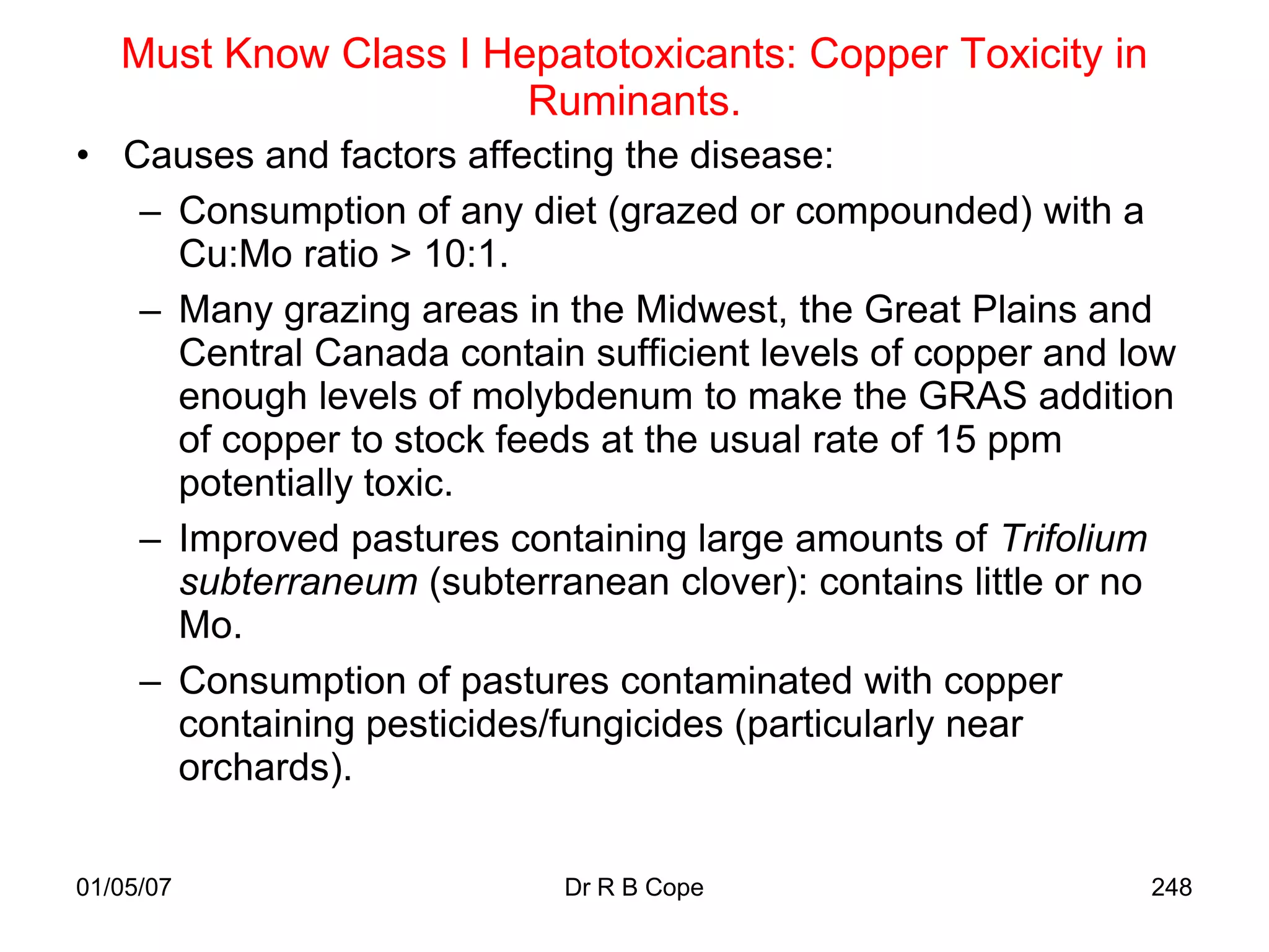
An Introduction to the Toxicology of the Liver
                  & Rodent Stomach.
           Rhian B. Cope BVSc BSc(Hon 1) PhD DABT ERT


01/05/07                    Dr R B Cope                 1
Yes, there is a lot of basic science.

It is included deliberately: if you do not understand
the fundamentals of how and why the liver reacts to
xenobiotics, you cannot really understand the
significance and human-relevance of the changes
that occur.

Understanding the mode of action is the key to just
about everything in toxicology and toxicological risk
assessment.

Please bear with me.

 01/05/07               Dr R B Cope                     2
Sections.
Section 1: A Revision of the Basic Anatomy and Physiology of the Liver,
Reasons for the Susceptibility of the Liver to Toxic Injury and Classical
Clinical Signs of Hepatic Disease.

Section 2: Responses of the Liver to Toxic Injury

Section 3: Interpretation of Rodent Hepatic Tumour Data: The Human-
Relevance Framework

Section 4: Detection/ Measurement/Assessment of Hepatic Toxicity.

Section 5: The Two Basic Classes of Hepatic Toxicants, and Classical “Must
Know” Agents Causing Hepatic Damage.

Section 6: Interpretation of Rodent Stomach Tumour Data: The Human-
Relevance Framework.

Section 7: Case Studies.


01/05/07                           Dr R B Cope                               3
Section 1.

 A Revision of the Basic Anatomy and Physiology of the
 Liver, Reasons for the Susceptibility of the Liver to Toxic
 Injury and Classical Clinical Signs of Hepatic Disease.


01/05/07                 Dr R B Cope                    4
Learning Tasks Section 1.
1.    Describe and understand the toxicologically significant features of the
      hepatic circulation.
2.    Describe and understand the structure and toxicologically significant
      features of the liver lobule.
3.    Describe and understand the structure and toxicologically significant
      features of the liver acinus.
4.    Understand the toxicological significance of Kupffer, Pit and Ito cells.
5.    Describe and understand the key physiological roles of the liver and the
      potential effects of disrupting these functions.
6.    Describe and understand the toxicologically significant features of bile
      formation/excretion and excretion of bilirubin.
7.    Describe and understand the basis for the susceptibility of the liver as a
      toxic target organ.
8.    Describe and understand the classical clinical signs of hepatic disease.

     01/05/07                        Dr R B Cope                                 5
Hepatic Circulation and Blood Supply.


                              •Key points:
                                   Liver receives blood via two routes:
                                   high oxygen blood from the hepatic
                                   artery (30%) and low oxygen blood
                                   from the portal vein (70%).
                                   Blood leaves the liver only by the
                                   hepatic vein.
                                   Liver is placed between venous
                                   blood returning from the bulk of the
                                   GI and peritoneal cavity and the
                                   venous arm of the systemic
                                   circulation.
                                   WHAT ARE THE TOXICOLOGICAL
                                   

                                   CONSEQUENCES OF THIS?

01/05/07                   Dr R B Cope                              6
Structure of the Liver Lobule.


     Low magnification view ofBthe a liver lobule in the pig
01/05/07                   Dr R Cope                           7
Structure of the Liver Lobule.


01/05/07   Low magnification view B Cope human liver lobule
                               Dr R of the                    8
Structure of the Liver Lobule.


01/05/07               Dr R B Cope          9
Structure of the Liver Lobule.


01/05/07              Dr R B Cope           10
Structure of the Liver Lobule.


Note the lack of an
endothelial basement
membrane, large
endothelial pores and
large endocytic vacuoles.
What are the key
toxicological
consequences of these
features?


  01/05/07                  Dr R B Cope        11
Structure of the Liver Acinus.


01/05/07              Dr R B Cope           12
Structure of the Liver Acinus.


01/05/07               Dr R B Cope          13
Structure of the Liver Acinus.


01/05/07              Dr R B Cope           14
Structure of the Liver Acinus.


01/05/07               Dr R B Cope          15
01/05/07   Dr R B Cope   16
Structure of the Liver Acinus.


• Acinar zone 1 approximates “Periportal” using the
  “Lobular” system.


• Acinar zone 3 approximates “Centrilobular” using the
  “Lobular” system.


01/05/07                 Dr R B Cope                     17
Describe the distribution of damage (necrosis) in this liver
01/05/07                     Dr R B Cope                      18
         section using the “lobular” and “acinar” system.
?


   Describe the distributionDr R B Cope
01/05/07
                             of damage (necrosis) in this liver
                                                              19
       section using the “lobular” and “acinar” system.
Central Vein


   Describe the distributionDr R B Cope
01/05/07
                             of damage (necrosis) in this liver
                                                              20
       section using the “lobular” and “acinar” system.
Central Vein


01/05/07
           Centrilobular orB Zone 3 Necrosis.
                       Dr R Cope                21
Structure of the Liver Acinus.
• Hepatocytes are generated in zone 1 from their primordial stem
  cell and migrate from zone 1 to zone 3 before undergoing
  senescence/apoptosis in zone 3.

   – The youngest hepatocytes occur in zone 1, the oldest occur
     in zone 3.

   – The hepatocyte cycle in the rat is approximately 200 days.


   01/05/07                 Dr R B Cope                     22
Structure of the Liver Acinus.


• All hepatocytes are NOT equal. Important
  functional/physiological differences occur between
  hepatocytes in different acinar zones.


 01/05/07                  Dr R B Cope                 23
Hepatocyte Zonal Specialization.
        Parameter                Zone 1          Zone 2             Zone 3
 Oxygen tension and level         High         Intermediate           Low
of nutrients in blood supply
 Exposure to portal blood      First site of   Intermediate   Last site of exposure
                                exposure
    Glutathione levels            High         Intermediate           Low
    Bile acid excretion           High         Intermediate           Low
 Overall balance between       Relatively      Intermediate   Phase I predominates
  Phase I and Phase II         balanced                          over Phase II
       metabolism
CYP level (particularly Cyp      Lower         Intermediate           High
           2E1)
    Level of fatty acid           High         Intermediate           Low
oxidation, gluconeogeneis,
     and ureagenesis
Concentration of materials        High         Intermediate           Low
   (bile salts, bilirubin,
 excreted compounds) in
 adjacent bile canaliculus

 Number of mitochondria           High         Intermediate           Low
   Glycogen and other             High         Intermediate           Low
     nutrient stores
Toxicological Consequences of Hepatocyte Zonal Specialization.
         Parameter              Zone 1   Zone 2    Zone 3
  Oxygen tension and level
 of nutrients in blood supply
  Exposure to portal blood
     Glutathione levels
     Bile acid excretion
  Overall balance between
   Phase I and Phase II
        metabolism
 CYP level (particularly Cyp
            2E1)
     Level of fatty acid
 oxidation, gluconeogeneis,
      and ureagenesis
 Concentration of materials
    (bile salts, bilirubin,
  excreted compounds) in
  adjacent bile canaliculus

  Number of mitochondria
    Glycogen and other
      nutrient stores
Kupffer Cells.
• Kupffer cells are the resident tissue macrophage of the
  liver. Located in the sinusoids.

• Large number of Kupffer cells are present in the liver:
  80% of body‟s resident tissue macrophages.

• Fully functional macrophage: can trigger inflammation
  and act as antigen presenting cells.


01/05/07                   Dr R B Cope                      26
Kupffer Cells.
• Of considerable importance in hepatic toxicology:

    – Activation during inflammation results in the
      generation of various free radicals e.g. superoxide
      anion, peroxynitrite, nitrogen oxides

    – Triggering and participation in inflammation.

    – Accumulation of iron (hemosiderin, ferritin).

    – Degradation of heme.


01/05/07                    Dr R B Cope                     27
Pigment accumulation within Kupffer cells.
01/05/07                    Dr R B Cope                 28
Pit Cells.
• Located in the space of Disse.

• Function as NK or LAK cells.

• Important in inflammation.


01/05/07                  Dr R B Cope   29
Ito Cells.
• Synonyms = “fat cells”, stellate cells.

    – Two major roles:

           • Storage of Vitamin A.

           • During inflammation or liver damage, produce
             collagen i.e. responsible for hepatic fibrosis.


01/05/07                       Dr R B Cope                     30
Congestive cirrhosis (replacement of hepatocytes with fibrous tissue)
secondary to right sided heart failure, trichrome stain. Remember: Ito cells
are responsible for the laying down of new collagen within the liver. WHAT
ARE THE CRITICAL FUNCTIONAL CONSEQUENCES OF SUCH A
REACTION IN THE LIVER?

 01/05/07                         Dr R B Cope                            31
A Concise Summary of Key Hepatic Functions


01/05/07                                  32
Consequences of Disruption of Hepatic Function
                                    Consequences


                                              33
01/05/07
Bile Formation and Hepatic Excretion.
•      Bile formation involves both hepatocytes and
       cholangiocytes

•      Bile formation involves 8 basic processes:

      1. Materials that undergo biliary excretion move from the sinusoid
         through the space of Disse and through the basolateral
         hepatocyte cell membrane via diffusion, active transport or
         endocytosis.

      2. The materials for excretion are transported across the
         hepatocyte with or without metabolism and storage and then
         actively transported into the canaliculi.

      3. Vesiclular transport involves the detachment of lipid vesicles
         from the apical hepatocyte membrane to form bile micelles.
         Bile micelles contain lipophilic compounds, bile salts,
         cholesterol, phospholipids, and high molecular compounds


01/05/07                        Dr R B Cope                               34
Bile Formation and Hepatic Excretion.
   4. Excretion of compounds is sufficient to generate osmotic water
      flow into the bile canaliculi.

   5. Forward movement of bile within the canaliculi occurs by ATP-
      dependent peristaltic contraction of the actin-myosin web
      located underneath the apical membrane of the hepatocytes.

   6. Within the bile ductules and common hepatic duct, bile
      composition and volume are modified by cholangiocytes:

   7. Volume increases due to the osmotic gradient created by the
      active excretion of HCO3- in exchange for Cl- by cholangiocytes;
      ~ 40% of bile volume is due to this excretion mechanism.

   8. Cholangiocyte re-uptake of some constituents (some bile acids)
      occurs.


01/05/07                       Dr R B Cope                             35
Bile Formation and Hepatic Excretion.


•     Molecules with a molecular weight of ≤ 300 Da are
      more efficiently excreted in bile than molecules with a
      greater molecular weight.


01/05/07                    Dr R B Cope                         36
Major Hepatocyte and Cholangiocyte
           Transporters involved in Bile Formation


01/05/07                   Dr R B Cope               37
Major Hepatocyte Involved in Bile Formation
Basolateral Transporters               Function

Na+-taurocholate-co-transporting       Uptake of conjugated bile acids,
peptide (NTCP)                         estrogens

Organic anion transporter              Uptake of amphiphilic compounds,
polypeptide (OATP)                     steroid conjugates, neutral steroids,
                                       sulfobromophthalein (OATP2),
                                       bilirubin (OATP2), glutathione
                                       conjugates, leukotriene s, C4
                                       organic cations, small peptides,
                                       digoxin

Organic cation transporter I (OCT I)   Divalent lipophilic cations,
                                       xenobiotics that contain a tertiary or
                                       quarternary amine group
Bilitranslocase                        Bilirubin, sulfobromophthalein;
                                       inhibited by phenylmethyl-sulphonyl
                                       fluoride; exists in two metastable
                                       forms: high and low affinity.
Major Hepatocyte Transporters Involved in Bile Formation.
BasolateralTransporters     Function
Organic anion transporter 2 (OAT2)   Uptake of indocyanine green, and
                                     nonsteroidal anti-inflammatory
                                     drugs, such as ketoprofen,
                                     indomethacin, and salicylates
                                      through the basolateral hepatocyte
                                     cell
                                     membrane


 01/05/07                       Dr R B Cope                           39
Major Hepatocyte Transporters Involved in Bile Formation.
Apical Transporters         Function
Multidrug resistance proteins        Excretion of cationic and lipophilic
(MDR), particularly MDR1             compounds. MDR1 has no physiological
                                     substrate in non-ruminants; function is
(Note: MDR1 = p-glycoprotein,        the secretion of amphiphilic cationic
which has now been renamed the       xenobiotics, steroid hormones,
ATP-binding cassette sub-family B    hydrophobic pesticides and glycolipids;
member 1 transporter, or ABCB1)      responsible for phyloerythrin excretion
                                     in ruminants!

SPGP = bile salt export pump         Transports monoanionic bile salts.
(BSEP)
Multidrug resistance-associated      Excretion of glucuronic acid, sulfate
proteins (MRP); MRP2 = canalicular   and glutathione (anionic)
multispecific organic anion          conjugates, phospholipids;
transporter (cMOAT)                  Excretion of mono- and diglucuronic
                                     acid bilirubin conjugates (MRP2)
                                     and glutathione-
                                     sulfobromophthalein conjugates
                                     (MRP2)
Hepatic Bilirubin Excretion.
Heme containing proteins (Hb,
Mb, CYP450)                                                                                                                   Hepatocyte


                                                                                        Sinusoid
                                  Reticuloendothelial system
                                                                                                   Alb


                                                                                                                                                                  Bile canaliculus
                                                               Spleen, Kupffer cells,
Free heme (red)


                                                                                                                                              UDP-glucuronide
Heme
                                                                                                                     OATP *
oxygenase
                                                                                                   Br                                    Br
Biliverdin (green)
Biliverdin
                                                                                        Alb-Br
                                                                                                                     BT   *
reductase


                                                                                                                               UGT-1A1
Bilirubin (Br;brown)                                                                                                                                            MRP2           *
                     Albumen


                                                                                                    Space of Disse
                     (ALB)

                                                                                                                          Conjugated Br
  Alb-Br                        Systemic                                                                                                                          Gluc-Br
                                                                                                                          (Gluc-Br)                               in Bile
                                Circulation
  (“Free” or unconjugated Br)


*Organic anion transport protein; *Bilitranslocase; * Rate limiting step for bilirubin excretion
Extrahepatic Aspects of Bilirubin Excretion.

 • Conjugated bilirubin excreted in the bile is converted by
   bacterial action within the ileum and colon into
   urobilinogen which undergoes enterohepatic circulation.

 • Urobilinogen that is not taken up and re-excreted by the
   liver passes into the systemic circulation and is excreted
   by the glomerular filtration in the kidneys


01/05/07                  Dr R B Cope                      42
Extrahepatic Aspects of Bilirubin Excretion.

  • The amount of urobilinogen formed, and thus excreted
    by the kidneys increases dramatically with increased
    formation of bilirubin (e.g. hemolysis).

  • The amount of urobilinogen in urine will decrease with:
     – Severe cholestasis (failure of conjugated bilirubin
       excretion).
     – Bile duct obstruction.
     – Severe disruption of the GI microflora (antibiotics).


01/05/07                  Dr R B Cope                      43
Important Aspects of Bilirubin Excretion.
• The excretion of conjugated bilirubin is inhibited by the
  administration of sulfobromophthalein due to competition for
  the MRP2 transporter.

• Impaired hepatic sulfobromophthalein excretion (i.e.
  increased or delayed retention) has at least three potential
  causes:

   – Cholestasis due to impaired apical excretion.
   – Inhibition of glutathione-S-transferases (requires
     conjugation to glutathione for excretion).
   – Impaired basloateral bilitranslocase and OATP function.

   * note: bromosulfonphthalein (BSP) was a commercial brand name for
       sulfobromophthalein. Older literature will often refer to a BSP test
       which simply means a test for plasma clearance of
       sulfobromophthalein.
 01/05/07                         Dr R B Cope                             44
Important Aspects of Bilirubin Excretion.
• Bilirubin in plasma is measured by the van den Bergh
  assay which makes two different measurements: total
  bilirubin and direct bilirubin.

• Classically, the direct bilirubin is regarded as a measure
  of conjugated bilirubin in plasma.

• Indirect bilirubin (unconjugated) = total bilirubin – direct
  bilirubin.


01/05/07                   Dr R B Cope                           45
Important Aspects of Bilirubin Excretion.
• Modern analytical methods have now demonstrated that
  plasma from normal individuals contains virtually no
  conjugated (i.e. “direct”) bilirubin.

• Elevations of plasma direct or conjugated bilirubin
  primarily occur with:
   – Obstruction of the bile ducts or canaliculi.
   – Decreased canalicular contraction.
   – Inhibition of MRP2.
   – Hepatocellular disease.


01/05/07                  Dr R B Cope                   46
Bilirubin Excretion in the Neonate.
• Bilirubin excretion, like most hepatic excretion, takes
  time to develop in neonates.

• Bilirubin produced by the fetus is cleared by the placenta
  and eliminated by the maternal liver.

• After birth, the neonatal liver slowly develops the
  capacity for bilirubin clearance and excretion.

• Levels of UGT1A1 in neonatal hepatocytes are low and
  unconjugated bilirubin is excreted into the gut.


01/05/07                  Dr R B Cope                       47
Bilirubin Excretion in the Neonate.

• The neonatal gut lacks the microflora to convert bilirubin
  to urobilinogen and bilirubin undergoes enterohepatic
  cycling.

• Levels of MRP2 are also low in the neonate. Remember
  transport of conjugated bilirubin across the hepatocyte
  apical cell membrane is the rate-limiting step for bilirubin
  excretion.

• Neonates typically have elevated free bilirubin in their
  plasma due to impaired excretion by MRP2 and
  enterohepatic cycling.


01/05/07                  Dr R B Cope                        48
Bilirubin Excretion in the Neonate.
• Any xenobiotic that increases the production of
  bilirubin in the neonate will produce rapid, large
  increases in plasma bilirubin.
      – Any agent that produces hemolysis or defective
        erythrogenesis.
      – Any agent that produces hemorrhage.
      – Any agent that produces cholestasis.

• This results in a condition called kernicterus
  (bilirubin encephalopathy) in which bilirubin crosses
  the blood-brain barrier and precipitates within the
  basal ganglia and other sites in the brain resulting in
  CNS damage. Yellow staining of brain nuclei due to
  bilirubin precipitates is the classical pathology
  associated with kernicterus.

01/05/07                     Dr R B Cope                 49
Globus pallidus staining with bilirubin
01/05/07                  Dr R B Cope                50
Basis for the Susceptibility of the Liver to Toxicity.

 • Position within the circulatory system: high exposure to
   xenobiotics absorbed via the GI (also peritoneum) i.e.
   first pass effect.

 • High level of biotransformation, and therefore, significant
   risk of generating reactive metabolites.

 • Susceptibility to oxidant injury.

 • Susceptibility to hypoxic injury (centrilobular).

 • Critical biosynthetic/homeostatic functions.

 01/05/07                   Dr R B Cope                       51
Basis for the Susceptibility of the Liver to Toxicity.
 • Ability to concentrate xenobiotics within the biliary tree,

 • Large tissue macrophage population: inflammation and
   oxidative injury.

 • Little or no selectivity of sinusoidal endothelium (large
   pores).

 • Capacity to separate xenbiotics from albumen and other
   carrier proteins.

 • Capacity to accumulate metals, vitamin A and other
   xenobiotics.
 • Liver has high energy consumption and Is susceptible to
   agents that affect mitochondrial function.
 01/05/07                    Dr R B Cope                         52
Basis for the Susceptibility of the Liver to Toxicity.
•     Enterohepatic circulation can result in sustained exposure to
      xenobiotics.

•     Lipophilic xenobiotics tend to concentrate within the liver
      since it is relatively rich in cell membranes

•     Substrates for the transporter systems of the basolateral
      hepatocyte membrane also tend to selectively accumulate in
      the liver e.g. phalloidin, microcystin.

•     Compounds that have hepatic storage can cause toxicity e.g.
      iron (stored as ferritin), cadmium (stored as a Cd-
      metallothionine complex), vitamin A (selectively stored in Ito
      cells)
     01/05/07                   Dr R B Cope                         53
Patients showing clear evidence of
               jaundice: yellow discoloration of the skin
               and sclera.
               Important differential is high dietary beta
               carotene – tissues and skin are stained
               yellow, but the sclera remains white!

01/05/07   Dr R B Cope                                  54
Clinical Signs of Acute Hepatocellular Disease.

• Markers of malaise i.e. fatigue, weakness, nausea, poor
  appetite.

• Icterus/jaundice: probably the best clinical marker of
  severity. Indicates bilirubin level > 2.5 mg/dl.

• Spider angiomata and palmar erythema.

• Itching (self mutilation in animals).


01/05/07                   Dr R B Cope                     55
Clinical Signs of Acute Hepatocellular Disease.

• Right upper quadrant abdominal pain.

• Abdominal distention.

• Intestinal bleeding.

• ± Heatomegaly.

• Bilirubinuria: dark characteristically colored urine

• In many cases of hepatocellular disease, there are no
  clinical signs. Cases are recognized by biochemical liver
  tests.


01/05/07                   Dr R B Cope                   56
01/05/07   Dr R B Cope   57
Clinical Signs of Advanced or Chronic
                   Hepatocellular Disease.

• Weight loss, muscle wasting.

• Evidence of hemorrhage and coagulopathy.

                                              Evidence of
• Ascites.                                    inadequate serum
                                              protein synthesis.
• Edema of the extremities.

• Fetor hepaticus = typical sweet ammoniacal odour of
  patients with hepatic failure (failure of ammonia
  clearance/metabolism).

01/05/07                  Dr R B Cope                     58
Ascites following severe liver disease. Note the eversion of
                       the umbilicus.
01/05/07                  Dr R B Cope                     59
Mid-level abdominal CT scans. Left = normal; Right = ascites
                 secondary to liver failure.

 01/05/07                 Dr R B Cope                    60
Clinical Signs of Advanced or Chronic
                   Hepatocellular Disease.

 • Hepatic encephalopathy (change in sleep patterns,
   change in personality, irritability, mental dullness,
   disorientation, stupor, asterixis*, flapping tremors of body
   and tongue, coma).

 • Caput medusa = development of prominent collateral
   veins radiating from the umbilicus due to the
   recanulation of the umbilical vein and its tributaries due
   to portal hypertension and porto-systemic shunting.

* Asterixis = a motor disturbance marked by intermittent lapse of an assumed
posture due to intermittent sustained contraction of muscle groups;
characteristic of hepatic coma; often assessed by asking the patient to write or
draw simple pictures (e.g. draw a clock face).
01/05/07                            Dr R B Cope                                    61
Caput medusae associated with portal hypertension,
       portosystemic shunting and severe liver disease.
01/05/07                   Dr R B Cope                     62
Clinical Signs of Advanced or Chronic
                   Hepatocellular Disease.

• Hepatorenal syndrome: characterized by progressive
  renal failure that develops following chronic liver disease
  + ascites and other evidence of liver failure. Mechanism
  is unknown but the syndrome is associated with altered
  renal hemodynamics and altered prostaglandin levels
  are implicated.

• Portal hypertension, portosystemic shunting and acute
  venous hemorrhage due to rupture of abdominal veins.

• Spontaneous bacterial peritonitis (failure of bacterial
  opsonization due to low albumen and other opsonizers).

01/05/07                  Dr R B Cope                      63
Clinical Signs of Advanced or Chronic
                   Hepatocellular Disease.

• Hepatopulmonary syndrome: development of right to left
  intrapulmonary shunts in advanced liver disease.
  Mechanism is unknown but involves altered pulmonary
  nitric oxide levels.


01/05/07                  Dr R B Cope                 64
Clinical Signs of Advanced Hepatocellular or Cholestatic
  Disease in Ruminants: Secondary Photosensization.
 In ruminants:

                  Rumen bacteria
Chlorophyll                         Phylloerythrin


                                         Absorbed


                                         Hepatocyte


                               Transported across the apical
       Excreted in bile        hepatocyte cell membrane by ATP-
                               binding cassette transporter B1 [p-
                               glycoprotein or MDR 1)

01/05/07                           Dr R B Cope                       65
Clinical Signs of Advanced Hepatocellular or Cholestatic
 Disease in Ruminants: Secondary Photosensitization.

 • Prolonged inhibition of ABCB1, cholestasis or
   hepatocelular disease in ruminants results in an
   accumulation of phylloerythrin within the circulation and
   tissues.

 • Phylloerythrin absorbs light and acts as a photosensitizer
   within the skin resulting in severe skin inflammation and
   sloughing.

 • Disease in sheep (particularly associated with
   sporodesmin-induced liver disease) is colloquially called
   “facial eczema.”

01/05/07                   Dr R B Cope                     66
Secondary photosensitization of the face due to
01/05/07      sporodesmin poisoning in a sheep
                           Dr R B Cope                     67
Severe secondary photosensitzation of the udder of a cow
     with advanced hepatic disease (again due to sporodesmin)
01/05/07                    Dr R B Cope                         68
Section 2:

           Responses of the Liver to Toxic Injury.


01/05/07                 Dr R B Cope                 69
Learning Tasks Section 2.

1.     Describe and understand the stereotypical cellular
       responses of the liver to xenobiotic injury.

2.     Describe and understand the processes involved in the
       development of cholestasis.


01/05/07                    Dr R B Cope                     70
Stereotypical Responses of the Liver to
                          Toxicant Injury.
• The patterns of the hepatocellular response to toxicant injury
  are generally stereotypical and not toxicant specific (although
  there are exceptions to this rule).

• The hallmark of the liver’s response to toxicant injury is its
  large functional reserve and large capacity for healing,
  often with no significant sequelae!
   – For example, a 2/3 hepatectomy is survivable and both
     normal liver function and size will be restored within weeks!

   – This will occur provided significant fibrosis or massive
     necrosis of the lobules does not occur and the source of
     injury is removed i.e. exposure is not chronic.

   01/05/07                   Dr R B Cope                     71
Hepatocellular Adaptive Responses.

•     These changes are generally reversible once
      xenobiotic exposure stops.

•     In terms of a toxicology study, ideally this propensity
      for reversal should be tested by the inclusion of an
      adequate post-exposure recovery period in the study.

•     This inevitably involves inclusion of additional
      experimental groups i.e. groups that is euthanitized at
      the end of exposure (necropsy + histology) plus groups
      that are euthanitized 14 to 30 days post exposure +
      appropriate control groups.

01/05/07                    Dr R B Cope                         72
Hepatocellular Adaptive Responses.


•     Sadly this is rarely done despite the provision for this in
      the OECD guidelines.

•     My personal view is that histological discrimination of
      the types of lesion present is not sufficient to claim
      reversibility; must have actual documented study
      evidence of the reversibility of hepatic adaptive
      responses!


01/05/07                     Dr R B Cope                        73
Hepatocellular Adaptive Responses.


•     Represent adaptive responses to xenobiotic response
      rather than hepatocellular damage per se.

•     Used as histological markers of xenobiotic exposure.


01/05/07                   Dr R B Cope                       74
Hepatocellular Adaptive Responses.

•     Do not result in disease per se but are often of
      significance for the toxicokinetics/toxicodynamics of
      drugs and other xenobiotics and thus may significantly
      influence the toxicity of particular toxins/toxicants.


•     Usually detected histologically but may be visible
      grossly as hepatomegaly and/or increased liver weight.


01/05/07                    Dr R B Cope                    75
Hepatocellular Adaptive Responses:
     Centrilobular Hepatocellular Hypertrophy.

•      Due to ↑ smooth endoplasmic reticulum content in
       centrilobular/Zone 3 hepatocytes.

•      Associated with chemical induction of CYP, particularly
       CYP2E1.

•      Associated with massive increases in the amount of
       smooth endoplasmic reticulum.


01/05/07                    Dr R B Cope                     76
Hepatocellular Adaptive Responses:
     Centrilobular Hepatocellular Hypertrophy.

•     Reversible following removal of the initiating agent.

•     Example initiating agents: phenobarbital and other
      oxybarbiturates, Ah receptor agonists (TCDD, PCDFs).


01/05/07                    Dr R B Cope                       77
Centrilobular hepatocyte hypertrophy in a mouse treated
              with phenobarbital for 8 months.
01/05/07                 Dr R B Cope                    78
Centrilobular hepatocyte hypertrophy in a mouse treated with
phenobarbital for 8 months Note the eosinophilic cytoplasm due to
 the large increase in smooth endoplasmic reticulum as a result of
 01/05/07
                CYP (particularly CYP2E1) induction.             79
Hepatocellular Adaptive Responses:
Eosinophilic Centrilobular Hepatocellular Hypertrophy.
 •     Due to ↑ peroxisomes in centrilobular hepatocytes.

 •     Prolonged eosinophilic centrilobular hypertrophy is
       associated with pericanalicular lipofuscin pigment
       deposition.

 •     Prolonged exposure to chemicals that induce
       peroxisome induction may result in hepatocellular
       neoplasia in rodents.


 01/05/07                   Dr R B Cope                      80
Hepatocellular Adaptive Responses:
Eosinophilic Centrilobular Hepatocellular Hypertrophy.
 •      Reversible following removal of the initiating agent.

 •      Classical agents: phthalate plasticizers.

 •      Rodent-specific response.

 •      Relevance to humans is controversial!

 •      Currently regarded as not relevant to humans in many
        jurisdictions, however this is an area of considerable
        scientific challenge

 01/05/07                     Dr R B Cope                       81
Centrilobular eosinophilic hepatocyte hypertrophy (left) in a mouse due to chronic
   exposure to phthalates. Right image shows immunohistochemical staining for
   peroxisomes. Note that chronic exposure to peroxisome proliferators is
   carcinogenic in rodents but not humans.
01/05/07                                                                           82
                                       Dr R B Cope
Hepatocellular Adaptive Responses:
     Xenobiotic-Induced Hepatocyte Hyperplasia.

•     Usually accompanied by CYP induction,
      hepatomegaly, and hepatocyte hypertrophy.

•     Never continues for more than a few days.

•     Reversible following removal of the initiating agent.
      Reversion is associated with ↑ hepatocyte apoptosis.


01/05/07                   Dr R B Cope                        83
Derived from the UK PSD guideline
                         (included as an appendix to the
                         notes)


01/05/07   Dr R B Cope                              84
Early Markers of Hepatocellular Damage:
           Hepatocyte Nucleolar Lesions.


•     Due to changes in RNA synthesis.

•     Changes include: ↓ size, ↑ size, nucleolar
      fragmentation, nucleolar segregation.

•     ↓ Nucleolar size is usually an acute lesion that
      occurs within hours of hepatotoxin exposure;
      often the first identifiable toxic hepatic lesion.

•     ↑ Nucleolar size is commonly associated with hepatic
      neoplasia.

01/05/07                   Dr R B Cope                       85
Early Markers of Hepatocellular Damage:
         Hepatocyte Polysome Breakdown.

•     In normal protein synthesis, ribosomes are evenly
      spaced along single strands of mRNA forming a
      structure called a polysome.

•     ↓ RNA synthesis  ↓ polysomes  loss of basophilic
      granules in hepatocyte cytoplasm.

•     Loss of basophilic granules in hepatocyte
      cytoplasm implies ↓ cellular protein synthesis and
      is an early marker of hepatocellular injury.


01/05/07                   Dr R B Cope                     86
Reversible Hepatocellular Injury:
                Hydropic Degeneration.

•      Accumulation of water in the cytosol or rough
       endoplasmic reticulum.

•      Characterized histologically by pale-staining
       cytoplasm, narrowing of the sinusoids and space of
       Dissė.

•      Typically reversible.

•      Due to failure of hepatocytes to maintain intracellular
       Na+ balance.


01/05/07                       Dr R B Cope                       87
Hepatocyte hydropic degeneration.

01/05/07                Dr R B Cope            88
Reversible Hepatocellular Injury:
           Hepatic Lipidosis (“Fatty Liver”).

•     Two basic forms: Accumulation of triglycerides or
      accumulation of phospholipids.

•     Responses are non-specific: many other
      conditions cause fatty liver and it is NOT
      pathognomonic for hepatotoxicity.

•     Accumulation of triglycerides within membrane-bound
      vesicles in hepatocytes


01/05/07                   Dr R B Cope                      89
Reversible Hepatocellular Injury:
           Hepatic Lipidosis (“Fatty Liver”).

•      Occurs due to an imbalance in the uptake of fatty acids
       and their excretion as very low density lipoproteins
       (VLDL) due either to impaired VLDL synthesis or
       secretion.

•      Typically associated with acute exposure to many
       hepatotoxins.

•      Typically reversible and usually does not involve
       hepatocellular death.


01/05/07                    Dr R B Cope                     90
Reversible Hepatocellular Injury:
             Hepatic Lipidosis (“Fatty Liver”).
Fatty liver due to triglyceride accumulation.

     –     Triglycerides are located within membrane-bound
           cytoplasmic vesicles.

     –     Occurs due to an imbalance in the uptake of fatty
           acids and their excretion as very low density
           lipoproteins (VLDL) due either to impaired VLDL
           synthesis or secretion.

     –     Typically associated with acute exposure to many
           hepatotoxins.

     –     Typically reversible and usually does not involve
           hepatocellular death.
01/05/07                      Dr R B Cope                      91
Reversible Hepatocellular Injury:
             Hepatic Lipidosis (“Fatty Liver”).
Fatty liver due to phospholipid accumulation.

     –     Caused by toxins that bind to phosopholipids and
           block their catabolism.

     –     Phosopholipids accumulate in hepatocytes, Kupffer
           cells and extrahepatic cells.

     –     Affected cells have foamy cytoplasm.

     –     Lesion is reversible and does not involve cell death.

01/05/07                      Dr R B Cope                      92
Human liver. Fatty change due to alcohol. Note the color.
                     Surface will feel “greasy”.
01/05/07                                                          93
Hepatocyte fatty change due to ethanol exposure. Note: fat
  droplets appear clear due to their extraction during tissue
01/05/07
                         processing.                         94
Fine needle aspirates of hepatocytes. Normal on
01/05/07           the left, fatty change on the right.      95
Hepatocellular Death:
     Hepatocellular Apoptosis and/or Necrosis.
•     Both apoptosis and necrosis occur and these
      endpoints can often be regarded as points on a dose
      response curve i.e. apoptosis for low exposures,
      necrosis for high exposures.

•     Toxins are generally specific for a single area or zone
      within the hepatic lobule, although this pattern can
      be altered by dose and duration of exposure.

•     The significance of necrosis as an endpoint in the
      liver is that it almost always occurs with
      inflammation which tends to amplify the amount of
      damage that occurs.


01/05/07                    Dr R B Cope                         96
Hepatocellular Death:
    Centrilobular, Zone 3 or Periacinar Necrosis.
•     Most common reaction to toxic injury.

•     Lesion is usually uniformly distributed within the liver.

•     Typically, cellular injury is typically limited to
      hepatocytes but destruction of the endothelium and
      centrilobular hemorrhage may also occur.

•     Generally rapidly repaired with minimal fibrosis in the
      area surrounding the central vein.


01/05/07                     Dr R B Cope                          97
Hepatocellular Death:
    Centrilobular, Zone 3 or Periacinar Necrosis.


•      Centrilobular necrosis can be triggered by ↓ blood flow
       since this is the area of the lobule that receives blood
       last, is the most hypoxic and is the most nutrient-
       limited.


01/05/07                     Dr R B Cope                      98
Hepatocellular Death:
  Centrilobular, Zone 3 or Periacinar Necrosis.
• Metabolic basis for the pattern (i.e. metabolic zonation)
  is that the centrilobular hepatocytes have the highest
  levels of CYP and therefore the highest activation of
  xenobiotics to potentially toxic metabolites.
•
    –      In this area, phase I and phase II metabolism are out of
           balance.

    –      Phase I metabolism often converts xenobiotics to electrophilic
           metabolites. Phase II metabolites are usually stable and non-
           reactive.

    –      If phase I predominates over phase II metabolism, the
           tendency for production/accumulation of reactive electrophilic
           metabolites is higher, thus there is a greater tendency for
01/05/07   hepatocellular injury.                                        99
Centrilobular necrosis.
01/05/07          Dr R B Cope        100
Hepatic centrilobular necrosis.
01/05/07            Dr R B Cope              101
Hepatocellular Death:
             Periportal or Zone 1 Necrosis.
•      Less common than centrilobular necrosis.

•      Hemorrhage is rarely associated with periportal
       necrosis.

•      Inflammatory response is usually very limited or
       absent.

•      Repair is usually rapid with minimal fibrosis.

•      Repair is often accompanied by bile ductule
       proliferation which usually regresses over time.


01/05/07                     Dr R B Cope                  102
Hepatocellular Death:
               Periportal or Zone 1 Necrosis.

•       Pathophysiological basis for periportal necrosis.

    •      Periportal area receives blood first and is thus the
           first area to be exposed to xenobiotics and is also
           exposed to the highest concentration of xenobiotics.

    •      Metabolic zonation effects: area has the highest
           oxygen tension.


01/05/07                      Dr R B Cope                     103
Periportal degeneration and portal cirrhosis.
01/05/07                       Dr R B Cope                 104
Hepatocellular Death:
           Massive or Panacinar Necrosis.
•     Massive wide-spread death of hepatocytes with only a
      few or no survivors.

•     Involves the whole lobule; not all lobules are equally
      affected.

•     Necrosis extends from the central vein to the portal
      area (bridging necrosis).


01/05/07                    Dr R B Cope                        105
Hepatocellular Death:
            Massive or Panacinar Necrosis.
•      Severe panacinar necrosis and destruction of the
       supporting structures usually results in ineffective
       repair i.e. variably sized regenerative nodules that lack
       normal lobar structure; significant permanent fibrosis
       usually occurs.

•      Usually occurs following exposure to massive doses of
       hepatotoxins or when toxins are directly injected into
       the portal venous system.

•      In the case of intravascular injection of the toxin,
       massive necrosis may be confined to specific liver
       lobes due to incomplete mixing of the agent in the
       portal vascular supply.

01/05/07                     Dr R B Cope                      106
Hepatic massive necrosis. Note the periportal
                  accumulation of bile pigments.
01/05/07                      Dr R B Cope                  107
Hepatic massive necrosis.
01/05/07           Dr R B Cope         108
Cirrhosis.
•      Cirrhosis = hepatic fibrosis + nodular regeneration.

•     2 basic forms:
     – Centrilobular (i.e. inside  outside fibrosis).
         Usually occurs secondary to chronic right sided
         heart failure and/or hepatic vein hypertension.

     –     Periportal (i.e. outside  inside fibrosis). Usually
           occurs secondary to repeated episodes of
           hepatocellular necrosis or following an episode of
           massive necrosis or chronic/significant damage to
           the sinusoidal vasculature.


01/05/07                      Dr R B Cope                         109
Nodular regeneration and periportal cirrhosis following massive necrosis. Trichrome
     stain. Note that the regenerating liver nodules vary in size and are highly
disorganized. There is no regular lobular structure and extensive periportal fibrosis
 is present. What do you think the functional consequences this lesion are?

  01/05/07                           Dr R B Cope                                110
Cirrhosis.
•      Regenerating hepatocyte lobules nodules do not have
       the normal lobular structure and vary in size.
       Inevitably hepatic function is significantly
       compromised.

•      Irreversible, usually progressive and typically has a
       poor prognosis.


01/05/07                     Dr R B Cope                       111
Hepatocyte Megalocytosis.
•     Characterized by the appearance of large
      multinucleate hepatocytes in areas of hepatocellular
      regeneration.

•     Megalocytes are hepatocytes that have undergone cell
      division but cannot complete cell separation.

•     Sign of frustrated or ineffective hepatocyte proliferation
      i.e. suggests a blockage in the cell division process.

•     Classically associated with the pyrrolizidine alkaloids,
      but also occur with several hepatic carcinogens.


01/05/07                    Dr R B Cope                       112
Bile Duct Hyperplasia.
•      Common response to xenobiotics.

•      May be restricted to the periportal area or may extend
       beyond the periportal area.

•     Simple bile duct hyperplasia is not associated with
      cholangiofibrosis.
     – May remain static, regress or progress.


01/05/07                    Dr R B Cope                     113
Bile Duct Hyperplasia.

•     Cholangiofibrosis.
     – Characterized by proliferation of bile ducts
        surrounded by fibrous tissue.

     –     May regress over time following removal of the
           initiating agent but is generally regarded as a more
           serious type of injury due to the fibrosis.


01/05/07                      Dr R B Cope                     114
Periportal Bile duct hyperplasia.

01/05/07               Dr R B Cope             115
Hepatocellular Death: Focal Necrosis.

•     Randomly distributed death of single or small clusters
      of hepatocytes.

•     Uncommon.

•     Usually accompanied by mononuclear cell infiltration at
      the lesion site.

•     Pathophysiological basis for the lesion is poorly
      understood.


01/05/07                    Dr R B Cope                    116
Damage to the Sinusoidal Epithelium:
      Peliosis Hepatis and Related Syndromes.
•      Progressive damage to the sinusoidal endothelium
       results in eythrocyte adhesion, eventual blockage of
       the sinusoidal lumen and hepatic engorgement.

•      Typically associated with pyrrolizidine alkaloids.


01/05/07                     Dr R B Cope                      117
Damage to the Sinusoidal Epithelium:
      Peliosis Hepatis and Related Syndromes.


•      Peliosis hepatis: characterized by clusters of greatly
       dilated sinusoids that occur randomly through the liver
       parenchyma.

•      Occasionally associated with other toxins that damage
       the hepatic endothelium, but also occurs
       spontaneously in rodents


01/05/07                    Dr R B Cope                      118
Lesions of Ito Cells:
    Ito Cell Hyperplasia and Spongiosis Hepatis .
•      Enlargement is associated with hypervitaminosis A.

•      Ito cell proliferation is often associated with
       centrilobular injury; under these circumstances, Ito
       cells produce collagen and are responsible for inside
        outside cirrhosis.

•     Spongiosis hepatis.
     – Found only in rodents.
     – Due to proliferation of abnormal Ito cells.
     – Due to aging or exposure to hepatocarcinogens.


01/05/07                    Dr R B Cope                        119
Lesions of Kupffer Cells:
                Iron, Endotoxin and Ricin.

•      Kupffer are the primary site of iron storage in the liver
       and damage occurs with iron overload.

•      Kupffer cells are the primary site of uptake of
       endotoxin/LPS in the liver. This may result in Kupffer
       cell activation and secondary damage to hepatocytes
       due to inflammation or death of the Kupffer cells.

•      Kupffer cells are preferentially damaged by ricin.


01/05/07                      Dr R B Cope                      120
Hepatocellular Pigmentation.
•     Glycogen accumulation.
     – Appears as a clear cytoplasm with indistinct
        vacuoles; identifiable using periodic acid-Schiff
        (PAS) staining.
     – Due to either up-regulation of glycogen synthesis or
        impaired glycolysis.

•     Lipofuscin.
     – Normally accumulates with aging, but ↑ deposition
         occurs following exposure to peroxisome
         proliferators.
     – Stains brown with H & E; special stain is Schmorl's
         stain; autofluoresces under UV light.
     – Lipofuscin is due to the lysosomal accumulation of
         partially digested lipids.

01/05/07                   Dr R B Cope                   121
Hepatocellular Pigmentation.
•      Ferritin/hemosiderin.

     –     Excess iron is stored as ferritin (conjugate of iron +
           apoferritin) or hemosiderin (incomplete breakdown
           product of ferritin) in membrane bound granules
           (siderosomes) particularly in Kupffer cells.

     –     Appears as golden brown granules in H & E
           sections; special stain is Pearl‟s Prussian blue.

     –     Often has a pericanalicular distribution.

     –     Due to excessive iron intake, excessive erythrocyte
           destruction or some hepatotoxins.
01/05/07                       Dr R B Cope                      122
Hepatocellular Pigmentation.
•     Copper.

     –     Appears as enlarged hyperchromatic hepatocytes +
           necrosis + granulocytic/monocytic infiltrate.

     –     Special stains are rubeanic acid or rhodamine.

     –     May also be associated with Mallory body formation
           (Mallory bodies are red globular accumulations in
           the cytoplasm which are composed of cytoskeletal
           filaments).


01/05/07                     Dr R B Cope                    123
Oval Cell Hyperplasia.
•      Response is peculiar to rodents; Extensive oval cell
       hyperplasia is only rarely observed in non-rodent
       species.

•      Oval cells are presumed to be hepatocyte stem cells.

•      Occurs under two circumstances:

     –      Hepatocyte proliferation following hepatocyte
            necrosis.
           • Oval cells are most numerous when hepatocyte
              regeneration is partially or completely blocked
              e.g. with repeated insults or chronic exposure to
              a toxicant.

     –     Exposure to hepatic carcinogens.
01/05/07                      Dr R B Cope                     124
Oval Cell Hyperplasia.

•     Can occur independently or concurrently with bile duct
      hyperplasia.


•     Response is always regarded as potentially neoplastic.


01/05/07                   Dr R B Cope                    125
Oval cell hyperplasia in a mouse exposed to a hepatic
                        carcinogen.
01/05/07                  Dr R B Cope                     126
Hepatic Neoplasia.
•      Involves hepatocellular neoplasia, bile duct neoplasia,
       endothelial neoplasms and Kupffer cell neoplasms.

•     Very common reaction to many carcinogens in rodent
      toxicology models:
     – ~ 50% of carcinogens cause hepatic neoplasia in
         rodents.
     – This is significantly different from humans where
         hepatic neoplasia is relatively uncommon: this
         remains a significant area of controversy and
         concern in terms of risk analysis and regulatory
         toxicology. Are agents that produce rodent liver
         tumors really of great significance to humans??
         (Answer: depends on the mechanism)
01/05/07                     Dr R B Cope                     127
Hepatic Neoplasia.
•        Hepatocyte neoplasias.

     •     Marked strain difference in rate of spontaneous
           hepatocellular carcinomas in rodents (~ 30 – 50%
           incidence in C3H mice versus < 5% in male
           C57B1/6 mice)
     •     Malignant hepatocyte neoplasias = hepatocellular
           carcinomas.
     •     Benign hepatocyte neoplasias = hepatocellular
           adenoma.
     •     Nodular hyperplasia = benign hepatocyte
           proliferative lesion which is reversible once the
           initiating agent is removed in some (but not all)
           cases.


01/05/07                     Dr R B Cope                       128
Hepatic Neoplasia.
•     Bile duct neoplasia.
     • 3 types: cholangiocarcinoma (malignant),
         cholangiofibroma (benign), cholangioma (benign).

     •     The 3 different types represent a single continuous
           spectrum of lesions.

     •     Chemicals that induce bile duct hyperplasia usually
           fail to cause bile duct neoplasia i.e. bile duct
           hyperplasia is NOT a preneoplastic condition.


01/05/07                     Dr R B Cope                     129
Cholestasis


01/05/07     Dr R B Cope   130
Classification of Cholestasis.
•     Definable at 3 levels: biochemical, physiological and
      morphological.

•     Biochemical cholestasis.
     – Hallmark is ↑ level of bile constituents in serum i.e. ↑
         conjugated bilirubin, ↑ serum bile acids.

•     Physiological cholestasis.
     – ↓ bile flow due to decrease in canalicular
        contraction.


01/05/07                    Dr R B Cope                       131
Classification of Cholestasis.


•      Morphological cholestasis.

     –     Hallmark is the accumulation of bile pigment in
           canaliculi or hepatocytes, often accompanied by
           deformation and/or loss of canalicular microvilli.

     –     Typically has a centrilobular distribution.


01/05/07                       Dr R B Cope                      132
Morphological cholestasis in mice chronically treated with phenobarbital.
  Note the predominantly intracellular accumulation of bile pigments.
  What basic mechanism does this pathology suggest? What other
  changes are present? What is the distribution of this lesion?
01/05/07                       Dr R B Cope                           133
Gross morphology of human liver showing evidence of
               cholestasis: note the color.
01/05/07                 Dr R B Cope                     134
Classification of Cholestasis.
•     An alternative system of classification is based on the
      presence or absence of evidence of damage to bile
      ducts:
        • Canalicular cholestasis: not associated with
            destruction of cholangiocytes and therefore,
            serum alkaline phosphastase (ALP) levels are
            normal.
        • Cholangiodestructive cholestasis/Acute bile duct
            necrosis.
            – Associated with ↑ serum ALP.
            – Associated with destruction of cholangiocytes,
               portal inflammation, bile duct proliferation and
               portal fibrosis.
            – Usually associated with rapid replacement of
               the bile duct epithelium.
01/05/07                     Dr R B Cope                     135
Mechanisms of Cholestasis.
•     There are at least 6 potential mechanisms of
      cholestasis:

    –      Impaired uptake of bile precursors through the
           hepatocyte basolateral cell membrane. e.g.
           estrogens ↓ the Na+/K+ ATPase necessary for bile
           salt transport across the hepatocyte basolateral cell
           membrane.

    –      ↓ transcytosis of bile precursors through the
           hepatocyte cytoplasm. e.g. microcystin disrupts the
           hepatocyte cytoskeleton which ↓ transcytoplasmic
           vesicular transport and hepatocyte deformation.


01/05/07                      Dr R B Cope                      136
Mechanisms of Cholestasis.
    –      Impaired hepatocyte apical secretion. e.g.
           estrogens inhibit transport of glutathione conjugates
           and bile salts.

    –      ↓ Canaliculus contractility.

    –      ↓ Integrity of bile canalicular tight junctions.

    –      Concentration of reactive species in the bile
           canaliculus and resultant damage to cholangiocytes
           and/or hepatocytes. This mechanism is probably the
           most common.


01/05/07                        Dr R B Cope                   137
Section 3:

     Rodent Liver Tumours and Human Health
                Risk Assessment


01/05/07             Dr R B Cope             138
Learning Tasks Section 3.

1.     Understand and recognize the types of pre-neoplastic
       lesions present in the rodent liver and their implications
       in terms of carcinogenesis and risk assessment.
2.     Understand the fundamental differences between
       adenomas and carcinomas.
3.     Understand the mode of action of human hepatic
       carcinoma.
4.     Under the ILSI/HESI mode of action framework for
       interpretation of rodent liver tumour data for human risk
       assessment.


01/05/07                     Dr R B Cope                      139
Progression to Neoplasia: Dichloroacetic Acid (DCA)

                               (A)Low-power photomicrograph of
                                  an focus of hepatocellular
                                  alteration (FHA) in a control
                                  mouse, which is recognizable as
                                  dysplastic under higher power
                                  (magnification, 63; bar = 100
                                  µm).

                               (B)Higher magnification of FHA in
                                  (A) illustrating dysplasia
                                  including nuclear enlargement,
                                  increased nuclear/cytoplasmic
                                  ratio, nuclear hyperchromasia,
                                  variation in nuclear size and
                                  shape, irregular nuclear borders,
                                  and nucleoli that are increased in
                                  size and number with irregular
                                  borders (magnification, 250; bar
                                  = 100 µm).
 01/05/07               Dr R B Cope                            140
Progression to Neoplasia: Dichloroacetic Acid (DCA)

                               (C) Large FHA in a liver from
                                 a mouse treated with 1 g/L
                                 DCA; note irregular border
                                 and lack of compression at
                                 edge (magnification, 63;
                                 bar = 100 µm).

                               (D) Higher magnification of
                                 FHA in (C) illustrating a
                                 focus of dysplastic cells
                                 within the LFCA
                                 (magnification, 400; bar =
                                 100 µm).


01/05/07                Dr R B Cope                      141
Progression to Neoplasia: Dichloroacetic Acid (DCA)

                               (E) Edge of a large area of
                                 dysplasia (AD) from a
                                 mouse treated with 3.5 g/L
                                 DCA, demonstrating
                                 compression of adjacent
                                 parenchyma and "pushing"
                                 border of lesion
                                 (magnification, 63; bar =
                                 100 µm).

                               (F) Higher magnification of
                                  AD in (E) illustrating
                                  dysplastic cells
                                  (magnification, 400; bar =
                                  100 µm).

01/05/07                Dr R B Cope                       142
Progression to Neoplasia: Dichloroacetic Acid (DCA)


                              Carcinoma


01/05/07                Dr R B Cope                   143
01/05/07   Dr R B Cope   144
01/05/07   Dr R B Cope   145
Foci of Hepatocellular Alteration:
               “Pre-neoplastic” change
• Society of Toxicologic Pathology Classifications:

      • Foci of hepatocellular alteration:
         • Basophilic cell foci, tigroid type and homogenous type –
           increased RER and decreased cell glycogen;

           • Eosinophilic (acidophilic) cell foci – deficient in glucose-6-
             phosphatase; ground glass appearance;

           • Clear cell foci – large unstained cytoplasm with no
             vacuoles;

           • Amphiphilic cell foci – intensely eosinophilic cytoplasm;

           • Mixed cell foci.
01/05/07                           Dr R B Cope                                146
Basophilic FHA


01/05/07           Dr R B Cope   147
Eosinophilic FHA


01/05/07             Dr R B Cope   148
Clear Cell FHA


01/05/07           Dr R B Cope   149
Mixed FHA


01/05/07        Dr R B Cope   150
Foci of Hepatocellular Alteration:
               “Pre-neoplastic” change
• Occur spontaneously with age in rats; also
  occasionally in dogs & non-human primates;

• Type and number of spontaneous foci vary with strain;

• Have the characteristics of initiated ± promoted cells;

• Number increase with exposure to genotoxic
  carcinogens;

• Represent an “adaptation” of the hepatocytes to a
  hostile environment i.e. maladaptive response;

01/05/07                  Dr R B Cope                       151
Foci of Hepatocellular Alteration:
               “Pre-neoplastic” change
      • Often express placental glutathione S-transferase
        (GST-P) and are UDP-glucuronosyltransferase
        negative in rats. Variable expression patterns
        found in mouse foci;

      • Elevated replicative DNA synthesis;

      • Altered expression of various growth factors;

      • Over responsive to mitogens;


01/05/07                    Dr R B Cope                     152
Foci of Hepatocellular Alteration:
               “Pre-neoplastic” change

      • Over responsive to mitogens

      • Inherent defects in growth control (i.e. becoming
        autonomous in terms of growth)

      • Genomic instability

      • Aberrant methylation of p16 TSG


01/05/07                      Dr R B Cope                   153
Foci of Hepatocellular Alteration:
               “Pre-neoplastic” change

      • Mutations of ß-catenin

      • Decreased apoptosis;

      • Clonal origin demonstrable in vitro


01/05/07                    Dr R B Cope         154
GST-P Positive FHA


01/05/07             Dr R B Cope   155
Foci of Hepatocellular Alteration: “Pre-
                     neoplastic” change

• Relevance to humans:

    • Similar pre-neoplastic foci occur in humans exposed to
      hepatic carcinogens (both viral and chemical);

    • Also occur with non-genotoxic hepatocarcinogens i.e.
      anabolic steroids;

    • Potentially relevant to humans depending on the
      mechanism/mode of action!


01/05/07                   Dr R B Cope                   156
Foci of Hepatocellular Alteration:
               “Pre-neoplastic” change
• Reversibility:

     • In the case of chemically stimulated FHA‟s, a high
       proportion will partially or near-completely regress
       when the stimulus is removed;

     • Meet the criteria for “initiation + promotion”;

     • Initiation is irreversible, but initiation is not
       phenotypically detectable;


01/05/07                      Dr R B Cope                     157
FHA Versus Focal Nodular Regenerative Hyperplasia
      and Nodular Regenerative Hyperplasia

 • Key differences:

      • Cells phenotypically normal;

      • Circumscribed i.e. not invading surrounding normal
        tissue;


 01/05/07                   Dr R B Cope                      158
FHA Versus Focal Nodular Regenerative Hyperplasia
      and Nodular Regenerative Hyperplasia

 • Key differences:

      • May be divided into pseudolobules by fibrous tissue
        (focal nodular regenerative hyperplasia);

      • Not pre-neoplastic.

      – BUT: Can be very difficult to distinguish from
        FHA!


 01/05/07                     Dr R B Cope                 159
Foci of Pancreatic Tissue

• Metaplasia NOT neoplasia;

• Islands of seemingly “normal” exocrine pancreatic
  tissue within the liver;

• Induced by Arochlor1254 i.e. Ah-receptor mediated
  phemnomenon;


01/05/07                 Dr R B Cope                  160
Focal hepatocyte adenoma


01/05/07          Dr R B Cope   161
Adenoma Acinar Type
(An adenoma is a benign tumor (-oma) of glandular origin)


  01/05/07                 Dr R B Cope                      162
01/05/07   Dr R B Cope   163
Adenoma Trabecular Type
(An adenoma is a benign tumor (-oma) of glandular origin)


  01/05/07                 Dr R B Cope                      164
Adenoma – Human Vs Rodent
 • Rodent
    • Clearly distinguishable from regenerative
      hyperplasia;

      • Usually larger than one lobule;

      • Compress the surrounding tissue;

      • Loss of normal lobular architecture but portal triads
        may be present;

      • Usually multifocal;

      • Not encapsulated with fibrous tissue;
01/05/07                      Dr R B Cope                       165
Adenoma – Human Vs Rodent

 • Humans
    • Difficult to differentiate from regenerative
      hyperplasia

      • Usually solitary

      • Usually encapsulated


01/05/07                   Dr R B Cope               166
Carcinoma


Carcinoma: Carcinoma refers to an invasive malignant tumor
consisting of transformed epithelial cells. Alternatively, it refers to
a malignant tumor composed of transformed cells of unknown
histogenesis, but which possess specific molecular or histological
characteristics that are associated with epithelial cells, such as the
production of cytokeratins or intercellular bridges.


   01/05/07                    Dr R B Cope                       167
Carcinoma Trabecular Type (Malignant)


  01/05/07                Dr R B Cope   168
Carcinoma Acinar Type (Malignant)


                                        What is this??


  01/05/07                Dr R B Cope          169
Carcinoma Clear Cell Type (Malignant)


  01/05/07                Dr R B Cope   170
Carcinoma Scirrhous Type (Malignant)


  01/05/07                Dr R B Cope   171
Carcinoma Poorly Differentiated (Malignant)


  01/05/07                 Dr R B Cope        172
What is so important about this?


  01/05/07                  Dr R B Cope   173
Carcinoma – Human Vs Rodent

• Humans
   • Mixed cell tumors are relatively common;
   • Concurrent cirrhosis is common;
   • Usually associated with chronic hepatitis;
   • Rarely spontaneous – usually a history of viral
     exposure and/or aflatoxin exposure and/or alcohol
     exposure.


01/05/07                 Dr R B Cope                     174
Carcinoma – Human Vs Rodent
• Rodent
   • Classically metastasize to lung (why?)

   • Derive from oval cells (pluripotent stem cells) in the
     periportal area

   • Mixed cell tumors (i.e. hepatocyte plus bile duct cell
     carcinomas) do not occur


  01/05/07                   Dr R B Cope                      175
Carcinoma – Human Vs Rodent
• Rodent

  • Usually do not involve concurrent cirrhosis or chronic
    hepatitis

  • “Spontaneous” in older animals (also in hamsters and
    beagle dogs)

  • “Spontaneous” tumors are common, particularly in
    some strains.


 01/05/07                  Dr R B Cope                       176
So what sort of tumor is this?


01/05/07                 Dr R B Cope   177
ILSI/HESI MOA Framework

• Is the weight of evidence sufficient to establish the MOA
  in animals?

   • Genotoxic (classically mutagenic)?
      • Potentially relevant to humans, particularly if
        tumors at multiple sites;

   • Nongenotoxic (non-mutagenic)?
      • Relevance to humans is highly dependent on the
        mechanism!


  01/05/07                   Dr R B Cope                      178
ILSI/HESI MOA Framework
• Are the key events in the animal MOA plausible in
  humans?

   • Genotoxic
      • Do the mutations occur in human cells in vitro and
        in vivo?
      • Do the same spectrum of mutations occur?
      • Is the genotoxic progression similar?

   • Histopathology
      • Is the same histopathological life history present in
        rodents and humans?


  01/05/07                  Dr R B Cope                         179
ILSI/HESI MOA Framework
• Are the key events in the animal MOA plausible in humans?

   • Nongenotoxic?
      • Relevance is HIGHLY dependent on the mechanism;

        • Do the hyperplastic effect + antiapoptotic effect occur
          in humans?

        • If a receptor-mediated pathway is involved, is this
          pathway present in humans and of similar
          pathophysiological relevance?

        • Is there a clear dose threshold and what is its
          relationship to human exposure?

 01/05/07                    Dr R B Cope                        180
ILSI/HESI MOA Framework
• Taking into account kinetic and dynamic factors, are the key
  events in the animal MOA plausible in humans?

   • TK is sufficiently similar to result in relevant concentrations
     at the site of action?

   • Promutagens activated to the same extent in humans (i.e.
     TD issues)? (TD encompasses all mechanisms through
     which the concentration/amount at the site of action elicits
     the toxic effect);

   • If redox damage is critical, does similar metabolism/events
     occur in humans?

   • Do the tumors occur in a non-rodent species?
 01/05/07                    Dr R B Cope                      181
• Observation of tumours under different circumstances lends
support to the significance of the findings for animal
carcinogenicity. Significance is generally increased by the
observation of more of the following factors:

    •Uncommon tumour types
    •Tumours at multiple sites
    •Tumours by more than one route of administration
    •Tumours in multiple species, strains, or both sexes
    •Progression of lesions from preneoplastic to benign to
    malignant
    •Reduced latency of neoplastic lesions
    •Metastases (malignancy, severity of histopath)
    •Unusual magnitude of tumour response
    •Proportion of malignant tumours
    •Dose-related increases
    •Tumor promulgation following the cessation of exposure

  01/05/07                 Dr R B Cope                    182
01/05/07   Dr R B Cope   183
Relevance Depends on MOA


 01/05/07         Dr R B Cope   184
Section 4:

    Detection and Measurement of Liver Injury


01/05/07             Dr R B Cope                185
Learning Tasks Section 4.

1.    Describe and understand the methods for detection/
      measurement/assessment of hepatic toxicity and
      understand their advantages and limitations.


01/05/07                  Dr R B Cope                      186
Interpretation of Changes in Liver Absolute and
                Relative Weight.
•     Liver weight is strongly correlated with body weight.

•     When interpreting changes, it is important to use
      relative liver weight (i.e. liver to body weight ratios)
      rather than absolute liver weight

•     If you are using absolute liver weights, you must take
      into account any changes in body weight!


01/05/07                      Dr R B Cope                        187
Interpretation of Changes in Liver Absolute and
                Relative Weight.
•     Guidance in relation to biological significance of
      changes in liver weights:

•     UK PSD Guidance Document: Interpretation of Liver
      Enlargement in Regulatory Toxicology Studies 2006
      (http://www.pesticides.gov.uk/Resources/CRD/Migrate
      d-
      Resources/Documents/A/ACP_Paper_on_the_interpre
      tation_of_Liver_Enlargement.pdf)


01/05/07                     Dr R B Cope                   188
Interpretation of Changes in Liver Absolute and
                Relative Weight.
•     “The toxicological significance of a statistically
      significant increase in liver weight of ≥ 10% will be
      interpreted following consideration of the mechanism
      of action. Findings will be interpreted as potentially
      adverse, with the specific exceptions of peroxisome
      proliferators and „phenobarbitone-type‟ P450 inducers”


01/05/07                   Dr R B Cope                    189
General Aspects of Evaluation of Liver Function.

•     Tests of liver function can be used for the following:

    –      Detect the presence of liver disease.

    –      Distinguish among different types of liver disorders.

    –      Gauge the extent of known liver damage

    –      Follow the response to treatment


01/05/07                      Dr R B Cope                      190
General Aspects of Evaluation of Liver Function.


•       Limitations common to all tests of liver function:

      –        Normal results can occur in individuals with
               serious liver disease (particularly near end-stage
               disease).

      –        Liver function tests rarely provide a specific diagnosis;
               rather they suggest a category of liver disease e.g.
               hepatocellular or cholestatic.


    01/05/07                        Dr R B Cope                      191
General Aspects of Evaluation of Liver Function.

•       Limitations common to all tests of liver function:

      –        Functional tests only measure a limited number of
               hepatic functions (usually only those that are amenable
               to analysis from blood samples) where as the liver
               carries out thousands of biochemical functions.

      –        Many of the common tests do not measure liver
               function; they most commonly detect cell damage
               or disruption of bile flow.

      –        Many of the common tests are influenced by
               disease outside of the liver i.e. are not absolutely
               liver specific.

    01/05/07                       Dr R B Cope                    192
Classification of Tests of Liver Function.

•       Tests based on detoxification and excretory functions:

      –        Serum bilirubin.

      –        Urine bilirubin.

      –        Blood ammonia.

      –        Serum enzyme levels.

•       Tests that detect cellular damage:

      –        Serum enzyme levels.
    01/05/07                      Dr R B Cope                    193
Classification of Tests of Liver Function.
•       Tests that measure the biosynthetic function of the liver:

      –        Serum albumin.

      –        Coagulation factors.

      –        Blood ammonia.

      –        Serum enzyme levels.

•       Tests that examine liver function ex vivo.

      –        Liver slice cultures (experimental only).
      –        3D tissue cultures
      –        Primary hepatocyte cultures
    01/05/07                          Dr R B Cope               194
Serum Bilirubin Measurement.

•       Unconjugated (“indirect”) bilirubin.

      –        Elevation is rarely due to xenobiotic-induced primary
               hepatic disease although examples of this effect do
               exist.

      –        Mostly associated with diseases/xenobiotics that
               produce hemolysis. The exceptions are heritable
               defects of UDP-glucuronyltransferase and impaired
               bilirubin conjugation (e.g. Gilbert‟s syndrome, Crigler-
               Najjar syndrome).


    01/05/07                        Dr R B Cope                      195
Serum Bilirubin Measurement.
•       Unconjugated (“indirect”) bilirubin.

      –        Xenobiotics can produce an increase in serum
               unconjugated bilirubin without associated hepatic injury
               if they inhibit bilirubin uptake across the hepatocyte
               basolateral membrane (flavispidic acid, novobiocin) or
               inhibit UDP-glucuronyl transferase 1A1 (pregnanediol,
               chloramphenicol and gemtamicin).

      –        Remember: in normal adults, the rate limiting step for
               bilirubin excretion is NOT conjugation by UGT1A1. The
               rate limiting step is excretion into the bile canaliculi by
               MRP2! Disruption of the excretion of conjugated
               bilirubin or leaking back of conjugated bilirubin from
               damaged bile canaliculi/bile ducts is a far more
               common xenobiotic injury than disruption of
               conjugation.
    01/05/07                        Dr R B Cope                      196
Serum Bilirubin Measurement.
•       Unconjugated (“indirect”) bilirubin.

      –        As previously discussed the previous point is not true
               for neonates who have deficient UGA1A1 and are
               particularly prone to any agent that increases bilirubin
               production (e.g. hemolytic agents).


    01/05/07                        Dr R B Cope                      197
Serum Bilirubin Measurement.

•       Conjugated (“direct”) bilirubin.

      –        Elevated serum conjugated bilirubin almost always
               implies liver or biliary tract disease.

      –        Elevation of serum conjugated bilirubin almost always
               occurs with just about any type of liver disease.

      –        Prolonged elevations of serum conjugated bilirubin
               result in covalent rather than reversible binding to
               albumin which thus delays bilirubin clearance i.e. the
               decline in serum conjugated bilirubin may be slower
               than expected following severe or prolonged liver
               injury.

    01/05/07                       Dr R B Cope                      198
Serum Bilirubin Measurement.
•      Conjugated (“direct”) bilirubin.
      – There are at least 2 basic causes of this phenomenon:

           •   “Leaking back” of conjugated bilirubin from the bile
               canaliculi or bile ducts due to cholestasis, damage to
               hepatocytes or bile duct epithelium (loss of tight
               junctions). This is undoubtedly the most common
               mechanism.

           •   Blockage of transport of conjugated bilirubin across
               the apical hepatocyte membrane (i.e. inhibition of
               MRP2). THE classical cause of this is glutathione-
               conjugated sulfobromophthalein which competes for
               biliary export via MRP2 but this effect occurs with
               other xenobiotics. Neonates and people with Dubin-
               Johnson syndrome are particularly prone to these
               effects since they have relatively low levels of MRP2
    01/05/07   on their apical hepatocyte cell membranes.         199
Urine Bilirubin Measurement.
•       Unconjugated bilirubin is always found bound to albumin
        in serum and thus does not pass through the normal renal
        glomerulus. Any bilirubin found in urine is almost always
        conjugated (direct) bilirubin.

•       Can be measured very simply using a urine dipstick.

•       Theoretically, the urine dipstick test can provide the same
        information as serum bilirubin measurement, is less
        invasive and almost 100% accurate.


    01/05/07                   Dr R B Cope                     200
Blood Ammonia Measurement.

•       Ammonia produced is produced in the body by protein
        metabolism and by bacteria in the colon. It is detoxified by
        two routes:

      –        In the liver by conversion to urea and subsequently
               excreted by the kidneys.
      –        In striated muscle where it is conjugated to glutamic
               acid to produce glutamine.

•       Notably, patients with advanced liver disease typically
        have significant muscle wasting which, in addition to the
        liver failure, decreases the ability to detoxify ammonia.


    01/05/07                       Dr R B Cope                         201
Blood Ammonia Measurement.

•      Elevated blood ammonia occurs with:
      – Advanced liver disease.
      – Porto-systemic shunting.

•       Sometimes used as an indicator of hepatic
        encephalopathy.

•      Problems:
      – Blood ammonia levels are not correlated with the
         presence or severity of hepatic encephalopathy.

      –        Blood ammonia levels are poorly correlated with
               hepatic function.

    01/05/07                      Dr R B Cope                    202
Blood Enzymes that Reflect Hepatocellular Damage.

•       Serum enzyme assays assume that increased serum
        levels are due to cellular damage, i.e. increased release
        into the serum, rather than inhibition of enzyme
        catabolism. Current data suggests that this is a
        reasonable assumption.

•       Serum enzyme levels are insensitive indicators of
        hepatocellular damage.

•       The absolute level of serum enzymes is not a
        prognostic indicator in hepatocellular injury.


    01/05/07                   Dr R B Cope                     203
Blood Enzymes that Reflect Hepatocellular Damage:
     Alanine Aminotransferase (ALT [SGPT]).

•       Primarily found in hepatocytes.

•       Normally present in the serum in low concentrations and
        released in high amounts with hepatocellular damage.

•       Looking for a 2-3 times increase for biological
        significance.

•       Level is an indicator of hepatocellular membrane
        damage rather than hepatocellular necrosis. Serum level
        of ALT is poorly correlated with the degree of liver cell
        damage.

•       Usually not increased in purely cholestatic disease.
    01/05/07                   Dr R B Cope                     204
Blood Enzymes that Reflect Hepatocellular Damage:
    Aspartate Aminotransferase (AST [SGOT]).

•       Primarily found in hepatocytes, cardiac muscle, skeletal
        muscle, kidneys, brain, pancreas, lung, leukocytes and
        erythrocytes i.e. increased AST in the absence of an
        increased ALT suggests another source other than liver.

• Looking for a 2-3 times increase for biological significance

•       Other features are similar to ALT.

•       Level of AST in some species, e.g. horse, is of no
        meaningful value.


    01/05/07                   Dr R B Cope                    205
Blood Enzymes that Reflect Cholestasis:
                Alkaline Phosphatase (ALP).

•       Primarily found in or near the apical hepatocyte membranes
        (i.e. the canalicular membranes).

•       An increase of ALP > 4 times normal is almost always due
        to cholestasis.


    01/05/07                  Dr R B Cope                   206
Blood Enzymes that Reflect Cholestasis:
                Alkaline Phosphatase (ALP).

•       Serum ALP consists of several isoenzymes, each of which
        is tissue specific (liver, bone, placenta, small intestine).
        Liver-specific isoenzyme measurement is sometimes
        required, particularly if significant bone disease is present.

      –        Heat stability of the different isoenzmes varies: bone and
               liver ALP are heat sensitive where as placental ALP is
               heat stable.

      –        Increases in heat stable ALP strongly suggest placental
               injury or the presence of an ALP producing tumor.


    01/05/07                       Dr R B Cope                     207
Blood Enzymes that Reflect Cholestasis:
          Gamma Glutamyl Transpeptidase (GGT).

•       Located in hepatocyte endoplasmic reticulum and in bile
        duct epithelial cells.

•       Blood levels of this enzyme are considered specific for
        hepatic disease.

•       Because of its diffuse localization in the liver, GGT is
        considered less specific for cholestasis than ALP.

•       Elevated levels of GGT are often interpreted to be evidence
        of damage to bile duct epithelium.


    01/05/07                    Dr R B Cope                        208
Blood Enzymes that Reflect Cholestasis:
                     5’-nucleotidase.

•       Located in or near the apical (i.e. canalicular) hepatocyte
        cell membrane.

•       Rarely elevated in any condition other than cholestasis and
        therefore considered to be relatively specific.


    01/05/07                    Dr R B Cope                     209
Tests Relying on Hepatic Biosynthetic Function:
                   Serum Albumin.
•       T1/2 in serum of 15 – 20 days; 1st order kinetics with ~4%
        degraded per day.

•       Because of its long T1/2 and slow turnover, albumin is not
        a good indicator of acute or mild hepatic dysfunction.


    01/05/07                   Dr R B Cope                     210
Tests Relying on Hepatic Biosynthetic Function:
                   Serum Albumin.
•         Useful as an indicator of chronic liver disease, particularly
          cirrhosis where decreases in serum albumin usually reflect
          decreased albumin synthesis provided other causes of
          hypoalbuminemia have been ruled out!

      •        Causes of hypoalbuminemia: malnutrition, protein-
               loosing enteropathies and nephropathies and chronic
               infections associated with sustained increases in serum
               IL-1/TNF (IL-1 and TNF suppress albumin synthesis).

      •        Albumin measurement is only of clinical value in ~ 0.4%
               of patients with liver disease!


    01/05/07                       Dr R B Cope                    211
Tests Relying on Hepatic Biosynthetic Function:
                 Coagulation Factors.
•       With the exception of factor VIII, all functional clotting
        factors are synthesized by the liver.

•       Serum T1/2 for clotting factors ranges from 6 hours (factor
        VII) to 5 days for fibrinogen.

•       The most rapidly depleted clotting factor is factor VII which
        is critical for the conversion of prothrombin to thrombin
        during the clotting cascade (thrombin, in turn, converts
        fibrinogen to fibrin monomer, the basic building block of
        polymeric fibrin).

•       Evidence of coagulopathy that is attributable to liver disease
        is regarded as a poor prognostic sign.
    01/05/07                     Dr R B Cope                         212
Tests Relying on Hepatic Biosynthetic Function:
                 Coagulation Factors.

•       The earliest detectable defect is a decline in prothrombin
        time, followed sometime later by a decline in the activated
        prothrombin time.

•       The decline in PT is associated with the development of
        clinical evidence of hemorrhage e.g. bruising, ptechial
        hemorrhages etc.

•       Remember, production of active factors II, VII, IX and X
        require vitamin K i.e. an important differential diagnoses will
        be vitamin K deficiency, warfarin treatment and
        anticoagulant rodenticide poisoning.


    01/05/07                    Dr R B Cope                      213
Tests Relying on Hepatic Metabolic Clearance:
    Antipyrine, Caffeine and Galactose Clearance

•       More complex to perform and more expensive than
        conventional biochemical tests, but superior in monitoring
        the degree of liver dysfunction.

•       Involve IV injection of a compound that is mostly or
        exclusively metabolized by the liver and measuring its
        clearance from the circulation.

•       The antipyrine clearance test is the most common and
        correlates well with the degree of liver damage.


    01/05/07                   Dr R B Cope                       214
Tests Relying on Hepatic Metabolic Clearance:
    Antipyrine, Caffeine and Galactose Clearance

•       The caffeine clearance test is beneficial in severe liver
        lesions, but practically useless in the case of moderate
        liver damage.

•       The galactose clearance test can be used early in the
        clinical course of jaundice to distinguish between
        hepatocellular disease and biliary obstruction.


    01/05/07                    Dr R B Cope                     215
Tests That Measure Hepatic Excretion:
                    Sulfobromophthalein (BSP).
•       BSP is actively transported across the basolateral
        hepatocyte membrane by OATP and bilitranslocase.

•       BSP is conjugated to glutathione and then transported
        across the apical hepatocyte membrane by MRP2.

•       Competes with conjugated bilirubin for excretion by MRP2.
        For both bilirubin and sulfobromophthalein, this is the rate
        limiting step i.e. Like bilirubin, retention of BSP is mostly
        likely due to competition or inhibition of MRP2 and impaired
        transport across the hepatocyte apical cell membrane.


    01/05/07                   Dr R B Cope                      216
Tests That Measure Hepatic Excretion:
                    Sulfobromophthalein (BSP).

•       BSP tests have been largely abandoned in clinical medicine
        mostly because of cost and complexity.

•       They are still extensively used experimentally and are
        superior to the standard biochemical tests for monitoring the
        degree of liver dysfunction when significant liver damage is
        present.


    01/05/07                   Dr R B Cope                     217
Tests That Measure Hepatic Excretion:
                    Sulfobromophthalein (BSP).
•      Impaired hepatic sulfobromophthalein excretion (i.e.
       increased or delayed retention) has at least four potential
       causes:
      – Cholestasis due to impaired apical excretion (i.e.
          inhibition of MRP2). This is the most likely cause since
          MRP2 function is the rate-limiting step in bilirubin
          excretion.

      –        Inhibition of glutathione-S-transferases (requires
               conjugation to glutathione for excretion).

      –        Impaired basloateral OATP function.

      –        Impaired basolateral bilitranslocase function.

    01/05/07                        Dr R B Cope                     218
Tests That Measure Hepatic Excretion:
                  Indocyanine Green (ICG).

•ICG is a water-soluble inert compound that is injected
intravenously.
•It mainly binds to albumin in the plasma. ICG is then
selectively taken up by hepatocytes via the basolateral OAT2
transporter, and subsequently excreted unchanged into the
bile via an ATP-dependent transport system.
•ICG is not metabolized; it does not undergo enterohepatic
recirculation.


01/05/07                   Dr R B Cope                    219
Tests That Measure Hepatic Excretion:
                  Indocyanine Green (ICG).

•ICG excretion rate in bile reflects the hepatic excretory
function and hepatic energy status.
•ICG has been found to be useful to assess liver function in
liver donors and transplant recipients, in patients with chronic
liver failure and as a prognostic factor in critically ill patients.


01/05/07                     Dr R B Cope                        220
Tests That Measure Hepatic Excretion:
      Oral Cholecystographic Contrast Agents.
• Technique: a radiocontrast agent that is exclusively
excreted vial the biliary system is administered by mouth
which allows two forms of observation by CT or MRI:
    • Detailed imaging of the biliary tree, particularly the
      gall bladder.
    • Measurement of the time required for the material to
      appear in the biliary tree and to be completely cleared
      from the biliary tree (measurements of biliary
      excretory capacity).
•Common agents used are: iopanoic acid, sodium ipodate,
and sodium tyropanoate.
01/05/07                    Dr R B Cope                        221
Other Tests.
• Diagnostic imaging
     • Ultrasonography, CT and MRI: high sensitivity for
       detection of biliary duct changes, hepatic masses.
     • Doppler CT, doppler ultrasonography and MRI
       hepatic angiography can be used to assess
       vasculature and hepatic hemodynamics.
     • Endoscopic retrograde cholangiopancreatography
       (retrograde infiltration of the biliary tract with
       radiocontrast materials).


•Liver biopsy: remains the gold standard in
evaluation of liver disease in living patients.
01/05/07                   Dr R B Cope                      222
Section 5.

    The Two Basic Classes of Hepatic Toxicants,
    and Classical “Must Know” Agents Causing
    Hepatic Damage.


01/05/07             Dr R B Cope             223
Learning Tasks Section 5.

•       Describe and understand the two basic classes of
        hepatotoxicants and be able to provide examples.

•       Describe and intimately understand the mechanisms of
        classical Class I and Class II
        hepatotoxins/hepatotoxicants.


    01/05/07                  Dr R B Cope                  224
Class I Hepatotoxicants.
• Produce a predictable histologic pattern of hepatic
  damage in most individuals within a population.

• Severity of damage is dose related.

• Damage can be reliably reproduced experimentally.

• Damage is typically fatty change, necrosis or
  cholestasis.

• Damage occurs following a brief, but predictable, latent
  period.

01/05/07                 Dr R B Cope                     225
Classical Examples of Class I Hepatotoxicants.
 •   Acetominophen
 •   Aflatoxin
 •   Allyl alcohol
 •   Bromobenzene
 •   Carbon tetrachloride (CCl4)
 •   Chloroform (HCCl3)
 •   Dimethylnitrosamine
 •   Ethanol                                All produce fatty
 •   Ethionine                              change or necrosis
 •   Orotic acid
 •   Phosphorus
 •   Tannic acid
 •   Tetracycline
 •   Thioacetamide
 •   Valproic acid


01/05/07                           Dr R B Cope                   226
Classical Examples of Class I Hepatotoxicants.
• Carbutamide
• Chlorpropamide
• Chlorpromazine
• Cyclosporine
• Erythromycin                            All produce
• Lantadene A (from Lantana camara)       cholestasis
• Lithocholic acid.                       & bile duct
                                          damage
• α-naphthylisothiocyanate.
• Manganese-billirubin.
• Methylene dianiline (Epping jaundice)
• Methyltestosterone.
• Norethandrolone.
• Sporodesmin.
01/05/07                 Dr R B Cope               227
Class II Hepatotoxicants.

• Non-predictable effects on the liver.

• Idiosyncratic or hypersensitivity/immune-mediated
  reactions i.e. only affect a small % of the population.

• Severity of lesion is not related to dose.

• Onset of pathology bears no consistent time relationship
  to exposure.


01/05/07                  Dr R B Cope                       228
Class II Hepatotoxicants.

• Often signs systemic signs of allergic reactions are
  present (i.e. fever, malaise, arthralgia, eosinophilia,
  rash).

• Two basic mechanisms:
   – immune mediated e.g. halothane.
   – difference in biotransformation (rare genotype).

• Usually not detected by toxicology testing prior to the
  marketing of a drug.


01/05/07                   Dr R B Cope                      229
Classical Examples of Class II Hepatotoxicants.
 •   Cincophen (antiarrhythmic agent)
 •   Chlorpromazine.
 •   Erthyromycin.
 •   Diclofenac.
 •   Halothane.
 •   Hydrazine MAO inhibitors (iproniazid, iscocarboxazid,
     nialamic, isoniazid, phenylzine),
 • Methyl DOPA.
 • Indomethacin.
 • Isoniazid.
 • Phenylbutazone.
 • Phenytoin.
 • Tricrynafen (diuretic used in treatment of heart failure)
 • Trimethoprim-sulfamethoxazole.
 • Troglitazone (anti-diabeticDr R B Cope
 01/05/07                       drug).                         230
Must Know Class I Hepatotoxicants:
                    Acetominophen.
• Mechanism:

                       CYP2E1
Acetominophen                          N-acetyl-p-benzoquinone imine
                                                (nucleophile)

                   Conjugation to
Sulfonation            GSH                           Centrilobular Necrosis

                  Mercapturic acid

    Detoxified if Low Dose
Long-term alcohol abuse has been established as potentiating acetaminophen
toxicity via induction of CYP2E1 and depletion of glutathione. Alcoholic patients may
develop severe, even fatal, toxic liver injury after ingestion of standard therapeutic
doses of acetaminophen.
Must Know Class I Hepatotoxicants:
                   Acetominophen.
• How would the following factors affect acetominophen
  toxicity?
   – Prior exposure to isopropyl alcohol?
   – Prior exposure to ethanol?
   – Prior treatment with phenobarbital?
   – Prior exposure to CCl4
   – Concurrent diethylmaleate treatment (depletes GSH)?
   – Concurrent treatment with piperonyl butoxide (inhibits
     CYP2E1)?
   – Concurrent treatment with SKF 525a?
   – Exposure in cats?

01/05/07                 Dr R B Cope                    232
Must Know Class I Hepatotoxicants: Aflatoxin.

• Mycotoxin produced on stored grains by Aspergillus
  flavus and A. parasiticus on cereal grains.
• Acute exposure results in centrilobular necrosis; hepatic
  carcinogen in some species (including humans) with
  chronic exposure.
• Humans are particularly resistant to acute hepatic injury
  by aflatoxins; growth retardation in children and hepatic
  carcinoma are THE major problems in humans.
• Mechanism of acute hepatic injury (Periportal/Zone 1):
                          CYP2E1           Aflatoxin epoxide
           Aflatoxin B1
                                            (Aflatoxin M1 )

     Why is damage in Zone 1?
01/05/07                     Dr R B Cope                       233
Must Know Class I Hepatotoxicants: Allyl Alcohol.
 • Important chemical precursor and common byproduct of
   combustion of organic materials (including fuels).
 • Produces periportal necrosis.
 • Allyl alcohol is a metabolite of cyclophosphamide and is
   responsible for some of this drug's effects.
 • Mechanism:
                ADH
Allyl alcohol              Acrolein       Lipid peroxidation
                                          Depletion ofGSH
         NAD+          NADH               Protein damage
 Why do you think that the hepatic damage due to allyl
 alcohol occurs in zone I (perioportal area)?

 Allyl alcohol is a suicide substrate: what do you think that
 prior exposure would have on a second treatment with 234
 allyl alcohol?
Must Know Class I Hepatotoxicants:
    Amanita phalloides (Death Cap Mushroom).
• Toxins are amatoxins and phallotoxins (both are
  cyclopeptides).
• Amatoxins have a propensity to concentrate in hepatocytes
  because of active uptake by OAT1B3.
   – Competitive substrates for OAT1B3, such as rifampicin,
     have been theoretically suggested for treatment
• Amatoxins inhibit RNA polymerase II, therefore interfering
  with DNA and RNA transcription
• Phallotoxins interrupt the actin polymerization-
  depolymerization cycle and thus may contribute to the liver
  disease due to suppression of bile canalicular motility.
• Effect is centrilobular necrosis.


01/05/07                    Dr R B Cope                         235
Must Know Class I Hepatotoxicants: Bromobenzene (Phenylbromide).
 • ONE OF THE CLASSICAL EXAMPLES OF CYP2E1
   TOXICATION
 • Mechanism:


 Centrilobular/Z3
necrosis
 Toxicity inhibited by
inhibitors of CYP2E1
 Toxicity enhanced by
inducers of CYP2E1
 Toxicity enhanced by
depletors of GSH

 01/05/07                  Dr R B Cope                    236
Must Know Class I Hepatotoxicants: Carbon Tetrachloride.
 • Another of THE classical examples of CYP2E1 toxication
 • Mechanism:


                                 CYP2E1


Trichloromeithyl radical acts to:
     Reacts with hydrogen to form chloroform which is then metabolized to
    a radical
     Reacts with itself to form hexachloroethane
     Reacts with proteins -> SUICIDE SUBSTRATE FOR CYP2E1
     Peroxidizes the polyenoic lipids of the endoplasmic reticulum and
    triggers the subsequent generation of secondary free radicals derived
    from the lipids in the membrane --> destroys the endoplasmic reticulum
    resulting in decreased CYP activity and decreased protein synthesis
     Triggers fatty liver by blocking the binding of triglycerol to apoproteins
 01/05/07 blocking excretion of apolipoproteins from hepatocytes
    thus                            Dr R B Cope                             237
Must Know Class I Hepatotoxicants: Carbon Tetrachloride.
• Classical effect is centrilobular/zone 3 fatty change/necrosis
• CCl4 is the best studied example of the effects of modulation
  of CYP levels and tissue damage.
• Potentiators of CCl4 hepatotoxicity:
   – Prior exposure to any CYP2E1 inducer e.g. Ethanol, most
     ketones (acetone), diabetes mellitus, isopropyl alcohol
     (converted to acetone by ADH), phenobarbital
• Inhibitors of CCl4 hepatotoxicity:
   – Piperonyl butoxide inhibition of CYP2E1 (remember:
     initially inhibits CYP but then later induces it! Effect
     depends on timing!)
   – SKF525a inhibition of CYP2E1
   – Concurrent treatment with a CYP2E1 substrate e.g.
     concurrent treatment with ethanol, acetone

01/05/07                    Dr R B Cope                         238
Must Know Class I Hepatotoxicants: Chloroform.
• Classical effect is centrilobular/zone 3 fatty change/necrosis
• Mechanism:
NU = tissue nuleophile


                           Phosgene


01/05/07                     Dr R B Cope                           239
Must Know Class I Hepatotoxicants: Chloroform.

• Classical effect is centrilobular/zone 3 fatty change/necrosis:
  WHY?
• Common source of human exposure is chlorinated drinking
  water.
• Modulation of toxicity by modulation of CYP2E1 resembles
  that of CCl4.


01/05/07                     Dr R B Cope                        240
Must Know Class I Hepatotoxicants: Copper.

• Several distinct diseases that all involve damage to the liver
   – Acute copper poisoning.
   – Wilson's disease, the Long-Evans cinnamon rat and toxic
      milk mice.
   – Idiopathic childhood cirrhosis and copper storage disease
      in Bedlington Terriers.
   – Copper toxicity in ruminants.
   – Copper hepatotoxicity secondary to cholestatic defects:
      Tyrolean childhood cirrhosis, Indian childhood cirrhosis,
      North Ronaldsay sheep, Doberman Pinscher hepatitis, Sky
      Terrier hepatitis, & non-suppurative feline
      cholangioheptatitis complex.
• All require a little bit of knowledge about Cu metabolism,
  storage and excretion (next couple of slides).

01/05/07                    Dr R B Cope                      241
Must Know Class I Hepatotoxicants: Copper.
         GUT                            ENTEROCYTE              CIRCULATION


                       CuS


              S2-                ?
                       Cu2+    hCTR1
S2-+   MoO4   2-
                               DMT1                  ATB7A      Cu2+        Albumin
                                                                            RBCs
                                                                            Histidine
                                       Cu2+
Terththiomolybdate                                           Cu-Albumin
                                                              Cu-RBC
                                                             Cu-Histidine
                                     Storage?
                     Chelate


  01/05/07                             Dr R B Cope                              242
Must Know Class I Hepatotoxicants: Copper.


                                                                                             CuMoO4-protein complex
Space of Disse                          Hepatocyte                                    Bile

                     MoO42- + protein             Ruminants in particular


Cu-Albumin
                                                               Lysosome

          hCTR1          Cu2+      Cu-Atox1       ATP7b                Cu2+


  Albumin
                                                                                       Cu2+
                                                               Murr1     Cu2+
                 Cu-metallothionine
                                                                      Murr-1-linked
Ceruloplasmin
                                                                       endosome
                                      Ceruloplasmin

   ATP7b is defective in Wilson's Disease, LEC rat & toxic milk mice; Murr1
     01/05/07                                  Dr R B Cope                               243
      is defective in Bedlington terriers and idiopathic childhood cirrhosis    Exocytosis
Must Know Class I Hepatotoxicants: Acute Copper Toxicity.

• Remember: the primary target is the gut. Liver,
  hematlogical and kidney disease will occur in those that
  survive the initial GI syndrome.
• Produces centrilobular hepatic necrosis.


01/05/07                    Dr R B Cope                     244
Must Know Class I Hepatotoxicants: Wilson's Disease, LEC Rat,
                      Toxic Milk Mice.
• Humans with Wilson's disease, LEC rats and toxic milk
  mice lack ATP7b and cannot synthesize ceruloplasmin
  and thus accumulate copper in the liver, cornea and
  CNS.
• Untreated Wilson's disease is associated with chronic
  active centrilobular hepatitis and eventual cirrhosis due
  to hepatic copper accumulation, damage to the cornea
  and significant neuropsychiatric disease.
• Treatment is by provision of a low copper diet, the use of
  copper chelators such as tetramine or penicillamine,
  inclusion of ammonium tetrathiomolybdate in the diet
  and increased dietary Zn, which competes with copper
  for GI absorption.


01/05/07                  Dr R B Cope                      245
Must Know Class I Hepatotoxicants: Idiopathic Childhood
  Cirrhosis and Copper Storage Disease in Bedlington Terriers.
• Both diseases are due to a defect in Murr1 which
  prevents the exocytosis of endosomal copper into the
  bile.
• The diseases are characterized by centrilobular chronic
  active hepatitis, hepatic fibrosis and eventual death if
  untreated
• In idiopathic childhood cirrhosis, storage of food items in
  copper utensils, particularly the storage of acidic
  materials like milk in copper containers.
• Unlike Wilson's disease, Kayser-Fleischer lines, renal
  and neuropsychiatric disease does not occur in ICC or
  the copper storage disease in Bedlington Terriers.
• Treatment is similar to Wilson's disease.

01/05/07                    Dr R B Cope                      246
Must Know Class I Hepatotoxicants: Copper Toxicity in
                       Ruminants.
• Disease is a cause of major livestock losses, particularly in
  sheep.
• Disease is caused by a relative imbalance in the
  amounts of copper and molybdenum in the diet and the
  disease is better termed “chronic copper
  excess/molybdenum deficiency.”
• Disease is exactly the same as molybdenum deficiency.
• Cattle, goats, swine, dogs, chickens and turkeys are
  relatively resistant to this problem. The problem has
  never been recorded in horses.


01/05/07                     Dr R B Cope                          247
Must Know Class I Hepatotoxicants: Copper Toxicity in
                       Ruminants.
• Causes and factors affecting the disease:
   – Consumption of any diet (grazed or compounded) with a
     Cu:Mo ratio > 10:1.
   – Many grazing areas in the Midwest, the Great Plains and
     Central Canada contain sufficient levels of copper and low
     enough levels of molybdenum to make the GRAS addition
     of copper to stock feeds at the usual rate of 15 ppm
     potentially toxic.
   – Improved pastures containing large amounts of Trifolium
     subterraneum (subterranean clover): contains little or no
     Mo.
   – Consumption of pastures contaminated with copper
     containing pesticides/fungicides (particularly near
     orchards).

01/05/07                   Dr R B Cope                       248
Must Know Class I Hepatotoxicants: Copper Toxicity in
                       Ruminants.
• Causes and factors affecting the disease:
   – Anything that impairs liver function (even when diets
     containing safe levels of Cu are fed) may ↓ liver Cu
     metabolism and excretion, ↑ liver Cu accumulation and
     predispose to chronic Cu toxicity.
   – Pyrrolizidine alkaloids are particularly important in Australia/New
     Zealand.
      • Heliotropium sp, Echium sp (particularly E. plantagineum;
        Paterson’s curse), Senecio sp
 Lupine alkaloids


01/05/07                         Dr R B Cope                               249
Must Know Class I Hepatotoxicants: Copper Toxicity in
                       Ruminants.
        Pathogenesis
        Essentially a 2 phase disease:
        Phase I: Characterized by the absence of disease and
         chronic hepatic Cu accumulation (weeks to months).
        Phase II: Clinical disease phase characterized by
         hemolytic crisis, renal failure and liver damage.


01/05/07                      Dr R B Cope                       250
Must Know Class I Hepatotoxicants: Copper Toxicity in
                       Ruminants.
        Phase I: chronic hepatic Cu accumulation (weeks to
         months)
        Disease is completely subclinical at this phase, although it
         is detectable using specialized histology (see next few
         slides)
        Characterized by progressive lysosomal accumulation of
         copper in the liver


01/05/07                        Dr R B Cope                         251
Excessive copper accumulation in hepatocytes in ovine copper toxicity (rhodanine stain
 for copper); excessive copper is also usually present in large amounts in hepatic Kupffer
  cells. Copper can also be identified using the rubeanic acid stain. REMEMBER: LARGE
   AMOUNTS OF COPPER WILL ONLY BE PRESENT IN THE LIVERS OF ASYMPTOMATIC
01/05/07
                 ANIMALS (I.E. BEFOREDr R B Cope HEMOLYTIC PHASE)!
                                         THE ACUTE                                     252
Excessive hepatocyte coper in ovine copper toxicity (Victoria blue stain: stains
  copper-associated protein); excessive copper is also usually present in large
 amounts in hepatic Kupffer cells. REMEMBER: LARGE AMOUNTS OF COPPER
   WILL ONLY BE PRESENT IN THE LIVERS OF ASYMPTOMATIC ANIMALS (I.E.
01/05/07          BEFORE THE ACUTE HEMOLYTIC PHASE)!
                                   Dr R B Cope                                  253
Must Know Class I Hepatotoxicants: Copper Toxicity in
                       Ruminants.
        Phase II: “Acute disease phase.”
        Cu levels reach a crisis point beyond which the liver cannot
         excrete sufficient copper or store it in a safe manner ± other
         triggering factors  hepatocellular death  immature
         replacement hepatocytes are unable to rapidly absorb and
         clear the excess Cu  sudden release of large amounts of
         Cu into the circulation  oxidative erythrocyte cell
         membrane damage and oxidation of hemoglobin to
         methemoglobin  intravascular hemolysis and
         methemoglobinemia  ↓ blood O2 carrying capacity 
         centrilobular hepatic anoxia/necrosis  further Cu release.
        Triggers include: hepatic toxins, reduced food intake,
         handling, strenuous exercise, sudden intake of Cu
         containing foods, sudden cold weather or any other
         stressor.
01/05/07                         Dr R B Cope                          254
Pale, swollen friable livers associated with the acute phase of
                        copper toxicity in sheep.


01/05/07                            Dr R B Cope                        255
Liver: Necrosis, centrilobular to submassive, with hemorrhage
                     due to copper toxicity in sheep
01/05/07                      Dr R B Cope                        256
Classical “Gunmetal Blue” kidneys from sheep with copper toxicity
01/05/07                    Dr R B Cope                        257
Must Know Class I Hepatotoxicants: Copper Toxicity
                 Secondary to Cholestatic Defects.
      Importantly, hepatic copper accumulation an associated hepatic

       disease can occur secondary to just about any chronic cholestatic
       condition, be it toxicant-induced or genetic.
      Classical examples of this phenomenon, all of which combine a

       genetic cholestatic defect with environmental copper association
       are
        Humans: Tyrolean childhood cirrhosis, Indian childhood

         cirrhosis.
        Domestic animals: North Ronaldsay sheep, Doberman Pinscher

         hepatitis, Sky Terrier hepatitis, & non-suppurative feline
         cholangioheptatitis complex.


01/05/07                        Dr R B Cope                           258
Must Know Class I Hepatotoxicants: Cyclophosphamide.
                           • Cancer chemotherapeutic;
                             side-effect is severe liver
                             damage.
                           • Target is the liver
                             sinusoids.
                           • Toxicity is due to
                             metabolism to acrolein and
                             phosphoramide mustard:


                                                     259
Must Know Class I Hepatotoxicants: Endotoxin.
• Classical target is Kupffer cells due to selective accumulation.
• Endotoxins (e.g. LPS) trigger Kupffer cell activation and the
  release of cytokines and reactive oxygen species which, in
  turn, trigger inflammation and extensive parenchymal
  damage.
• Endotoxins and Kupffer cells appear to play a key role in
  ethnol-induced chronic liver disease:
   – Ethanol exposure results in increased endotoxin release
      and uptake by Kupffer cells.
   – Endotoxin exposure appears to “prime” the liver for
      damage by ethanol.
   – Endotoxin is an inducer of ADH and enhances free radical
      production associated with ethanol metabolism
   – Endotoxin depletes GSH content in hepatocytes, reducing
      the detoxification of free radicals
   – Endotoxin stimulates the laying down of collagen by Ito
      cells, thus favoring inappropriate repair over regeneration260
Must Know Class I Hepatotoxicants: Ethanol.
• Without question THE MAJOR CAUSE of toxic liver disease in
  humans.
• In almost all cases, ethanol consumption makes all other
  forms of liver disease (toxic or otherwise) worse.
                                             Important in
                                                              ADH occurs in 3
                                               addicts
                                                            isoforms in humans:
                                                            ADH1, ADH2, ADH3


               Predominates in non-addicts


                                             Important in
                                               addicts

01/05/07                          Dr R B Cope                             261
Must Know Class I Hepatotoxicants: Ethanol Fatty Liver.


• Pathogenesis of alcohol-induced fatty liver:
   – Can occur acutely after consumption of surprisingly low
     amounts of ethanol over a surprisingly short period! 90-
     100% of patients with alcohol hepatitis will also have
     alcohol-induced fatty liver.
   – There are 3 main theories regarding the pathophysiology
     of ethanol-induced fatty liver:
      • Decreased NAD/NADH ratio theory
      • Modulation of the hypothalamic-pituitary-adrenal axis
         by ethanol consumption theory.
      • Inhibition of the release of VLDL into the circulation
         theory.


01/05/07                    Dr R B Cope                      262
Must Know Class I Hepatotoxicants: Ethanol Fatty Liver.


• Pathogenesis of alcohol-induced fatty liver:
   – Decreased NAD/NADH ratio theory
      • Metabolism of EtOh results in reduced amounts of
        NAD+ in hepatocytes. This, in turn, is associated with
        inhibition of glycerol-3-phosphate dehydrogenase (NAD
        dependent), glycolysis and gluconeogenesis. Cellular
        accumulation of glycerol-3-phosphate ensues which
        results in enhanced esterification of fatty acids to form
        triacylglycerols (neutral fats) that accumulate in
        hepatocytes and a shift in metabolism towards
        ketogenesis.
      • Decreased NAD/NADH ratio results in decreased
        availability of NAD+ for β-oxidation of fatty acids.


01/05/07                    Dr R B Cope                       263
Must Know Class I Hepatotoxicants: Ethanol Fatty Liver.
• Pathogenesis of acute alcohol fatty liver:
   – Modulation of the hypothalamic-pituitary-adrenal axis by
     ethanol consumption theory.
       • GI irritation by ethanol results in the release of arginine
         vasopressin → stimulates release of ACTH due to
         activation of the V1b receptor in the anterior pituitary →
         ↑ cortisol → increased lipid mobilization →
         accumulation of fatty acids within the hepatocyte at a
         rate that exceeds the capacity for β-oxidation.
   – Inhibition of the release of VLDL into the circulation theory.
       • Chronic ethanol consumption results in inhibition of
         apolipoprotein B synthesis and decreased VLDL
         synthesis and secretion by hepatocytes.


01/05/07                     Dr R B Cope                          264
Must Know Class I Hepatotoxicants: Ethanol Fatty Liver.
• Centrilobular localization of steatosis results from decreased
  energy stores from relative hypoxia and a shift in lipid
  metabolism, along with a shift in the redox reaction caused by
  the preferential oxidation of alcohol in the central zone.


01/05/07                    Dr R B Cope                       265
Must Know Class I Hepatotoxicants: Ethanol Fatty Liver.


• Consequences of alcohol-induced fatty liver:
   – Increased collagen turnover within the liver.
   – increased propensity for cirrhosis and other fibrotic liver
     diseases.
   – Associated with an increased propensity for liver cancer in
     humans.
   – Large changes in drug metabolism by the liver, particularly
     due to induction of CYP2E1
   – Large changes in hormone catabolism by the liver.


01/05/07                    Dr R B Cope                       266
Must Know Class I Hepatotoxicants: Ethanol Hepatitis.
• Pathogenesis of alcohol hepatitis.
   – There are several theories regarding the pathophysiology:
      • Protein-energy malnutrition theory.
      • Alterations in cell membranes theory.
      • Indution of an altered metabolic state in hepatocytes
        theory.
      • Generation of free radicals and oxidative injury theory.
      • Acetaldehyde-associated damage theory.
      • The endotoxin-cytokine-Kupffer cell activation, cytokine
        release and inflammation theory.
      • Induction of autoimmune hepatitis theory.
      • Exacerbation of hepatitis viral infections.


01/05/07                    Dr R B Cope                       267
Must Know Class I Hepatotoxicants: Ethanol Hepatitis.
• Protein-energy malnutrition theory.
   – Most patients with alcoholic hepatitis exhibit evidence of
     protein-energy malnutrition (PEM). In the past, nutritional
     deficiencies were assumed to play a major role in the
     development of liver injury. This assumption was
     supported by several animal models in which susceptibility
     to alcohol-induced cirrhosis could be produced by diets
     deficient in choline and methionine.
   – This view changed in the early 1970s after key studies by
     Lieber and DiCarlo performed in baboons demonstrated
     that alcohol ingestion could lead to steatohepatitis and
     cirrhosis in the presence of a nutritionally complete diet.
   – However, recent studies suggest that enteral or parenteral
     nutritional supplementation in patients with alcoholic
     hepatitis may improve survival.


01/05/07                    Dr R B Cope                       268
Must Know Class I Hepatotoxicants:Ethanol Hepatitis.

• Altered cell membrane theory.
   – Ethanol and its metabolite, acetaldehyde, have been
     shown to damage liver cell membranes.
   – Ethanol can alter the fluidity of cell membranes, thereby
     altering the activity of membrane-bound enzymes and
     transport proteins.
   – Ethanol damage to mitochondrial membranes may be
     responsible for the giant mitochondria (megamitochondria)
     observed in patients with alcoholic hepatitis.
   – Acetaldehyde-modified proteins and lipids on the cell
     surface may behave as neoantigens and trigger
     immunologic injury.


01/05/07                   Dr R B Cope                      269
Must Know Class I Hepatotoxicants: Ethanol Hepatitis.
• Indution of an altered metabolic state in hepatocytes theory.
   – Hepatic injury in alcoholic hepatitis is most prominent in
     the centrilobular area (zone 3) of the hepatic lobule. This
     zone is known to be the most dependent on anerobic
     metabolism.
   – The reduced NAD/NADH ratio that occurs in hepatocytes
     exposed to ethanol results in inhibition of the energy-
     producing steps in glycolysis. The net result is decreased
     anaerobic generation of ATP in the area of the liver lobule
     that is most dependent on anerobic metabolism (i.e. Zone
     3).


01/05/07                    Dr R B Cope                       270
Must Know Class I Hepatotoxicants: Ethanol Hepatitis.
• Indution of an altered metabolic state in hepatocytes theory.
   – Chronic alcohol consumption depresses the activity of all
     mitochondrial complexes, except complex II.
   – Several abnormalities in mitochondrial respiratory chain
     have been described:
      • Decreased activity and heme content of cytochrome
         oxidase.
      • Impaired electron transport and proton translocation
         through complex I.
      • Cecreased cytochrome b content in complex III.
      • Reduced function in ATP synthase complex.
   – The net result is severe impairment of mitochodrial
     generation of ATP via oxidative phosphorylation.


01/05/07                    Dr R B Cope                       271
Must Know Class I Hepatotoxicants: Ethanol Hepatitis.
• Indution of an altered metabolic state in hepatocytes theory.
   – Ethanol induces a hypermetabolic state in the hepatocytes,
     partially because ethanol metabolism via CYP2E1 does
     not result in energy capture via formation of ATP via
     ethanol metabolism. Rather, this pathway leads to loss of
     energy in the form of excessive heat production.
   – Decreased NAD/NADH ratio also results in the inhibition of
     gluconeogenesis. and inhibition of energy generation by β-
     oxidation of fatty acids.


01/05/07                   Dr R B Cope                      272
Must Know Class I Hepatotoxicants: Ethanol Hepatitis.


• Generation of free radicals and oxidative injury theory.
   – Due to the decreased NAD/NADH ratio, there is an
     increased availability of reducing equivalents, such as
     NADH, which leads to their shunting into mitochondria,
     which induces the electron transport chain components to
     assume a reduced state. This facilitates the transfer of an
     electron to molecular oxygen to generate reactive species
     as superoxide anion.
   – Mitochondrial ROS generation can also derive from the
     ethanol-induced changes in the mitochondrial respiratory
     chain. These changes promote superoxide anion
     generation within the mitochondria which leads to cell
     damage and necrosis.


01/05/07                    Dr R B Cope                       273
Must Know Class I Hepatotoxicants: Ethanol Hepatitis.


• Generation of free radicals and oxidative injury theory.
   – Free radicals, superoxide and hydroperoxides, are
     generated as byproducts of ethanol metabolism via
     CYP2E1 and catalase pathways which become
     predominant in chronic alcoholism.
      • CYP2E1 interacts with cytochrome reductase, which
        leads to electron leaks in the respiratory chain and
        ROS production. The species produced in this cascade
        can interact with iron (Fenton reaction) generating even
        more potent hydroxyl, ferryl and perferryl radicals which
        perpetuate liver damage


01/05/07                    Dr R B Cope                       274
Must Know Class I Hepatotoxicants: Ethanol Hepatitis.


• Generation of free radicals and oxidative injury theory.
   – Acetaldehyde reacts with glutathione and depletes this key
     element of the hepatocytic defense against free radicals.
   – Other antioxidant defenses, including selenium, zinc, and
     vitamin E, are often reduced in individuals with alcoholism,
     possibly due to malnutrition.


01/05/07                    Dr R B Cope                        275
Must Know Class I Hepatotoxicants: Ethanol Hepatitis.

• Acetaldehyde associated damage theory.
   – Levels of acetaldehyde in the liver represent a balance
     between its rate of formation (determined by the alcohol
     load and activities of the 3 alcohol-dehydrogenating
     enzymes) and its rate of degradation by ALDH. ALDH is
     down-regulated by long-term ethanol abuse, with resultant
     acetaldehyde accumulation.


01/05/07                   Dr R B Cope                      276
Must Know Class I Hepatotoxicants: Ethanol Hepatitis.

• Acetaldehyde associated damage theory.
   – The deleterious effects of acetaldehyde accumulation in
     hepatocytes include:
      • Impaired β-oxidation of fatty acids → fatty liver and
        impaired energy metabolism
      • Acetaldehyde covalently binds with hepatic
        macromolecules, such as amines and thiols, in cell
        membranes, enzymes, and microtubules to form
        acetaldehyde adducts. This binding may trigger an
        immune response through formation of neoantigens,
        impair function of intracellular transport through
        precipitation of intermediate filaments and other
        cytoskeletal elements, and stimulate Ito cells to
        produce collagen.


01/05/07                    Dr R B Cope                         277
Must Know Class I Hepatotoxicants: Ethanol Hepatitis.
• The endotoxin-cytokine-Kupffer cell activation, cytokine
  release and inflammation theory.


   Increased endotoxin
   uptake from gut


 01/05/07                     Dr R B Cope                    278
Must Know Class I Hepatotoxicants: Ethanol Hepatitis.


01/05/07                        Dr R B Cope                        279
Must Know Class I Hepatotoxicants: Ethanol Hepatitis.

• Alcohol-induced autoimmune hepatitis theory.
   – Active alcoholic hepatitis often persists for months after
     cessation of drinking: in fact, its severity may worsen
     during the first few weeks of abstinence. This observation
     suggests that an immunologic mechanism may be
     responsible for perpetuation of the injury.
   – Levels of serum immunoglobulins, especially the
     immunoglobulin A class, are increased in persons with
     alcoholic hepatitis.
   – Antibodies directed against acetaldehyde-modified
     cytoskeletal proteins can be demonstrated in some
     individuals.
   – Autoantibodies, including antinuclear and anti–single-
     stranded or anti–double-stranded DNA antibodies, have
     also been detected in some patients with alcoholic liver
     disease.
01/05/07                    Dr R B Cope                       280
Must Know Class I Hepatotoxicants: Methylene Dianiline.

• Famous because of “Epping Jaundice”: an outbreak of acute
  cholestatic jaundice in the English city of Epping due to
  consumption of bread that became contaminated with
  methylene dianiline during transportation.
• MD specifically targets bile duct epithelial cells causing acute
  cholestatsis.
• Mechanism is uncertain:
   – Phase I N-acetylation of MD by both NAT1 and NAT2
     enhances the disease and individuals that have the fast
     N-acetylating NAT2 genotype are more susceptible to the
     poisoning.
   – Glutathione depletion enhances the disease, implying an
     oxidative or free-radical-dependent mechanism


01/05/07                     Dr R B Cope                         281
Normal rat portal triad for comparison with the next slide. Note the healthy bile
01/05/07                         duct epithelium.
                                    Dr R B Cope                                282
Portal triad of a rat treated with methylene dianiline. Note that the bile ducts
(marked with “*”) have lost their epithelium and the inflammatory infiltrate
(marked by the “►”).
01/05/07                            Dr R B Cope                                    283
Must Know Class I Hepatotoxicants:
                  Microcystins and Nodularins.
• Cyanobacterial toxins associated with cyanobacterial blooms in water
  (classically by Microcystis aeruginosa and Nodularia spumigena,, but they
  are also produced by other cyanobacterial species; classically, microcystin-
  LR is most associated with Microcystis cyanobacterial blooms).
• Cyclic peptides.
• Concentrate in the liver due to active uptake by OATP
• Inhibit protein phosphatases 1 and 2A which results in this leads to the
  rapid disaggregation of intermediate filaments (cytokeratins) that form the
  cellular scaffold. Microfilaments become detached from the cytoplasmic
  membrane, which results in cell cytoskeletal deformation and bleb
  formation. Cell lysis and apoptosis follow, depending on dose.
• Unusually, the necrosis is primarily midzonal (zone 2).


01/05/07                           Dr R B Cope                                  284
Must Know Class I Hepatotoxicants:
                      Pyrrolizidine Alkaloids.
• Probably THE most important of the plant hepatotoxicants.
• On a world-wide basis, cause $billions of losses to the animal industries.
• Major epidemics of PA poisoning in humans have occurred.
• PAs are notable food contaminants, particularly in honey from hives grazed
  on PA containing plants, and in grains contaminated with the seeds from PA
  producing plants.
• The principal families involved are the Asteraceae (Compositae),
  Boraginaceae and Leguminaceae (Fabaceae), while the main genera are
  Senecio (Asteraceae), Crotalaria (Leguminaceae), Heliotroprium, Echium,
  Trichodesma, and Symphytum (Boraginaceae).
• The most famous plant involved in PA poisoning of livestock in Oregon is
  Tansy Ragwort (Senecio jacobaea).


01/05/07                          Dr R B Cope                                  285
01/05/07   – Tansy Ragwort Cope
                     Dr R B (Senecio jacobaea).   286
Must Know Class I Hepatotoxicants:
                      Pyrrolizidine Alkaloids.
• PAs are metabolized within hepatocytes to a reactive pyrrole which react
  with cellular macromolecules at or near the site of formation.
   – They bind most strongly with sulphydryl groups but also with amino
      groups of proteins and nucleic acid bases.
• Many of the reactive pyrroles have a sufficiently long half-life to allow for
  damage to the structures surrounding the hepatocytes. The sinusoidal
  endothelium is particularly sensitive.
   – In the case of the PAs from Crotalaria spectabilis, the reactive pyrroles
     are long lived and are carried by RBCs to the lung where they induce
     damage to the pulmonary vasculature


01/05/07                           Dr R B Cope                               287
Must Know Class I Hepatotoxicants:
                      Pyrrolizidine Alkaloids.
• PA-induced hepatic disease usually takes 2 forms:
   – Acute hepatic disease characterized by centrilobular necrosis and acute
     hepatic failure.
   – More commonly: chronic liver disease characterized by cirrhosis, veno-
     occlusive disease (“peliosis hepatis”) and attempts at hepatic
     regeneration.
   – The development of abnormal hepatocyte megalocytes is is a
     characteristic feature of the liver pathology. PAs cause alkylation of
     DNA which impairs the proliferation of endogenous hepatocytes which
     results in hepatocytes that enlarge (megalocytes) but cannot complete
     cell division. The net result is that hepatic regeneration is ineffective.


01/05/07                          Dr R B Cope                               288
Must Know Class I Hepatotoxicants: Sporodesmin.

• Mycotoxin produced by Pithomyces chartarum that
  grows on perennial rye grass (Lolium perenne)
• Major disease of ruminants grazed on perennial rye
  grass pastures.
• Concentrates in the bile and undergoes futile redox
  cycling resulting in free radical damage to the canalicular
  hepatocyte cell membrane. Net result is
  cholangiohepatitis and secondary photosensitization due
  to phylloerythrin accumulation (facial eczema in sheep).
• Redox cycling of sporodesmin is strongly catalyzed by
  copper and agents that reduce copper absorption (e.g.
  Zinc) reduce the toxicity.


01/05/07                  Dr R B Cope                      289
Must Know Class II Hepatotoxicants: Halothane.


• Halothane is probably THE best studied human Type II
  hepatotoxicant.
• There are two types of halothane toxicity:
   – Type I: predominantes in rodents and is usually very
     mild in humans. This is due to the formation of a
     reactive metabolite.
   – Type II: only occurs in humans and is very severe.
     This is an autoimmune hepatitis due to neoantigen
     formation.


01/05/07                     Dr R B Cope                    290
Must Know Class II Hepatotoxicants: Halothane.


                           Trifluoroacetylchloride    Binds to protein


                                                        Neoantigen
                                                         formation


                                                        Autoimmune
                                                     hepatitis in humans


01/05/07                      Dr R B Cope                        291
Must Know Class II Hepatotoxicants: Diclofenac.


• Diclofenac is a NSAID. Similar forms of drug-induced
  autoimmune hepatitis occur with many NSAIDs.
• NSAIDs act as both Type I and Type II hepatotoxicants
   – Type I mechanism: due to dysregulation of
     hepatocyte mitochondrial function and futile REDOX
     cycling
   – Type II mechanism: diclofenac metabolites form
     protein adducts within the hepatocyte resulting in
     neoantigen formation and immune-mediated hepatitis.


 01/05/07                      Dr R B Cope                    292
Example of Unexpected Drug-Induced Liver Failure Detected
               During Post-Market Surveillance.
FDA Public Health Advisory

Ketek (telithromycin) Tablets

(Currently being updated)

      Today, January 20, 2006, Annals of Internal Medicine published an article reporting three patients who experienced serious liver toxicity following
      administration of Ketek (telithromycin). These cases have also been reported to FDA MedWatch. Telithromycin is marketed and used extensively in
      many other countries, including countries in Europe and Japan. While it is difficult to determine the actual frequency of adverse events from voluntary
      reporting systems such as the MedWatch program, the FDA is continuing to evaluate the issue of liver problems in association with use of
      telithromycin in order to determine if labeling changes or other actions are warranted. As a part of this, FDA is continuing to work to understand better
      the frequency of liver-related adverse events reported for approved antibiotics, including telithromycin. While FDA is continuing its investigation of this
      issue, we are providing the following recommendations to healthcare providers and patients:

        Healthcare providers should monitor patients taking telithromycin for signs or symptoms of liver problems. Telithromycin should be stopped
        in patients who develop signs or symptoms of liver problems.

        Patients who have been prescribed telithromycin and are not experiencing side effects such as jaundice should continue taking their              medicine
      as prescribed unless otherwise directed by their healthcare provider.

        Patients who notice any yellowing of their eyes or skin or other problems like blurry vision should contact their healthcare provider immediately.

        As with all antibiotics, telithromycin should only be used for infections caused by a susceptible microorganism. Telithromycin is not effective
        in treating viral infections, so a patient with a viral infection should not receive telithromycin since they would be exposed to the risk of side effects
      without any benefit.
      The case review in today‟s online publication by Annals of Internal Medicine reports three serious adverse events following administration of
      telithromycin. All three patients developed jaundice and abnormal liver function. One patient recovered, one required a transplant, and one died. When
      the livers of the latter two patients were examined in the laboratory, they showed massive tissue death. These two patients had reported some alcohol
      use. All three patients had previously been healthy and were not using other prescription drugs. The FDA is also aware that these patients were all
      treated by physicians in the same geographic area. The significance of this observation is not clear at the present time.

      In pre-marketing clinical studies, including a large safety trial and data from other countries, the occurrence of liver problems was infrequent and
      usually reversible. Based on the pre-marketing clinical data, it appeared that the risk of liver injury with telithromycin was similar to that of other
      marketed antibiotics. Nonetheless, the product label advises doctors about the potential for liver-related adverse events associated with the use of
      telithromycin.

      Telithromycin is an antibiotic of the ketolide class. It was the first antibiotic of this class to be approved by the FDA in April, 2004 for the treatment of
      respiratory infections in adults caused by several types of susceptible microorganisms including Streptococcus pneumoniae and Haemophilus
      influenzae.
    01/05/07                                                         Dr R B Cope                                                                    293
Example of Unexpected Drug-Induced Liver Failure Detected
            During Post-Market Surveillance.


Approx 50% of telithromycin is metabolized
 to an inactive metabolite by CYP3A4.

Question: is this yet another case of phase I
 toxication or is another mechanism
 involved?


01/05/07                Dr R B Cope                  294
Examples of Unexpected Drug-Induced Liver Failure Detected
             During Post-Market Surveillance.


 • Other recent examples (2006 and 2007 )of
   unexpected drug-induced liver toxicity that was
   detected by post-market surveillance:
      • Ketek (telithromycin)
      • Cymbalta (duloxetine hydrochloride)
      • Betaseron (interferon beta-1b)
      • Viramune (nevirapine)
      • Serzone (nefazodone hydrochloride)
      • Kava kava (“natural” health supplement i.e. regulated
        as a food and not a drug).
      • Arava (leflunomide)
01/05/07                    Dr R B Cope                    295
Unexpected Drug-Induced Liver Failure Detected During Post-
                   Market Surveillance.
 • Why so many?
      • Idiosyncratic reactions:
           • Currently there is no possible way to test a large enough
             number of animals or humans to ensure that every genotype
             is examined.
           • Use of inbred (i.e. syngenomic) strains in pre-market testing.
           • Economic pressure: tendency to ignore or “weasel word” a
             way around the one or two patients that have severe toxic
             reactions during the clinical trials (as was the case with
             telithromycin) due to the high costs of bringing a new drug to
             market (total development costs from concept to market is
             now approaching $US1 billion and 10 years of work for each
             new molecule!)
           • Toxicogenomics has reduced this problem, but it is not
             perfect: not every single allele has been sequenced let alone
             incorporated onto a gene chip.
01/05/07                         Dr R B Cope                            296
The Latest Lawsuit Due to Unexpected Drug-Induced Liver
     Failure Detected During Post-Market Surveillance.
• Why so many?
  • Immune-mediated reactions:
     • Current techniques are good at detecting strong
       sensitizers but are very poor at detecting marginal
       or weak sensitizers that sometimes take months
       or years of exposure before the immune reaction
       is manifested.
     • There is currently no way to test every Ig idiotype
       or T-cell receptor type present in the entire human
       population for reactivity with a new drug (although
       there are some obvious guidelines e.g. avoid
       molecules or metabolites that have covalent
       binding to host proteins i.e. behave like hapten-
       carriers).
01/05/07                                                297
                         Dr R B Cope
Section 6:

Mode of Action of Rodent Forestomach Tumours:
             Relevance to Humans.
Learning Tasks Section 6.

1.    Under the ILSI/HESI mode of action framework for
      interpretation of stomach tumour data for human risk
      assessment.


01/05/07                   Dr R B Cope                       299
Gross anatomy of murine forestomach after NMBA
(N-nitrosomethylbenzylamine) treatment.


                             Zanesi N et al. PNAS 2001;98:10250-10255


©2001 by The National Academy of Sciences
NMBA-induced histopathology of murine forestomach.


                                            Zanesi N et al. PNAS 2001;98:10250-10255


©2001 by The National Academy of Sciences
Introduction
• Forestomach tumors/pre-neoplastic lesions in rats and
  mice are a common finding in repeat-dose toxicology
  studies;

• Debate over the human relevance due to:
   • Dose and exposure differences between rodents and
     humans;
   • Substantial toxicokinetic differences (exposure);
   • Substantial anatomical differences;
   • Substantial physiological/metabolic differences of the
     forestomach epithelium;
   • Different mechanisms and tumor types in humans
     compared with rodents;
Dose and Exposure Problems
• Doses used in rodent oral carcinogenesis often far exceed
  normal human environmental exposure conditions
  (possible rare exception is some direct food additives);

• Doses that produce forestomach irritation in rodents really
  should be considered as exceeding the MTD – i.e. poor
  practice in rodent carcinogenesis studies and not according
  to GLP/test guidelines;
Dose and Exposure Problems

• Gavage can produce forestomach irritation and is not
  physiological:
   – Large volumes;
   – Damage to the mucosa;
   – Esophageal reflux;
   – Possibly replicates tablets (but not capsules);
Tissue specificity
• Forestomach carcinogens divisible into at least 3
  categories:

   – Produce forestomach tumors and tumors at other sites
     when administered by gavage;
   – Produce only forestomach tumors when administered by
     gavage;
   – Produce forestomach tumors and tumors when
     administered by non-oral routes;

• In terms of human relevance, forestomach + tumors at
  other sites is likely to be more important except in the case
  of site of first contact carcinogens.
Tissue concordance/anatomical issues
• Humans do not have a forestomach or a pars esophagea:

   – Roughly equivalent tissue in terms of histology is the
     esophagus;
   – Humans do not store food in the esophagus where as
     rodents store food in the forestomach;
   – Transit time through the human stomach is lower than
     transit time through the rodent stomach (forestomach)
      difference in tissue exposure;
   – Chemicals pass quickly through the human esophagus
     and thus the exposure is very limited compared with
     chemical exposure of the rodent forestomach.
Tissue concordance/anatomical issues
• Physiological issues:

   – Rodent forestomach does not have a protective mucous
     coating  increased tissue exposure to chemicals and
     more prone to irritant effects;

   – pH in rodent forestomach is higher than the pH of the
     human stomach  relevant to detoxification (e.g.
     hexavalent chromium to trivalent chromium in low pH of
     human stomach);

   – Potential metabolic differences of rodent forestomach
     epithelium  conversion of 2-butoxy ethanol to 2-
     butoxyacetic acid in rodent forestomach but not in
     human stomach;
Tumour types and biology issues
• Rodents
   – Predominant tumor types are papillomas (non-
     malignant) and squamous cell (low malignancy –
     regional metastasis) carcinomas;

   – Typically located at the limiting ridge;

   – Possibly have some relevance to human esophageal
     squamous cell carcinoma BUT chemical exposure of the
     human esophagus is much lower than in the rodent
     forestomach due to much lower transit time (no storage
     in esophagus);
   – Not relevant to human esophageal adenocarcinoma.
Tumour Types and Biology Issues

• Humans

  – All human stomach cancers are gastric
    adenocarcinomas and arise from the glandular
    epithelium;

  – Rodent forestomach tumors have a different
    histiogenesis and are not relevant to the human gastric
    tumors;
Genotoxicity Issues
• Forestomach carcinogens are divisible into 2 basic groups:

   – DNA reactive chemicals (classical in vivo genotoxic
     carcinogens)
      • Site of first contact carcinogens (generally direct
        acting carcinogens and are usually highly reactive
        chemicals; typically direct acting alkylating agents);
      • Classical pro-carcinogen DNA reactive chemicals;

   – Non-DNA reactive chemicals (classical non-genotoxic
     carcinogens);
      • Typically irritant chemicals or chemicals that produce
        local increased cell turnover.
Genotoxicity Issuses
• Site of first contact carcinogens:
   – Generally require no metabolism to be carcinogenic;
   – Generally will produce tumors at other sites if the route
     of administration is different  tumor location is the site
     of contact;
   – Generally only produce forestomach tumors in
     gavage/dietary studies because of limited/no systemic
     bioavailability;
   – Typically alkylating agents;
   – Typically genotoxicants in vitro and in vivo;

   – Forestomach tumours are potentially human
     relevant but only at the site of first contact in
     humans (e.g. dermal exposures)
Genotoxicity Issuses
• Classical pro-carcinogen DNA reactive chemicals;
   – Generally pro-carcinogens;
   – Often produce tumours at more than one anatomical site
     following oral dosing (at least one systemic site +
     forestomach);
   – Often other routes of administration also result in
     tumors;
   – Generally systemically bioavailable;

   – Human relevance of forestomach tumors depends
     on: (a) was there evidence of gastric irritation; (b)
     were the doses excessive (> MTD); (c) were the
     effects only seen with gavage dosing/diet studies
     and not with drinking water studies?
• Observation   of tumours under different circumstances lends support
to the significance of the findings for animal carcinogenicity.
Significance is generally increased by the observation of more of the
following factors:

   •Uncommon tumour types;
   •Tumours at multiple sites;
   •Tumours by more than one route of administration;
   •Tumours in multiple species, strains, or both sexes;
   •Progression of lesions from preneoplastic to benign to
   malignant;
   •Reduced latency of neoplastic lesions;
   •Metastases (malignancy, severity of histopath);
   •Unusual magnitude of tumour response;
   •Proportion of malignant tumours;
   •Dose-related increases;
   •Tumor promulgation following the cessation of exposure.
Benzo(a)pyrene (IARC 1)
Parameter
Genotoxicity in vivo that is relevant to humans                                  +
Forestomach cancers following oral dosing                                        +
Not observed in drinking water studies, only observed with gavage/diet studies   -
Only observed at doses that irritate the forestomach (> MTD)                     -
Uncommon tumour types;                                                           +
Tumours at multiple sites;                                                       +
Tumours by more than one route of administration;                                +
Tumours in multiple species, strains, or both sexes;                             +
Progression of lesions from preneoplastic to benign to malignant;                +
Reduced latency of neoplastic lesions;                                           +
Metastases (malignancy, severity of histopath);                                  +
Unusual magnitude of tumour response;                                            +
Proportion of malignant tumours;                                                 +
Dose-related increases;                                                          +
Tumour promulgation following the cessation of exposure.                         +
Ethyl Acrylate


•Oral gavage: dose related increases in the incidence of
squamous-cell papillomas and carcinomas of the
forestomach were observed in rats and mice. Exposure
caused gastric irritancy;

•Ethyl acrylate was tested by inhalation in the same
strains of mice and rats; no treatment-related neoplastic
lesions were observed;

•No treatment-related tumour was observed following
skin application of ethyl acrylate for lifespan to male
mice.
Ethyl Acrylate
Ethyl acrylate (IARC 2B)
Parameter
Genotoxicity in vivo that is relevant to humans                                  -
Forestomach cancers following oral dosing                                        +
Not observed in drinking water studies, only observed with gavage/diet studies   ?
Only observed at doses that irritate the forestomach (> MTD)                     +
Uncommon tumour types;                                                           -
Tumours at multiple sites;                                                       -
Tumours by more than one route of administration;                                -
Tumours in multiple species, strains, or both sexes;                             +
Progression of lesions from preneoplastic to benign to malignant;                +
Reduced latency of neoplastic lesions;                                           +
Metastases (malignancy, severity of histopath);                                  -
Unusual magnitude of tumour response;                                            -
Proportion of malignant tumours;                                                 -
Dose-related increases;                                                          -
Tumour promulgation following the cessation of exposure.                         +
Mercuric chloride (IARC 3)
Parameter
Genotoxicity in vivo that is relevant to humans                                          -
Forestomach cancers following oral dosing                                                +
Not observed in drinking water studies, only observed with gavage/diet                   ?
studies
Only observed at doses that irritate the forestomach (> MTD)                             +
Uncommon tumour types;                                                                   -
Tumours at multiple sites;                                                               -
Tumours by more than one route of administration;                        (thyroid follicular cell adenomas)

Tumours in multiple species, strains, or both sexes;                                     -
Progression of lesions from preneoplastic to benign to malignant;                        -
Reduced latency of neoplastic lesions;                                                   -
Metastases (malignancy, severity of histopath);                                          -
Unusual magnitude of tumour response;                                                    -
Proportion of malignant tumours;                                                         -
Dose-related increases;                                                                  -
Tumour promulgation following the cessation of exposure.                                 -
Section 7:

           Case Studies


01/05/07      Dr R B Cope   323

Liver nicnas-nov-2012

  • 1.
    An Introduction tothe Toxicology of the Liver & Rodent Stomach. Rhian B. Cope BVSc BSc(Hon 1) PhD DABT ERT 01/05/07 Dr R B Cope 1
  • 2.
    Yes, there isa lot of basic science. It is included deliberately: if you do not understand the fundamentals of how and why the liver reacts to xenobiotics, you cannot really understand the significance and human-relevance of the changes that occur. Understanding the mode of action is the key to just about everything in toxicology and toxicological risk assessment. Please bear with me. 01/05/07 Dr R B Cope 2
  • 3.
    Sections. Section 1: ARevision of the Basic Anatomy and Physiology of the Liver, Reasons for the Susceptibility of the Liver to Toxic Injury and Classical Clinical Signs of Hepatic Disease. Section 2: Responses of the Liver to Toxic Injury Section 3: Interpretation of Rodent Hepatic Tumour Data: The Human- Relevance Framework Section 4: Detection/ Measurement/Assessment of Hepatic Toxicity. Section 5: The Two Basic Classes of Hepatic Toxicants, and Classical “Must Know” Agents Causing Hepatic Damage. Section 6: Interpretation of Rodent Stomach Tumour Data: The Human- Relevance Framework. Section 7: Case Studies. 01/05/07 Dr R B Cope 3
  • 4.
    Section 1. ARevision of the Basic Anatomy and Physiology of the Liver, Reasons for the Susceptibility of the Liver to Toxic Injury and Classical Clinical Signs of Hepatic Disease. 01/05/07 Dr R B Cope 4
  • 5.
    Learning Tasks Section1. 1. Describe and understand the toxicologically significant features of the hepatic circulation. 2. Describe and understand the structure and toxicologically significant features of the liver lobule. 3. Describe and understand the structure and toxicologically significant features of the liver acinus. 4. Understand the toxicological significance of Kupffer, Pit and Ito cells. 5. Describe and understand the key physiological roles of the liver and the potential effects of disrupting these functions. 6. Describe and understand the toxicologically significant features of bile formation/excretion and excretion of bilirubin. 7. Describe and understand the basis for the susceptibility of the liver as a toxic target organ. 8. Describe and understand the classical clinical signs of hepatic disease. 01/05/07 Dr R B Cope 5
  • 6.
    Hepatic Circulation andBlood Supply. •Key points: Liver receives blood via two routes: high oxygen blood from the hepatic artery (30%) and low oxygen blood from the portal vein (70%). Blood leaves the liver only by the hepatic vein. Liver is placed between venous blood returning from the bulk of the GI and peritoneal cavity and the venous arm of the systemic circulation. WHAT ARE THE TOXICOLOGICAL  CONSEQUENCES OF THIS? 01/05/07 Dr R B Cope 6
  • 7.
    Structure of theLiver Lobule. Low magnification view ofBthe a liver lobule in the pig 01/05/07 Dr R Cope 7
  • 8.
    Structure of theLiver Lobule. 01/05/07 Low magnification view B Cope human liver lobule Dr R of the 8
  • 9.
    Structure of theLiver Lobule. 01/05/07 Dr R B Cope 9
  • 10.
    Structure of theLiver Lobule. 01/05/07 Dr R B Cope 10
  • 11.
    Structure of theLiver Lobule. Note the lack of an endothelial basement membrane, large endothelial pores and large endocytic vacuoles. What are the key toxicological consequences of these features? 01/05/07 Dr R B Cope 11
  • 12.
    Structure of theLiver Acinus. 01/05/07 Dr R B Cope 12
  • 13.
    Structure of theLiver Acinus. 01/05/07 Dr R B Cope 13
  • 14.
    Structure of theLiver Acinus. 01/05/07 Dr R B Cope 14
  • 15.
    Structure of theLiver Acinus. 01/05/07 Dr R B Cope 15
  • 16.
    01/05/07 Dr R B Cope 16
  • 17.
    Structure of theLiver Acinus. • Acinar zone 1 approximates “Periportal” using the “Lobular” system. • Acinar zone 3 approximates “Centrilobular” using the “Lobular” system. 01/05/07 Dr R B Cope 17
  • 18.
    Describe the distributionof damage (necrosis) in this liver 01/05/07 Dr R B Cope 18 section using the “lobular” and “acinar” system.
  • 19.
    ? Describe the distributionDr R B Cope 01/05/07 of damage (necrosis) in this liver 19 section using the “lobular” and “acinar” system.
  • 20.
    Central Vein Describe the distributionDr R B Cope 01/05/07 of damage (necrosis) in this liver 20 section using the “lobular” and “acinar” system.
  • 21.
    Central Vein 01/05/07 Centrilobular orB Zone 3 Necrosis. Dr R Cope 21
  • 22.
    Structure of theLiver Acinus. • Hepatocytes are generated in zone 1 from their primordial stem cell and migrate from zone 1 to zone 3 before undergoing senescence/apoptosis in zone 3. – The youngest hepatocytes occur in zone 1, the oldest occur in zone 3. – The hepatocyte cycle in the rat is approximately 200 days. 01/05/07 Dr R B Cope 22
  • 23.
    Structure of theLiver Acinus. • All hepatocytes are NOT equal. Important functional/physiological differences occur between hepatocytes in different acinar zones. 01/05/07 Dr R B Cope 23
  • 24.
    Hepatocyte Zonal Specialization. Parameter Zone 1 Zone 2 Zone 3 Oxygen tension and level High Intermediate Low of nutrients in blood supply Exposure to portal blood First site of Intermediate Last site of exposure exposure Glutathione levels High Intermediate Low Bile acid excretion High Intermediate Low Overall balance between Relatively Intermediate Phase I predominates Phase I and Phase II balanced over Phase II metabolism CYP level (particularly Cyp Lower Intermediate High 2E1) Level of fatty acid High Intermediate Low oxidation, gluconeogeneis, and ureagenesis Concentration of materials High Intermediate Low (bile salts, bilirubin, excreted compounds) in adjacent bile canaliculus Number of mitochondria High Intermediate Low Glycogen and other High Intermediate Low nutrient stores
  • 25.
    Toxicological Consequences ofHepatocyte Zonal Specialization. Parameter Zone 1 Zone 2 Zone 3 Oxygen tension and level of nutrients in blood supply Exposure to portal blood Glutathione levels Bile acid excretion Overall balance between Phase I and Phase II metabolism CYP level (particularly Cyp 2E1) Level of fatty acid oxidation, gluconeogeneis, and ureagenesis Concentration of materials (bile salts, bilirubin, excreted compounds) in adjacent bile canaliculus Number of mitochondria Glycogen and other nutrient stores
  • 26.
    Kupffer Cells. • Kupffercells are the resident tissue macrophage of the liver. Located in the sinusoids. • Large number of Kupffer cells are present in the liver: 80% of body‟s resident tissue macrophages. • Fully functional macrophage: can trigger inflammation and act as antigen presenting cells. 01/05/07 Dr R B Cope 26
  • 27.
    Kupffer Cells. • Ofconsiderable importance in hepatic toxicology: – Activation during inflammation results in the generation of various free radicals e.g. superoxide anion, peroxynitrite, nitrogen oxides – Triggering and participation in inflammation. – Accumulation of iron (hemosiderin, ferritin). – Degradation of heme. 01/05/07 Dr R B Cope 27
  • 28.
    Pigment accumulation withinKupffer cells. 01/05/07 Dr R B Cope 28
  • 29.
    Pit Cells. • Locatedin the space of Disse. • Function as NK or LAK cells. • Important in inflammation. 01/05/07 Dr R B Cope 29
  • 30.
    Ito Cells. • Synonyms= “fat cells”, stellate cells. – Two major roles: • Storage of Vitamin A. • During inflammation or liver damage, produce collagen i.e. responsible for hepatic fibrosis. 01/05/07 Dr R B Cope 30
  • 31.
    Congestive cirrhosis (replacementof hepatocytes with fibrous tissue) secondary to right sided heart failure, trichrome stain. Remember: Ito cells are responsible for the laying down of new collagen within the liver. WHAT ARE THE CRITICAL FUNCTIONAL CONSEQUENCES OF SUCH A REACTION IN THE LIVER? 01/05/07 Dr R B Cope 31
  • 32.
    A Concise Summaryof Key Hepatic Functions 01/05/07 32
  • 33.
    Consequences of Disruptionof Hepatic Function Consequences 33 01/05/07
  • 34.
    Bile Formation andHepatic Excretion. • Bile formation involves both hepatocytes and cholangiocytes • Bile formation involves 8 basic processes: 1. Materials that undergo biliary excretion move from the sinusoid through the space of Disse and through the basolateral hepatocyte cell membrane via diffusion, active transport or endocytosis. 2. The materials for excretion are transported across the hepatocyte with or without metabolism and storage and then actively transported into the canaliculi. 3. Vesiclular transport involves the detachment of lipid vesicles from the apical hepatocyte membrane to form bile micelles. Bile micelles contain lipophilic compounds, bile salts, cholesterol, phospholipids, and high molecular compounds 01/05/07 Dr R B Cope 34
  • 35.
    Bile Formation andHepatic Excretion. 4. Excretion of compounds is sufficient to generate osmotic water flow into the bile canaliculi. 5. Forward movement of bile within the canaliculi occurs by ATP- dependent peristaltic contraction of the actin-myosin web located underneath the apical membrane of the hepatocytes. 6. Within the bile ductules and common hepatic duct, bile composition and volume are modified by cholangiocytes: 7. Volume increases due to the osmotic gradient created by the active excretion of HCO3- in exchange for Cl- by cholangiocytes; ~ 40% of bile volume is due to this excretion mechanism. 8. Cholangiocyte re-uptake of some constituents (some bile acids) occurs. 01/05/07 Dr R B Cope 35
  • 36.
    Bile Formation andHepatic Excretion. • Molecules with a molecular weight of ≤ 300 Da are more efficiently excreted in bile than molecules with a greater molecular weight. 01/05/07 Dr R B Cope 36
  • 37.
    Major Hepatocyte andCholangiocyte Transporters involved in Bile Formation 01/05/07 Dr R B Cope 37
  • 38.
    Major Hepatocyte Involvedin Bile Formation Basolateral Transporters Function Na+-taurocholate-co-transporting Uptake of conjugated bile acids, peptide (NTCP) estrogens Organic anion transporter Uptake of amphiphilic compounds, polypeptide (OATP) steroid conjugates, neutral steroids, sulfobromophthalein (OATP2), bilirubin (OATP2), glutathione conjugates, leukotriene s, C4 organic cations, small peptides, digoxin Organic cation transporter I (OCT I) Divalent lipophilic cations, xenobiotics that contain a tertiary or quarternary amine group Bilitranslocase Bilirubin, sulfobromophthalein; inhibited by phenylmethyl-sulphonyl fluoride; exists in two metastable forms: high and low affinity.
  • 39.
    Major Hepatocyte TransportersInvolved in Bile Formation. BasolateralTransporters Function Organic anion transporter 2 (OAT2) Uptake of indocyanine green, and nonsteroidal anti-inflammatory drugs, such as ketoprofen, indomethacin, and salicylates through the basolateral hepatocyte cell membrane 01/05/07 Dr R B Cope 39
  • 40.
    Major Hepatocyte TransportersInvolved in Bile Formation. Apical Transporters Function Multidrug resistance proteins Excretion of cationic and lipophilic (MDR), particularly MDR1 compounds. MDR1 has no physiological substrate in non-ruminants; function is (Note: MDR1 = p-glycoprotein, the secretion of amphiphilic cationic which has now been renamed the xenobiotics, steroid hormones, ATP-binding cassette sub-family B hydrophobic pesticides and glycolipids; member 1 transporter, or ABCB1) responsible for phyloerythrin excretion in ruminants! SPGP = bile salt export pump Transports monoanionic bile salts. (BSEP) Multidrug resistance-associated Excretion of glucuronic acid, sulfate proteins (MRP); MRP2 = canalicular and glutathione (anionic) multispecific organic anion conjugates, phospholipids; transporter (cMOAT) Excretion of mono- and diglucuronic acid bilirubin conjugates (MRP2) and glutathione- sulfobromophthalein conjugates (MRP2)
  • 41.
    Hepatic Bilirubin Excretion. Hemecontaining proteins (Hb, Mb, CYP450) Hepatocyte Sinusoid Reticuloendothelial system Alb Bile canaliculus Spleen, Kupffer cells, Free heme (red) UDP-glucuronide Heme OATP * oxygenase Br Br Biliverdin (green) Biliverdin Alb-Br BT * reductase UGT-1A1 Bilirubin (Br;brown) MRP2 * Albumen Space of Disse (ALB) Conjugated Br Alb-Br Systemic Gluc-Br (Gluc-Br) in Bile Circulation (“Free” or unconjugated Br) *Organic anion transport protein; *Bilitranslocase; * Rate limiting step for bilirubin excretion
  • 42.
    Extrahepatic Aspects ofBilirubin Excretion. • Conjugated bilirubin excreted in the bile is converted by bacterial action within the ileum and colon into urobilinogen which undergoes enterohepatic circulation. • Urobilinogen that is not taken up and re-excreted by the liver passes into the systemic circulation and is excreted by the glomerular filtration in the kidneys 01/05/07 Dr R B Cope 42
  • 43.
    Extrahepatic Aspects ofBilirubin Excretion. • The amount of urobilinogen formed, and thus excreted by the kidneys increases dramatically with increased formation of bilirubin (e.g. hemolysis). • The amount of urobilinogen in urine will decrease with: – Severe cholestasis (failure of conjugated bilirubin excretion). – Bile duct obstruction. – Severe disruption of the GI microflora (antibiotics). 01/05/07 Dr R B Cope 43
  • 44.
    Important Aspects ofBilirubin Excretion. • The excretion of conjugated bilirubin is inhibited by the administration of sulfobromophthalein due to competition for the MRP2 transporter. • Impaired hepatic sulfobromophthalein excretion (i.e. increased or delayed retention) has at least three potential causes: – Cholestasis due to impaired apical excretion. – Inhibition of glutathione-S-transferases (requires conjugation to glutathione for excretion). – Impaired basloateral bilitranslocase and OATP function. * note: bromosulfonphthalein (BSP) was a commercial brand name for sulfobromophthalein. Older literature will often refer to a BSP test which simply means a test for plasma clearance of sulfobromophthalein. 01/05/07 Dr R B Cope 44
  • 45.
    Important Aspects ofBilirubin Excretion. • Bilirubin in plasma is measured by the van den Bergh assay which makes two different measurements: total bilirubin and direct bilirubin. • Classically, the direct bilirubin is regarded as a measure of conjugated bilirubin in plasma. • Indirect bilirubin (unconjugated) = total bilirubin – direct bilirubin. 01/05/07 Dr R B Cope 45
  • 46.
    Important Aspects ofBilirubin Excretion. • Modern analytical methods have now demonstrated that plasma from normal individuals contains virtually no conjugated (i.e. “direct”) bilirubin. • Elevations of plasma direct or conjugated bilirubin primarily occur with: – Obstruction of the bile ducts or canaliculi. – Decreased canalicular contraction. – Inhibition of MRP2. – Hepatocellular disease. 01/05/07 Dr R B Cope 46
  • 47.
    Bilirubin Excretion inthe Neonate. • Bilirubin excretion, like most hepatic excretion, takes time to develop in neonates. • Bilirubin produced by the fetus is cleared by the placenta and eliminated by the maternal liver. • After birth, the neonatal liver slowly develops the capacity for bilirubin clearance and excretion. • Levels of UGT1A1 in neonatal hepatocytes are low and unconjugated bilirubin is excreted into the gut. 01/05/07 Dr R B Cope 47
  • 48.
    Bilirubin Excretion inthe Neonate. • The neonatal gut lacks the microflora to convert bilirubin to urobilinogen and bilirubin undergoes enterohepatic cycling. • Levels of MRP2 are also low in the neonate. Remember transport of conjugated bilirubin across the hepatocyte apical cell membrane is the rate-limiting step for bilirubin excretion. • Neonates typically have elevated free bilirubin in their plasma due to impaired excretion by MRP2 and enterohepatic cycling. 01/05/07 Dr R B Cope 48
  • 49.
    Bilirubin Excretion inthe Neonate. • Any xenobiotic that increases the production of bilirubin in the neonate will produce rapid, large increases in plasma bilirubin. – Any agent that produces hemolysis or defective erythrogenesis. – Any agent that produces hemorrhage. – Any agent that produces cholestasis. • This results in a condition called kernicterus (bilirubin encephalopathy) in which bilirubin crosses the blood-brain barrier and precipitates within the basal ganglia and other sites in the brain resulting in CNS damage. Yellow staining of brain nuclei due to bilirubin precipitates is the classical pathology associated with kernicterus. 01/05/07 Dr R B Cope 49
  • 50.
    Globus pallidus stainingwith bilirubin 01/05/07 Dr R B Cope 50
  • 51.
    Basis for theSusceptibility of the Liver to Toxicity. • Position within the circulatory system: high exposure to xenobiotics absorbed via the GI (also peritoneum) i.e. first pass effect. • High level of biotransformation, and therefore, significant risk of generating reactive metabolites. • Susceptibility to oxidant injury. • Susceptibility to hypoxic injury (centrilobular). • Critical biosynthetic/homeostatic functions. 01/05/07 Dr R B Cope 51
  • 52.
    Basis for theSusceptibility of the Liver to Toxicity. • Ability to concentrate xenobiotics within the biliary tree, • Large tissue macrophage population: inflammation and oxidative injury. • Little or no selectivity of sinusoidal endothelium (large pores). • Capacity to separate xenbiotics from albumen and other carrier proteins. • Capacity to accumulate metals, vitamin A and other xenobiotics. • Liver has high energy consumption and Is susceptible to agents that affect mitochondrial function. 01/05/07 Dr R B Cope 52
  • 53.
    Basis for theSusceptibility of the Liver to Toxicity. • Enterohepatic circulation can result in sustained exposure to xenobiotics. • Lipophilic xenobiotics tend to concentrate within the liver since it is relatively rich in cell membranes • Substrates for the transporter systems of the basolateral hepatocyte membrane also tend to selectively accumulate in the liver e.g. phalloidin, microcystin. • Compounds that have hepatic storage can cause toxicity e.g. iron (stored as ferritin), cadmium (stored as a Cd- metallothionine complex), vitamin A (selectively stored in Ito cells) 01/05/07 Dr R B Cope 53
  • 54.
    Patients showing clearevidence of jaundice: yellow discoloration of the skin and sclera. Important differential is high dietary beta carotene – tissues and skin are stained yellow, but the sclera remains white! 01/05/07 Dr R B Cope 54
  • 55.
    Clinical Signs ofAcute Hepatocellular Disease. • Markers of malaise i.e. fatigue, weakness, nausea, poor appetite. • Icterus/jaundice: probably the best clinical marker of severity. Indicates bilirubin level > 2.5 mg/dl. • Spider angiomata and palmar erythema. • Itching (self mutilation in animals). 01/05/07 Dr R B Cope 55
  • 56.
    Clinical Signs ofAcute Hepatocellular Disease. • Right upper quadrant abdominal pain. • Abdominal distention. • Intestinal bleeding. • ± Heatomegaly. • Bilirubinuria: dark characteristically colored urine • In many cases of hepatocellular disease, there are no clinical signs. Cases are recognized by biochemical liver tests. 01/05/07 Dr R B Cope 56
  • 57.
    01/05/07 Dr R B Cope 57
  • 58.
    Clinical Signs ofAdvanced or Chronic Hepatocellular Disease. • Weight loss, muscle wasting. • Evidence of hemorrhage and coagulopathy. Evidence of • Ascites. inadequate serum protein synthesis. • Edema of the extremities. • Fetor hepaticus = typical sweet ammoniacal odour of patients with hepatic failure (failure of ammonia clearance/metabolism). 01/05/07 Dr R B Cope 58
  • 59.
    Ascites following severeliver disease. Note the eversion of the umbilicus. 01/05/07 Dr R B Cope 59
  • 60.
    Mid-level abdominal CTscans. Left = normal; Right = ascites secondary to liver failure. 01/05/07 Dr R B Cope 60
  • 61.
    Clinical Signs ofAdvanced or Chronic Hepatocellular Disease. • Hepatic encephalopathy (change in sleep patterns, change in personality, irritability, mental dullness, disorientation, stupor, asterixis*, flapping tremors of body and tongue, coma). • Caput medusa = development of prominent collateral veins radiating from the umbilicus due to the recanulation of the umbilical vein and its tributaries due to portal hypertension and porto-systemic shunting. * Asterixis = a motor disturbance marked by intermittent lapse of an assumed posture due to intermittent sustained contraction of muscle groups; characteristic of hepatic coma; often assessed by asking the patient to write or draw simple pictures (e.g. draw a clock face). 01/05/07 Dr R B Cope 61
  • 62.
    Caput medusae associatedwith portal hypertension, portosystemic shunting and severe liver disease. 01/05/07 Dr R B Cope 62
  • 63.
    Clinical Signs ofAdvanced or Chronic Hepatocellular Disease. • Hepatorenal syndrome: characterized by progressive renal failure that develops following chronic liver disease + ascites and other evidence of liver failure. Mechanism is unknown but the syndrome is associated with altered renal hemodynamics and altered prostaglandin levels are implicated. • Portal hypertension, portosystemic shunting and acute venous hemorrhage due to rupture of abdominal veins. • Spontaneous bacterial peritonitis (failure of bacterial opsonization due to low albumen and other opsonizers). 01/05/07 Dr R B Cope 63
  • 64.
    Clinical Signs ofAdvanced or Chronic Hepatocellular Disease. • Hepatopulmonary syndrome: development of right to left intrapulmonary shunts in advanced liver disease. Mechanism is unknown but involves altered pulmonary nitric oxide levels. 01/05/07 Dr R B Cope 64
  • 65.
    Clinical Signs ofAdvanced Hepatocellular or Cholestatic Disease in Ruminants: Secondary Photosensization. In ruminants: Rumen bacteria Chlorophyll Phylloerythrin Absorbed Hepatocyte Transported across the apical Excreted in bile hepatocyte cell membrane by ATP- binding cassette transporter B1 [p- glycoprotein or MDR 1) 01/05/07 Dr R B Cope 65
  • 66.
    Clinical Signs ofAdvanced Hepatocellular or Cholestatic Disease in Ruminants: Secondary Photosensitization. • Prolonged inhibition of ABCB1, cholestasis or hepatocelular disease in ruminants results in an accumulation of phylloerythrin within the circulation and tissues. • Phylloerythrin absorbs light and acts as a photosensitizer within the skin resulting in severe skin inflammation and sloughing. • Disease in sheep (particularly associated with sporodesmin-induced liver disease) is colloquially called “facial eczema.” 01/05/07 Dr R B Cope 66
  • 67.
    Secondary photosensitization ofthe face due to 01/05/07 sporodesmin poisoning in a sheep Dr R B Cope 67
  • 68.
    Severe secondary photosensitzationof the udder of a cow with advanced hepatic disease (again due to sporodesmin) 01/05/07 Dr R B Cope 68
  • 69.
    Section 2: Responses of the Liver to Toxic Injury. 01/05/07 Dr R B Cope 69
  • 70.
    Learning Tasks Section2. 1. Describe and understand the stereotypical cellular responses of the liver to xenobiotic injury. 2. Describe and understand the processes involved in the development of cholestasis. 01/05/07 Dr R B Cope 70
  • 71.
    Stereotypical Responses ofthe Liver to Toxicant Injury. • The patterns of the hepatocellular response to toxicant injury are generally stereotypical and not toxicant specific (although there are exceptions to this rule). • The hallmark of the liver’s response to toxicant injury is its large functional reserve and large capacity for healing, often with no significant sequelae! – For example, a 2/3 hepatectomy is survivable and both normal liver function and size will be restored within weeks! – This will occur provided significant fibrosis or massive necrosis of the lobules does not occur and the source of injury is removed i.e. exposure is not chronic. 01/05/07 Dr R B Cope 71
  • 72.
    Hepatocellular Adaptive Responses. • These changes are generally reversible once xenobiotic exposure stops. • In terms of a toxicology study, ideally this propensity for reversal should be tested by the inclusion of an adequate post-exposure recovery period in the study. • This inevitably involves inclusion of additional experimental groups i.e. groups that is euthanitized at the end of exposure (necropsy + histology) plus groups that are euthanitized 14 to 30 days post exposure + appropriate control groups. 01/05/07 Dr R B Cope 72
  • 73.
    Hepatocellular Adaptive Responses. • Sadly this is rarely done despite the provision for this in the OECD guidelines. • My personal view is that histological discrimination of the types of lesion present is not sufficient to claim reversibility; must have actual documented study evidence of the reversibility of hepatic adaptive responses! 01/05/07 Dr R B Cope 73
  • 74.
    Hepatocellular Adaptive Responses. • Represent adaptive responses to xenobiotic response rather than hepatocellular damage per se. • Used as histological markers of xenobiotic exposure. 01/05/07 Dr R B Cope 74
  • 75.
    Hepatocellular Adaptive Responses. • Do not result in disease per se but are often of significance for the toxicokinetics/toxicodynamics of drugs and other xenobiotics and thus may significantly influence the toxicity of particular toxins/toxicants. • Usually detected histologically but may be visible grossly as hepatomegaly and/or increased liver weight. 01/05/07 Dr R B Cope 75
  • 76.
    Hepatocellular Adaptive Responses: Centrilobular Hepatocellular Hypertrophy. • Due to ↑ smooth endoplasmic reticulum content in centrilobular/Zone 3 hepatocytes. • Associated with chemical induction of CYP, particularly CYP2E1. • Associated with massive increases in the amount of smooth endoplasmic reticulum. 01/05/07 Dr R B Cope 76
  • 77.
    Hepatocellular Adaptive Responses: Centrilobular Hepatocellular Hypertrophy. • Reversible following removal of the initiating agent. • Example initiating agents: phenobarbital and other oxybarbiturates, Ah receptor agonists (TCDD, PCDFs). 01/05/07 Dr R B Cope 77
  • 78.
    Centrilobular hepatocyte hypertrophyin a mouse treated with phenobarbital for 8 months. 01/05/07 Dr R B Cope 78
  • 79.
    Centrilobular hepatocyte hypertrophyin a mouse treated with phenobarbital for 8 months Note the eosinophilic cytoplasm due to the large increase in smooth endoplasmic reticulum as a result of 01/05/07 CYP (particularly CYP2E1) induction. 79
  • 80.
    Hepatocellular Adaptive Responses: EosinophilicCentrilobular Hepatocellular Hypertrophy. • Due to ↑ peroxisomes in centrilobular hepatocytes. • Prolonged eosinophilic centrilobular hypertrophy is associated with pericanalicular lipofuscin pigment deposition. • Prolonged exposure to chemicals that induce peroxisome induction may result in hepatocellular neoplasia in rodents. 01/05/07 Dr R B Cope 80
  • 81.
    Hepatocellular Adaptive Responses: EosinophilicCentrilobular Hepatocellular Hypertrophy. • Reversible following removal of the initiating agent. • Classical agents: phthalate plasticizers. • Rodent-specific response. • Relevance to humans is controversial! • Currently regarded as not relevant to humans in many jurisdictions, however this is an area of considerable scientific challenge 01/05/07 Dr R B Cope 81
  • 82.
    Centrilobular eosinophilic hepatocytehypertrophy (left) in a mouse due to chronic exposure to phthalates. Right image shows immunohistochemical staining for peroxisomes. Note that chronic exposure to peroxisome proliferators is carcinogenic in rodents but not humans. 01/05/07 82 Dr R B Cope
  • 83.
    Hepatocellular Adaptive Responses: Xenobiotic-Induced Hepatocyte Hyperplasia. • Usually accompanied by CYP induction, hepatomegaly, and hepatocyte hypertrophy. • Never continues for more than a few days. • Reversible following removal of the initiating agent. Reversion is associated with ↑ hepatocyte apoptosis. 01/05/07 Dr R B Cope 83
  • 84.
    Derived from theUK PSD guideline (included as an appendix to the notes) 01/05/07 Dr R B Cope 84
  • 85.
    Early Markers ofHepatocellular Damage: Hepatocyte Nucleolar Lesions. • Due to changes in RNA synthesis. • Changes include: ↓ size, ↑ size, nucleolar fragmentation, nucleolar segregation. • ↓ Nucleolar size is usually an acute lesion that occurs within hours of hepatotoxin exposure; often the first identifiable toxic hepatic lesion. • ↑ Nucleolar size is commonly associated with hepatic neoplasia. 01/05/07 Dr R B Cope 85
  • 86.
    Early Markers ofHepatocellular Damage: Hepatocyte Polysome Breakdown. • In normal protein synthesis, ribosomes are evenly spaced along single strands of mRNA forming a structure called a polysome. • ↓ RNA synthesis  ↓ polysomes  loss of basophilic granules in hepatocyte cytoplasm. • Loss of basophilic granules in hepatocyte cytoplasm implies ↓ cellular protein synthesis and is an early marker of hepatocellular injury. 01/05/07 Dr R B Cope 86
  • 87.
    Reversible Hepatocellular Injury: Hydropic Degeneration. • Accumulation of water in the cytosol or rough endoplasmic reticulum. • Characterized histologically by pale-staining cytoplasm, narrowing of the sinusoids and space of Dissė. • Typically reversible. • Due to failure of hepatocytes to maintain intracellular Na+ balance. 01/05/07 Dr R B Cope 87
  • 88.
  • 89.
    Reversible Hepatocellular Injury: Hepatic Lipidosis (“Fatty Liver”). • Two basic forms: Accumulation of triglycerides or accumulation of phospholipids. • Responses are non-specific: many other conditions cause fatty liver and it is NOT pathognomonic for hepatotoxicity. • Accumulation of triglycerides within membrane-bound vesicles in hepatocytes 01/05/07 Dr R B Cope 89
  • 90.
    Reversible Hepatocellular Injury: Hepatic Lipidosis (“Fatty Liver”). • Occurs due to an imbalance in the uptake of fatty acids and their excretion as very low density lipoproteins (VLDL) due either to impaired VLDL synthesis or secretion. • Typically associated with acute exposure to many hepatotoxins. • Typically reversible and usually does not involve hepatocellular death. 01/05/07 Dr R B Cope 90
  • 91.
    Reversible Hepatocellular Injury: Hepatic Lipidosis (“Fatty Liver”). Fatty liver due to triglyceride accumulation. – Triglycerides are located within membrane-bound cytoplasmic vesicles. – Occurs due to an imbalance in the uptake of fatty acids and their excretion as very low density lipoproteins (VLDL) due either to impaired VLDL synthesis or secretion. – Typically associated with acute exposure to many hepatotoxins. – Typically reversible and usually does not involve hepatocellular death. 01/05/07 Dr R B Cope 91
  • 92.
    Reversible Hepatocellular Injury: Hepatic Lipidosis (“Fatty Liver”). Fatty liver due to phospholipid accumulation. – Caused by toxins that bind to phosopholipids and block their catabolism. – Phosopholipids accumulate in hepatocytes, Kupffer cells and extrahepatic cells. – Affected cells have foamy cytoplasm. – Lesion is reversible and does not involve cell death. 01/05/07 Dr R B Cope 92
  • 93.
    Human liver. Fattychange due to alcohol. Note the color. Surface will feel “greasy”. 01/05/07 93
  • 94.
    Hepatocyte fatty changedue to ethanol exposure. Note: fat droplets appear clear due to their extraction during tissue 01/05/07 processing. 94
  • 95.
    Fine needle aspiratesof hepatocytes. Normal on 01/05/07 the left, fatty change on the right. 95
  • 96.
    Hepatocellular Death: Hepatocellular Apoptosis and/or Necrosis. • Both apoptosis and necrosis occur and these endpoints can often be regarded as points on a dose response curve i.e. apoptosis for low exposures, necrosis for high exposures. • Toxins are generally specific for a single area or zone within the hepatic lobule, although this pattern can be altered by dose and duration of exposure. • The significance of necrosis as an endpoint in the liver is that it almost always occurs with inflammation which tends to amplify the amount of damage that occurs. 01/05/07 Dr R B Cope 96
  • 97.
    Hepatocellular Death: Centrilobular, Zone 3 or Periacinar Necrosis. • Most common reaction to toxic injury. • Lesion is usually uniformly distributed within the liver. • Typically, cellular injury is typically limited to hepatocytes but destruction of the endothelium and centrilobular hemorrhage may also occur. • Generally rapidly repaired with minimal fibrosis in the area surrounding the central vein. 01/05/07 Dr R B Cope 97
  • 98.
    Hepatocellular Death: Centrilobular, Zone 3 or Periacinar Necrosis. • Centrilobular necrosis can be triggered by ↓ blood flow since this is the area of the lobule that receives blood last, is the most hypoxic and is the most nutrient- limited. 01/05/07 Dr R B Cope 98
  • 99.
    Hepatocellular Death: Centrilobular, Zone 3 or Periacinar Necrosis. • Metabolic basis for the pattern (i.e. metabolic zonation) is that the centrilobular hepatocytes have the highest levels of CYP and therefore the highest activation of xenobiotics to potentially toxic metabolites. • – In this area, phase I and phase II metabolism are out of balance. – Phase I metabolism often converts xenobiotics to electrophilic metabolites. Phase II metabolites are usually stable and non- reactive. – If phase I predominates over phase II metabolism, the tendency for production/accumulation of reactive electrophilic metabolites is higher, thus there is a greater tendency for 01/05/07 hepatocellular injury. 99
  • 100.
  • 101.
  • 102.
    Hepatocellular Death: Periportal or Zone 1 Necrosis. • Less common than centrilobular necrosis. • Hemorrhage is rarely associated with periportal necrosis. • Inflammatory response is usually very limited or absent. • Repair is usually rapid with minimal fibrosis. • Repair is often accompanied by bile ductule proliferation which usually regresses over time. 01/05/07 Dr R B Cope 102
  • 103.
    Hepatocellular Death: Periportal or Zone 1 Necrosis. • Pathophysiological basis for periportal necrosis. • Periportal area receives blood first and is thus the first area to be exposed to xenobiotics and is also exposed to the highest concentration of xenobiotics. • Metabolic zonation effects: area has the highest oxygen tension. 01/05/07 Dr R B Cope 103
  • 104.
    Periportal degeneration andportal cirrhosis. 01/05/07 Dr R B Cope 104
  • 105.
    Hepatocellular Death: Massive or Panacinar Necrosis. • Massive wide-spread death of hepatocytes with only a few or no survivors. • Involves the whole lobule; not all lobules are equally affected. • Necrosis extends from the central vein to the portal area (bridging necrosis). 01/05/07 Dr R B Cope 105
  • 106.
    Hepatocellular Death: Massive or Panacinar Necrosis. • Severe panacinar necrosis and destruction of the supporting structures usually results in ineffective repair i.e. variably sized regenerative nodules that lack normal lobar structure; significant permanent fibrosis usually occurs. • Usually occurs following exposure to massive doses of hepatotoxins or when toxins are directly injected into the portal venous system. • In the case of intravascular injection of the toxin, massive necrosis may be confined to specific liver lobes due to incomplete mixing of the agent in the portal vascular supply. 01/05/07 Dr R B Cope 106
  • 107.
    Hepatic massive necrosis.Note the periportal accumulation of bile pigments. 01/05/07 Dr R B Cope 107
  • 108.
  • 109.
    Cirrhosis. • Cirrhosis = hepatic fibrosis + nodular regeneration. • 2 basic forms: – Centrilobular (i.e. inside  outside fibrosis). Usually occurs secondary to chronic right sided heart failure and/or hepatic vein hypertension. – Periportal (i.e. outside  inside fibrosis). Usually occurs secondary to repeated episodes of hepatocellular necrosis or following an episode of massive necrosis or chronic/significant damage to the sinusoidal vasculature. 01/05/07 Dr R B Cope 109
  • 110.
    Nodular regeneration andperiportal cirrhosis following massive necrosis. Trichrome stain. Note that the regenerating liver nodules vary in size and are highly disorganized. There is no regular lobular structure and extensive periportal fibrosis is present. What do you think the functional consequences this lesion are? 01/05/07 Dr R B Cope 110
  • 111.
    Cirrhosis. • Regenerating hepatocyte lobules nodules do not have the normal lobular structure and vary in size. Inevitably hepatic function is significantly compromised. • Irreversible, usually progressive and typically has a poor prognosis. 01/05/07 Dr R B Cope 111
  • 112.
    Hepatocyte Megalocytosis. • Characterized by the appearance of large multinucleate hepatocytes in areas of hepatocellular regeneration. • Megalocytes are hepatocytes that have undergone cell division but cannot complete cell separation. • Sign of frustrated or ineffective hepatocyte proliferation i.e. suggests a blockage in the cell division process. • Classically associated with the pyrrolizidine alkaloids, but also occur with several hepatic carcinogens. 01/05/07 Dr R B Cope 112
  • 113.
    Bile Duct Hyperplasia. • Common response to xenobiotics. • May be restricted to the periportal area or may extend beyond the periportal area. • Simple bile duct hyperplasia is not associated with cholangiofibrosis. – May remain static, regress or progress. 01/05/07 Dr R B Cope 113
  • 114.
    Bile Duct Hyperplasia. • Cholangiofibrosis. – Characterized by proliferation of bile ducts surrounded by fibrous tissue. – May regress over time following removal of the initiating agent but is generally regarded as a more serious type of injury due to the fibrosis. 01/05/07 Dr R B Cope 114
  • 115.
    Periportal Bile ducthyperplasia. 01/05/07 Dr R B Cope 115
  • 116.
    Hepatocellular Death: FocalNecrosis. • Randomly distributed death of single or small clusters of hepatocytes. • Uncommon. • Usually accompanied by mononuclear cell infiltration at the lesion site. • Pathophysiological basis for the lesion is poorly understood. 01/05/07 Dr R B Cope 116
  • 117.
    Damage to theSinusoidal Epithelium: Peliosis Hepatis and Related Syndromes. • Progressive damage to the sinusoidal endothelium results in eythrocyte adhesion, eventual blockage of the sinusoidal lumen and hepatic engorgement. • Typically associated with pyrrolizidine alkaloids. 01/05/07 Dr R B Cope 117
  • 118.
    Damage to theSinusoidal Epithelium: Peliosis Hepatis and Related Syndromes. • Peliosis hepatis: characterized by clusters of greatly dilated sinusoids that occur randomly through the liver parenchyma. • Occasionally associated with other toxins that damage the hepatic endothelium, but also occurs spontaneously in rodents 01/05/07 Dr R B Cope 118
  • 119.
    Lesions of ItoCells: Ito Cell Hyperplasia and Spongiosis Hepatis . • Enlargement is associated with hypervitaminosis A. • Ito cell proliferation is often associated with centrilobular injury; under these circumstances, Ito cells produce collagen and are responsible for inside  outside cirrhosis. • Spongiosis hepatis. – Found only in rodents. – Due to proliferation of abnormal Ito cells. – Due to aging or exposure to hepatocarcinogens. 01/05/07 Dr R B Cope 119
  • 120.
    Lesions of KupfferCells: Iron, Endotoxin and Ricin. • Kupffer are the primary site of iron storage in the liver and damage occurs with iron overload. • Kupffer cells are the primary site of uptake of endotoxin/LPS in the liver. This may result in Kupffer cell activation and secondary damage to hepatocytes due to inflammation or death of the Kupffer cells. • Kupffer cells are preferentially damaged by ricin. 01/05/07 Dr R B Cope 120
  • 121.
    Hepatocellular Pigmentation. • Glycogen accumulation. – Appears as a clear cytoplasm with indistinct vacuoles; identifiable using periodic acid-Schiff (PAS) staining. – Due to either up-regulation of glycogen synthesis or impaired glycolysis. • Lipofuscin. – Normally accumulates with aging, but ↑ deposition occurs following exposure to peroxisome proliferators. – Stains brown with H & E; special stain is Schmorl's stain; autofluoresces under UV light. – Lipofuscin is due to the lysosomal accumulation of partially digested lipids. 01/05/07 Dr R B Cope 121
  • 122.
    Hepatocellular Pigmentation. • Ferritin/hemosiderin. – Excess iron is stored as ferritin (conjugate of iron + apoferritin) or hemosiderin (incomplete breakdown product of ferritin) in membrane bound granules (siderosomes) particularly in Kupffer cells. – Appears as golden brown granules in H & E sections; special stain is Pearl‟s Prussian blue. – Often has a pericanalicular distribution. – Due to excessive iron intake, excessive erythrocyte destruction or some hepatotoxins. 01/05/07 Dr R B Cope 122
  • 123.
    Hepatocellular Pigmentation. • Copper. – Appears as enlarged hyperchromatic hepatocytes + necrosis + granulocytic/monocytic infiltrate. – Special stains are rubeanic acid or rhodamine. – May also be associated with Mallory body formation (Mallory bodies are red globular accumulations in the cytoplasm which are composed of cytoskeletal filaments). 01/05/07 Dr R B Cope 123
  • 124.
    Oval Cell Hyperplasia. • Response is peculiar to rodents; Extensive oval cell hyperplasia is only rarely observed in non-rodent species. • Oval cells are presumed to be hepatocyte stem cells. • Occurs under two circumstances: – Hepatocyte proliferation following hepatocyte necrosis. • Oval cells are most numerous when hepatocyte regeneration is partially or completely blocked e.g. with repeated insults or chronic exposure to a toxicant. – Exposure to hepatic carcinogens. 01/05/07 Dr R B Cope 124
  • 125.
    Oval Cell Hyperplasia. • Can occur independently or concurrently with bile duct hyperplasia. • Response is always regarded as potentially neoplastic. 01/05/07 Dr R B Cope 125
  • 126.
    Oval cell hyperplasiain a mouse exposed to a hepatic carcinogen. 01/05/07 Dr R B Cope 126
  • 127.
    Hepatic Neoplasia. • Involves hepatocellular neoplasia, bile duct neoplasia, endothelial neoplasms and Kupffer cell neoplasms. • Very common reaction to many carcinogens in rodent toxicology models: – ~ 50% of carcinogens cause hepatic neoplasia in rodents. – This is significantly different from humans where hepatic neoplasia is relatively uncommon: this remains a significant area of controversy and concern in terms of risk analysis and regulatory toxicology. Are agents that produce rodent liver tumors really of great significance to humans?? (Answer: depends on the mechanism) 01/05/07 Dr R B Cope 127
  • 128.
    Hepatic Neoplasia. • Hepatocyte neoplasias. • Marked strain difference in rate of spontaneous hepatocellular carcinomas in rodents (~ 30 – 50% incidence in C3H mice versus < 5% in male C57B1/6 mice) • Malignant hepatocyte neoplasias = hepatocellular carcinomas. • Benign hepatocyte neoplasias = hepatocellular adenoma. • Nodular hyperplasia = benign hepatocyte proliferative lesion which is reversible once the initiating agent is removed in some (but not all) cases. 01/05/07 Dr R B Cope 128
  • 129.
    Hepatic Neoplasia. • Bile duct neoplasia. • 3 types: cholangiocarcinoma (malignant), cholangiofibroma (benign), cholangioma (benign). • The 3 different types represent a single continuous spectrum of lesions. • Chemicals that induce bile duct hyperplasia usually fail to cause bile duct neoplasia i.e. bile duct hyperplasia is NOT a preneoplastic condition. 01/05/07 Dr R B Cope 129
  • 130.
    Cholestasis 01/05/07 Dr R B Cope 130
  • 131.
    Classification of Cholestasis. • Definable at 3 levels: biochemical, physiological and morphological. • Biochemical cholestasis. – Hallmark is ↑ level of bile constituents in serum i.e. ↑ conjugated bilirubin, ↑ serum bile acids. • Physiological cholestasis. – ↓ bile flow due to decrease in canalicular contraction. 01/05/07 Dr R B Cope 131
  • 132.
    Classification of Cholestasis. • Morphological cholestasis. – Hallmark is the accumulation of bile pigment in canaliculi or hepatocytes, often accompanied by deformation and/or loss of canalicular microvilli. – Typically has a centrilobular distribution. 01/05/07 Dr R B Cope 132
  • 133.
    Morphological cholestasis inmice chronically treated with phenobarbital. Note the predominantly intracellular accumulation of bile pigments. What basic mechanism does this pathology suggest? What other changes are present? What is the distribution of this lesion? 01/05/07 Dr R B Cope 133
  • 134.
    Gross morphology ofhuman liver showing evidence of cholestasis: note the color. 01/05/07 Dr R B Cope 134
  • 135.
    Classification of Cholestasis. • An alternative system of classification is based on the presence or absence of evidence of damage to bile ducts: • Canalicular cholestasis: not associated with destruction of cholangiocytes and therefore, serum alkaline phosphastase (ALP) levels are normal. • Cholangiodestructive cholestasis/Acute bile duct necrosis. – Associated with ↑ serum ALP. – Associated with destruction of cholangiocytes, portal inflammation, bile duct proliferation and portal fibrosis. – Usually associated with rapid replacement of the bile duct epithelium. 01/05/07 Dr R B Cope 135
  • 136.
    Mechanisms of Cholestasis. • There are at least 6 potential mechanisms of cholestasis: – Impaired uptake of bile precursors through the hepatocyte basolateral cell membrane. e.g. estrogens ↓ the Na+/K+ ATPase necessary for bile salt transport across the hepatocyte basolateral cell membrane. – ↓ transcytosis of bile precursors through the hepatocyte cytoplasm. e.g. microcystin disrupts the hepatocyte cytoskeleton which ↓ transcytoplasmic vesicular transport and hepatocyte deformation. 01/05/07 Dr R B Cope 136
  • 137.
    Mechanisms of Cholestasis. – Impaired hepatocyte apical secretion. e.g. estrogens inhibit transport of glutathione conjugates and bile salts. – ↓ Canaliculus contractility. – ↓ Integrity of bile canalicular tight junctions. – Concentration of reactive species in the bile canaliculus and resultant damage to cholangiocytes and/or hepatocytes. This mechanism is probably the most common. 01/05/07 Dr R B Cope 137
  • 138.
    Section 3: Rodent Liver Tumours and Human Health Risk Assessment 01/05/07 Dr R B Cope 138
  • 139.
    Learning Tasks Section3. 1. Understand and recognize the types of pre-neoplastic lesions present in the rodent liver and their implications in terms of carcinogenesis and risk assessment. 2. Understand the fundamental differences between adenomas and carcinomas. 3. Understand the mode of action of human hepatic carcinoma. 4. Under the ILSI/HESI mode of action framework for interpretation of rodent liver tumour data for human risk assessment. 01/05/07 Dr R B Cope 139
  • 140.
    Progression to Neoplasia:Dichloroacetic Acid (DCA) (A)Low-power photomicrograph of an focus of hepatocellular alteration (FHA) in a control mouse, which is recognizable as dysplastic under higher power (magnification, 63; bar = 100 µm). (B)Higher magnification of FHA in (A) illustrating dysplasia including nuclear enlargement, increased nuclear/cytoplasmic ratio, nuclear hyperchromasia, variation in nuclear size and shape, irregular nuclear borders, and nucleoli that are increased in size and number with irregular borders (magnification, 250; bar = 100 µm). 01/05/07 Dr R B Cope 140
  • 141.
    Progression to Neoplasia:Dichloroacetic Acid (DCA) (C) Large FHA in a liver from a mouse treated with 1 g/L DCA; note irregular border and lack of compression at edge (magnification, 63; bar = 100 µm). (D) Higher magnification of FHA in (C) illustrating a focus of dysplastic cells within the LFCA (magnification, 400; bar = 100 µm). 01/05/07 Dr R B Cope 141
  • 142.
    Progression to Neoplasia:Dichloroacetic Acid (DCA) (E) Edge of a large area of dysplasia (AD) from a mouse treated with 3.5 g/L DCA, demonstrating compression of adjacent parenchyma and "pushing" border of lesion (magnification, 63; bar = 100 µm). (F) Higher magnification of AD in (E) illustrating dysplastic cells (magnification, 400; bar = 100 µm). 01/05/07 Dr R B Cope 142
  • 143.
    Progression to Neoplasia:Dichloroacetic Acid (DCA) Carcinoma 01/05/07 Dr R B Cope 143
  • 144.
    01/05/07 Dr R B Cope 144
  • 145.
    01/05/07 Dr R B Cope 145
  • 146.
    Foci of HepatocellularAlteration: “Pre-neoplastic” change • Society of Toxicologic Pathology Classifications: • Foci of hepatocellular alteration: • Basophilic cell foci, tigroid type and homogenous type – increased RER and decreased cell glycogen; • Eosinophilic (acidophilic) cell foci – deficient in glucose-6- phosphatase; ground glass appearance; • Clear cell foci – large unstained cytoplasm with no vacuoles; • Amphiphilic cell foci – intensely eosinophilic cytoplasm; • Mixed cell foci. 01/05/07 Dr R B Cope 146
  • 147.
    Basophilic FHA 01/05/07 Dr R B Cope 147
  • 148.
  • 149.
    Clear Cell FHA 01/05/07 Dr R B Cope 149
  • 150.
    Mixed FHA 01/05/07 Dr R B Cope 150
  • 151.
    Foci of HepatocellularAlteration: “Pre-neoplastic” change • Occur spontaneously with age in rats; also occasionally in dogs & non-human primates; • Type and number of spontaneous foci vary with strain; • Have the characteristics of initiated ± promoted cells; • Number increase with exposure to genotoxic carcinogens; • Represent an “adaptation” of the hepatocytes to a hostile environment i.e. maladaptive response; 01/05/07 Dr R B Cope 151
  • 152.
    Foci of HepatocellularAlteration: “Pre-neoplastic” change • Often express placental glutathione S-transferase (GST-P) and are UDP-glucuronosyltransferase negative in rats. Variable expression patterns found in mouse foci; • Elevated replicative DNA synthesis; • Altered expression of various growth factors; • Over responsive to mitogens; 01/05/07 Dr R B Cope 152
  • 153.
    Foci of HepatocellularAlteration: “Pre-neoplastic” change • Over responsive to mitogens • Inherent defects in growth control (i.e. becoming autonomous in terms of growth) • Genomic instability • Aberrant methylation of p16 TSG 01/05/07 Dr R B Cope 153
  • 154.
    Foci of HepatocellularAlteration: “Pre-neoplastic” change • Mutations of ß-catenin • Decreased apoptosis; • Clonal origin demonstrable in vitro 01/05/07 Dr R B Cope 154
  • 155.
  • 156.
    Foci of HepatocellularAlteration: “Pre- neoplastic” change • Relevance to humans: • Similar pre-neoplastic foci occur in humans exposed to hepatic carcinogens (both viral and chemical); • Also occur with non-genotoxic hepatocarcinogens i.e. anabolic steroids; • Potentially relevant to humans depending on the mechanism/mode of action! 01/05/07 Dr R B Cope 156
  • 157.
    Foci of HepatocellularAlteration: “Pre-neoplastic” change • Reversibility: • In the case of chemically stimulated FHA‟s, a high proportion will partially or near-completely regress when the stimulus is removed; • Meet the criteria for “initiation + promotion”; • Initiation is irreversible, but initiation is not phenotypically detectable; 01/05/07 Dr R B Cope 157
  • 158.
    FHA Versus FocalNodular Regenerative Hyperplasia and Nodular Regenerative Hyperplasia • Key differences: • Cells phenotypically normal; • Circumscribed i.e. not invading surrounding normal tissue; 01/05/07 Dr R B Cope 158
  • 159.
    FHA Versus FocalNodular Regenerative Hyperplasia and Nodular Regenerative Hyperplasia • Key differences: • May be divided into pseudolobules by fibrous tissue (focal nodular regenerative hyperplasia); • Not pre-neoplastic. – BUT: Can be very difficult to distinguish from FHA! 01/05/07 Dr R B Cope 159
  • 160.
    Foci of PancreaticTissue • Metaplasia NOT neoplasia; • Islands of seemingly “normal” exocrine pancreatic tissue within the liver; • Induced by Arochlor1254 i.e. Ah-receptor mediated phemnomenon; 01/05/07 Dr R B Cope 160
  • 161.
  • 162.
    Adenoma Acinar Type (Anadenoma is a benign tumor (-oma) of glandular origin) 01/05/07 Dr R B Cope 162
  • 163.
    01/05/07 Dr R B Cope 163
  • 164.
    Adenoma Trabecular Type (Anadenoma is a benign tumor (-oma) of glandular origin) 01/05/07 Dr R B Cope 164
  • 165.
    Adenoma – HumanVs Rodent • Rodent • Clearly distinguishable from regenerative hyperplasia; • Usually larger than one lobule; • Compress the surrounding tissue; • Loss of normal lobular architecture but portal triads may be present; • Usually multifocal; • Not encapsulated with fibrous tissue; 01/05/07 Dr R B Cope 165
  • 166.
    Adenoma – HumanVs Rodent • Humans • Difficult to differentiate from regenerative hyperplasia • Usually solitary • Usually encapsulated 01/05/07 Dr R B Cope 166
  • 167.
    Carcinoma Carcinoma: Carcinoma refersto an invasive malignant tumor consisting of transformed epithelial cells. Alternatively, it refers to a malignant tumor composed of transformed cells of unknown histogenesis, but which possess specific molecular or histological characteristics that are associated with epithelial cells, such as the production of cytokeratins or intercellular bridges. 01/05/07 Dr R B Cope 167
  • 168.
    Carcinoma Trabecular Type(Malignant) 01/05/07 Dr R B Cope 168
  • 169.
    Carcinoma Acinar Type(Malignant) What is this?? 01/05/07 Dr R B Cope 169
  • 170.
    Carcinoma Clear CellType (Malignant) 01/05/07 Dr R B Cope 170
  • 171.
    Carcinoma Scirrhous Type(Malignant) 01/05/07 Dr R B Cope 171
  • 172.
    Carcinoma Poorly Differentiated(Malignant) 01/05/07 Dr R B Cope 172
  • 173.
    What is soimportant about this? 01/05/07 Dr R B Cope 173
  • 174.
    Carcinoma – HumanVs Rodent • Humans • Mixed cell tumors are relatively common; • Concurrent cirrhosis is common; • Usually associated with chronic hepatitis; • Rarely spontaneous – usually a history of viral exposure and/or aflatoxin exposure and/or alcohol exposure. 01/05/07 Dr R B Cope 174
  • 175.
    Carcinoma – HumanVs Rodent • Rodent • Classically metastasize to lung (why?) • Derive from oval cells (pluripotent stem cells) in the periportal area • Mixed cell tumors (i.e. hepatocyte plus bile duct cell carcinomas) do not occur 01/05/07 Dr R B Cope 175
  • 176.
    Carcinoma – HumanVs Rodent • Rodent • Usually do not involve concurrent cirrhosis or chronic hepatitis • “Spontaneous” in older animals (also in hamsters and beagle dogs) • “Spontaneous” tumors are common, particularly in some strains. 01/05/07 Dr R B Cope 176
  • 177.
    So what sortof tumor is this? 01/05/07 Dr R B Cope 177
  • 178.
    ILSI/HESI MOA Framework •Is the weight of evidence sufficient to establish the MOA in animals? • Genotoxic (classically mutagenic)? • Potentially relevant to humans, particularly if tumors at multiple sites; • Nongenotoxic (non-mutagenic)? • Relevance to humans is highly dependent on the mechanism! 01/05/07 Dr R B Cope 178
  • 179.
    ILSI/HESI MOA Framework •Are the key events in the animal MOA plausible in humans? • Genotoxic • Do the mutations occur in human cells in vitro and in vivo? • Do the same spectrum of mutations occur? • Is the genotoxic progression similar? • Histopathology • Is the same histopathological life history present in rodents and humans? 01/05/07 Dr R B Cope 179
  • 180.
    ILSI/HESI MOA Framework •Are the key events in the animal MOA plausible in humans? • Nongenotoxic? • Relevance is HIGHLY dependent on the mechanism; • Do the hyperplastic effect + antiapoptotic effect occur in humans? • If a receptor-mediated pathway is involved, is this pathway present in humans and of similar pathophysiological relevance? • Is there a clear dose threshold and what is its relationship to human exposure? 01/05/07 Dr R B Cope 180
  • 181.
    ILSI/HESI MOA Framework •Taking into account kinetic and dynamic factors, are the key events in the animal MOA plausible in humans? • TK is sufficiently similar to result in relevant concentrations at the site of action? • Promutagens activated to the same extent in humans (i.e. TD issues)? (TD encompasses all mechanisms through which the concentration/amount at the site of action elicits the toxic effect); • If redox damage is critical, does similar metabolism/events occur in humans? • Do the tumors occur in a non-rodent species? 01/05/07 Dr R B Cope 181
  • 182.
    • Observation oftumours under different circumstances lends support to the significance of the findings for animal carcinogenicity. Significance is generally increased by the observation of more of the following factors: •Uncommon tumour types •Tumours at multiple sites •Tumours by more than one route of administration •Tumours in multiple species, strains, or both sexes •Progression of lesions from preneoplastic to benign to malignant •Reduced latency of neoplastic lesions •Metastases (malignancy, severity of histopath) •Unusual magnitude of tumour response •Proportion of malignant tumours •Dose-related increases •Tumor promulgation following the cessation of exposure 01/05/07 Dr R B Cope 182
  • 183.
    01/05/07 Dr R B Cope 183
  • 184.
    Relevance Depends onMOA 01/05/07 Dr R B Cope 184
  • 185.
    Section 4: Detection and Measurement of Liver Injury 01/05/07 Dr R B Cope 185
  • 186.
    Learning Tasks Section4. 1. Describe and understand the methods for detection/ measurement/assessment of hepatic toxicity and understand their advantages and limitations. 01/05/07 Dr R B Cope 186
  • 187.
    Interpretation of Changesin Liver Absolute and Relative Weight. • Liver weight is strongly correlated with body weight. • When interpreting changes, it is important to use relative liver weight (i.e. liver to body weight ratios) rather than absolute liver weight • If you are using absolute liver weights, you must take into account any changes in body weight! 01/05/07 Dr R B Cope 187
  • 188.
    Interpretation of Changesin Liver Absolute and Relative Weight. • Guidance in relation to biological significance of changes in liver weights: • UK PSD Guidance Document: Interpretation of Liver Enlargement in Regulatory Toxicology Studies 2006 (http://www.pesticides.gov.uk/Resources/CRD/Migrate d- Resources/Documents/A/ACP_Paper_on_the_interpre tation_of_Liver_Enlargement.pdf) 01/05/07 Dr R B Cope 188
  • 189.
    Interpretation of Changesin Liver Absolute and Relative Weight. • “The toxicological significance of a statistically significant increase in liver weight of ≥ 10% will be interpreted following consideration of the mechanism of action. Findings will be interpreted as potentially adverse, with the specific exceptions of peroxisome proliferators and „phenobarbitone-type‟ P450 inducers” 01/05/07 Dr R B Cope 189
  • 190.
    General Aspects ofEvaluation of Liver Function. • Tests of liver function can be used for the following: – Detect the presence of liver disease. – Distinguish among different types of liver disorders. – Gauge the extent of known liver damage – Follow the response to treatment 01/05/07 Dr R B Cope 190
  • 191.
    General Aspects ofEvaluation of Liver Function. • Limitations common to all tests of liver function: – Normal results can occur in individuals with serious liver disease (particularly near end-stage disease). – Liver function tests rarely provide a specific diagnosis; rather they suggest a category of liver disease e.g. hepatocellular or cholestatic. 01/05/07 Dr R B Cope 191
  • 192.
    General Aspects ofEvaluation of Liver Function. • Limitations common to all tests of liver function: – Functional tests only measure a limited number of hepatic functions (usually only those that are amenable to analysis from blood samples) where as the liver carries out thousands of biochemical functions. – Many of the common tests do not measure liver function; they most commonly detect cell damage or disruption of bile flow. – Many of the common tests are influenced by disease outside of the liver i.e. are not absolutely liver specific. 01/05/07 Dr R B Cope 192
  • 193.
    Classification of Testsof Liver Function. • Tests based on detoxification and excretory functions: – Serum bilirubin. – Urine bilirubin. – Blood ammonia. – Serum enzyme levels. • Tests that detect cellular damage: – Serum enzyme levels. 01/05/07 Dr R B Cope 193
  • 194.
    Classification of Testsof Liver Function. • Tests that measure the biosynthetic function of the liver: – Serum albumin. – Coagulation factors. – Blood ammonia. – Serum enzyme levels. • Tests that examine liver function ex vivo. – Liver slice cultures (experimental only). – 3D tissue cultures – Primary hepatocyte cultures 01/05/07 Dr R B Cope 194
  • 195.
    Serum Bilirubin Measurement. • Unconjugated (“indirect”) bilirubin. – Elevation is rarely due to xenobiotic-induced primary hepatic disease although examples of this effect do exist. – Mostly associated with diseases/xenobiotics that produce hemolysis. The exceptions are heritable defects of UDP-glucuronyltransferase and impaired bilirubin conjugation (e.g. Gilbert‟s syndrome, Crigler- Najjar syndrome). 01/05/07 Dr R B Cope 195
  • 196.
    Serum Bilirubin Measurement. • Unconjugated (“indirect”) bilirubin. – Xenobiotics can produce an increase in serum unconjugated bilirubin without associated hepatic injury if they inhibit bilirubin uptake across the hepatocyte basolateral membrane (flavispidic acid, novobiocin) or inhibit UDP-glucuronyl transferase 1A1 (pregnanediol, chloramphenicol and gemtamicin). – Remember: in normal adults, the rate limiting step for bilirubin excretion is NOT conjugation by UGT1A1. The rate limiting step is excretion into the bile canaliculi by MRP2! Disruption of the excretion of conjugated bilirubin or leaking back of conjugated bilirubin from damaged bile canaliculi/bile ducts is a far more common xenobiotic injury than disruption of conjugation. 01/05/07 Dr R B Cope 196
  • 197.
    Serum Bilirubin Measurement. • Unconjugated (“indirect”) bilirubin. – As previously discussed the previous point is not true for neonates who have deficient UGA1A1 and are particularly prone to any agent that increases bilirubin production (e.g. hemolytic agents). 01/05/07 Dr R B Cope 197
  • 198.
    Serum Bilirubin Measurement. • Conjugated (“direct”) bilirubin. – Elevated serum conjugated bilirubin almost always implies liver or biliary tract disease. – Elevation of serum conjugated bilirubin almost always occurs with just about any type of liver disease. – Prolonged elevations of serum conjugated bilirubin result in covalent rather than reversible binding to albumin which thus delays bilirubin clearance i.e. the decline in serum conjugated bilirubin may be slower than expected following severe or prolonged liver injury. 01/05/07 Dr R B Cope 198
  • 199.
    Serum Bilirubin Measurement. • Conjugated (“direct”) bilirubin. – There are at least 2 basic causes of this phenomenon: • “Leaking back” of conjugated bilirubin from the bile canaliculi or bile ducts due to cholestasis, damage to hepatocytes or bile duct epithelium (loss of tight junctions). This is undoubtedly the most common mechanism. • Blockage of transport of conjugated bilirubin across the apical hepatocyte membrane (i.e. inhibition of MRP2). THE classical cause of this is glutathione- conjugated sulfobromophthalein which competes for biliary export via MRP2 but this effect occurs with other xenobiotics. Neonates and people with Dubin- Johnson syndrome are particularly prone to these effects since they have relatively low levels of MRP2 01/05/07 on their apical hepatocyte cell membranes. 199
  • 200.
    Urine Bilirubin Measurement. • Unconjugated bilirubin is always found bound to albumin in serum and thus does not pass through the normal renal glomerulus. Any bilirubin found in urine is almost always conjugated (direct) bilirubin. • Can be measured very simply using a urine dipstick. • Theoretically, the urine dipstick test can provide the same information as serum bilirubin measurement, is less invasive and almost 100% accurate. 01/05/07 Dr R B Cope 200
  • 201.
    Blood Ammonia Measurement. • Ammonia produced is produced in the body by protein metabolism and by bacteria in the colon. It is detoxified by two routes: – In the liver by conversion to urea and subsequently excreted by the kidneys. – In striated muscle where it is conjugated to glutamic acid to produce glutamine. • Notably, patients with advanced liver disease typically have significant muscle wasting which, in addition to the liver failure, decreases the ability to detoxify ammonia. 01/05/07 Dr R B Cope 201
  • 202.
    Blood Ammonia Measurement. • Elevated blood ammonia occurs with: – Advanced liver disease. – Porto-systemic shunting. • Sometimes used as an indicator of hepatic encephalopathy. • Problems: – Blood ammonia levels are not correlated with the presence or severity of hepatic encephalopathy. – Blood ammonia levels are poorly correlated with hepatic function. 01/05/07 Dr R B Cope 202
  • 203.
    Blood Enzymes thatReflect Hepatocellular Damage. • Serum enzyme assays assume that increased serum levels are due to cellular damage, i.e. increased release into the serum, rather than inhibition of enzyme catabolism. Current data suggests that this is a reasonable assumption. • Serum enzyme levels are insensitive indicators of hepatocellular damage. • The absolute level of serum enzymes is not a prognostic indicator in hepatocellular injury. 01/05/07 Dr R B Cope 203
  • 204.
    Blood Enzymes thatReflect Hepatocellular Damage: Alanine Aminotransferase (ALT [SGPT]). • Primarily found in hepatocytes. • Normally present in the serum in low concentrations and released in high amounts with hepatocellular damage. • Looking for a 2-3 times increase for biological significance. • Level is an indicator of hepatocellular membrane damage rather than hepatocellular necrosis. Serum level of ALT is poorly correlated with the degree of liver cell damage. • Usually not increased in purely cholestatic disease. 01/05/07 Dr R B Cope 204
  • 205.
    Blood Enzymes thatReflect Hepatocellular Damage: Aspartate Aminotransferase (AST [SGOT]). • Primarily found in hepatocytes, cardiac muscle, skeletal muscle, kidneys, brain, pancreas, lung, leukocytes and erythrocytes i.e. increased AST in the absence of an increased ALT suggests another source other than liver. • Looking for a 2-3 times increase for biological significance • Other features are similar to ALT. • Level of AST in some species, e.g. horse, is of no meaningful value. 01/05/07 Dr R B Cope 205
  • 206.
    Blood Enzymes thatReflect Cholestasis: Alkaline Phosphatase (ALP). • Primarily found in or near the apical hepatocyte membranes (i.e. the canalicular membranes). • An increase of ALP > 4 times normal is almost always due to cholestasis. 01/05/07 Dr R B Cope 206
  • 207.
    Blood Enzymes thatReflect Cholestasis: Alkaline Phosphatase (ALP). • Serum ALP consists of several isoenzymes, each of which is tissue specific (liver, bone, placenta, small intestine). Liver-specific isoenzyme measurement is sometimes required, particularly if significant bone disease is present. – Heat stability of the different isoenzmes varies: bone and liver ALP are heat sensitive where as placental ALP is heat stable. – Increases in heat stable ALP strongly suggest placental injury or the presence of an ALP producing tumor. 01/05/07 Dr R B Cope 207
  • 208.
    Blood Enzymes thatReflect Cholestasis: Gamma Glutamyl Transpeptidase (GGT). • Located in hepatocyte endoplasmic reticulum and in bile duct epithelial cells. • Blood levels of this enzyme are considered specific for hepatic disease. • Because of its diffuse localization in the liver, GGT is considered less specific for cholestasis than ALP. • Elevated levels of GGT are often interpreted to be evidence of damage to bile duct epithelium. 01/05/07 Dr R B Cope 208
  • 209.
    Blood Enzymes thatReflect Cholestasis: 5’-nucleotidase. • Located in or near the apical (i.e. canalicular) hepatocyte cell membrane. • Rarely elevated in any condition other than cholestasis and therefore considered to be relatively specific. 01/05/07 Dr R B Cope 209
  • 210.
    Tests Relying onHepatic Biosynthetic Function: Serum Albumin. • T1/2 in serum of 15 – 20 days; 1st order kinetics with ~4% degraded per day. • Because of its long T1/2 and slow turnover, albumin is not a good indicator of acute or mild hepatic dysfunction. 01/05/07 Dr R B Cope 210
  • 211.
    Tests Relying onHepatic Biosynthetic Function: Serum Albumin. • Useful as an indicator of chronic liver disease, particularly cirrhosis where decreases in serum albumin usually reflect decreased albumin synthesis provided other causes of hypoalbuminemia have been ruled out! • Causes of hypoalbuminemia: malnutrition, protein- loosing enteropathies and nephropathies and chronic infections associated with sustained increases in serum IL-1/TNF (IL-1 and TNF suppress albumin synthesis). • Albumin measurement is only of clinical value in ~ 0.4% of patients with liver disease! 01/05/07 Dr R B Cope 211
  • 212.
    Tests Relying onHepatic Biosynthetic Function: Coagulation Factors. • With the exception of factor VIII, all functional clotting factors are synthesized by the liver. • Serum T1/2 for clotting factors ranges from 6 hours (factor VII) to 5 days for fibrinogen. • The most rapidly depleted clotting factor is factor VII which is critical for the conversion of prothrombin to thrombin during the clotting cascade (thrombin, in turn, converts fibrinogen to fibrin monomer, the basic building block of polymeric fibrin). • Evidence of coagulopathy that is attributable to liver disease is regarded as a poor prognostic sign. 01/05/07 Dr R B Cope 212
  • 213.
    Tests Relying onHepatic Biosynthetic Function: Coagulation Factors. • The earliest detectable defect is a decline in prothrombin time, followed sometime later by a decline in the activated prothrombin time. • The decline in PT is associated with the development of clinical evidence of hemorrhage e.g. bruising, ptechial hemorrhages etc. • Remember, production of active factors II, VII, IX and X require vitamin K i.e. an important differential diagnoses will be vitamin K deficiency, warfarin treatment and anticoagulant rodenticide poisoning. 01/05/07 Dr R B Cope 213
  • 214.
    Tests Relying onHepatic Metabolic Clearance: Antipyrine, Caffeine and Galactose Clearance • More complex to perform and more expensive than conventional biochemical tests, but superior in monitoring the degree of liver dysfunction. • Involve IV injection of a compound that is mostly or exclusively metabolized by the liver and measuring its clearance from the circulation. • The antipyrine clearance test is the most common and correlates well with the degree of liver damage. 01/05/07 Dr R B Cope 214
  • 215.
    Tests Relying onHepatic Metabolic Clearance: Antipyrine, Caffeine and Galactose Clearance • The caffeine clearance test is beneficial in severe liver lesions, but practically useless in the case of moderate liver damage. • The galactose clearance test can be used early in the clinical course of jaundice to distinguish between hepatocellular disease and biliary obstruction. 01/05/07 Dr R B Cope 215
  • 216.
    Tests That MeasureHepatic Excretion: Sulfobromophthalein (BSP). • BSP is actively transported across the basolateral hepatocyte membrane by OATP and bilitranslocase. • BSP is conjugated to glutathione and then transported across the apical hepatocyte membrane by MRP2. • Competes with conjugated bilirubin for excretion by MRP2. For both bilirubin and sulfobromophthalein, this is the rate limiting step i.e. Like bilirubin, retention of BSP is mostly likely due to competition or inhibition of MRP2 and impaired transport across the hepatocyte apical cell membrane. 01/05/07 Dr R B Cope 216
  • 217.
    Tests That MeasureHepatic Excretion: Sulfobromophthalein (BSP). • BSP tests have been largely abandoned in clinical medicine mostly because of cost and complexity. • They are still extensively used experimentally and are superior to the standard biochemical tests for monitoring the degree of liver dysfunction when significant liver damage is present. 01/05/07 Dr R B Cope 217
  • 218.
    Tests That MeasureHepatic Excretion: Sulfobromophthalein (BSP). • Impaired hepatic sulfobromophthalein excretion (i.e. increased or delayed retention) has at least four potential causes: – Cholestasis due to impaired apical excretion (i.e. inhibition of MRP2). This is the most likely cause since MRP2 function is the rate-limiting step in bilirubin excretion. – Inhibition of glutathione-S-transferases (requires conjugation to glutathione for excretion). – Impaired basloateral OATP function. – Impaired basolateral bilitranslocase function. 01/05/07 Dr R B Cope 218
  • 219.
    Tests That MeasureHepatic Excretion: Indocyanine Green (ICG). •ICG is a water-soluble inert compound that is injected intravenously. •It mainly binds to albumin in the plasma. ICG is then selectively taken up by hepatocytes via the basolateral OAT2 transporter, and subsequently excreted unchanged into the bile via an ATP-dependent transport system. •ICG is not metabolized; it does not undergo enterohepatic recirculation. 01/05/07 Dr R B Cope 219
  • 220.
    Tests That MeasureHepatic Excretion: Indocyanine Green (ICG). •ICG excretion rate in bile reflects the hepatic excretory function and hepatic energy status. •ICG has been found to be useful to assess liver function in liver donors and transplant recipients, in patients with chronic liver failure and as a prognostic factor in critically ill patients. 01/05/07 Dr R B Cope 220
  • 221.
    Tests That MeasureHepatic Excretion: Oral Cholecystographic Contrast Agents. • Technique: a radiocontrast agent that is exclusively excreted vial the biliary system is administered by mouth which allows two forms of observation by CT or MRI: • Detailed imaging of the biliary tree, particularly the gall bladder. • Measurement of the time required for the material to appear in the biliary tree and to be completely cleared from the biliary tree (measurements of biliary excretory capacity). •Common agents used are: iopanoic acid, sodium ipodate, and sodium tyropanoate. 01/05/07 Dr R B Cope 221
  • 222.
    Other Tests. • Diagnosticimaging • Ultrasonography, CT and MRI: high sensitivity for detection of biliary duct changes, hepatic masses. • Doppler CT, doppler ultrasonography and MRI hepatic angiography can be used to assess vasculature and hepatic hemodynamics. • Endoscopic retrograde cholangiopancreatography (retrograde infiltration of the biliary tract with radiocontrast materials). •Liver biopsy: remains the gold standard in evaluation of liver disease in living patients. 01/05/07 Dr R B Cope 222
  • 223.
    Section 5. The Two Basic Classes of Hepatic Toxicants, and Classical “Must Know” Agents Causing Hepatic Damage. 01/05/07 Dr R B Cope 223
  • 224.
    Learning Tasks Section5. • Describe and understand the two basic classes of hepatotoxicants and be able to provide examples. • Describe and intimately understand the mechanisms of classical Class I and Class II hepatotoxins/hepatotoxicants. 01/05/07 Dr R B Cope 224
  • 225.
    Class I Hepatotoxicants. •Produce a predictable histologic pattern of hepatic damage in most individuals within a population. • Severity of damage is dose related. • Damage can be reliably reproduced experimentally. • Damage is typically fatty change, necrosis or cholestasis. • Damage occurs following a brief, but predictable, latent period. 01/05/07 Dr R B Cope 225
  • 226.
    Classical Examples ofClass I Hepatotoxicants. • Acetominophen • Aflatoxin • Allyl alcohol • Bromobenzene • Carbon tetrachloride (CCl4) • Chloroform (HCCl3) • Dimethylnitrosamine • Ethanol All produce fatty • Ethionine change or necrosis • Orotic acid • Phosphorus • Tannic acid • Tetracycline • Thioacetamide • Valproic acid 01/05/07 Dr R B Cope 226
  • 227.
    Classical Examples ofClass I Hepatotoxicants. • Carbutamide • Chlorpropamide • Chlorpromazine • Cyclosporine • Erythromycin All produce • Lantadene A (from Lantana camara) cholestasis • Lithocholic acid. & bile duct damage • α-naphthylisothiocyanate. • Manganese-billirubin. • Methylene dianiline (Epping jaundice) • Methyltestosterone. • Norethandrolone. • Sporodesmin. 01/05/07 Dr R B Cope 227
  • 228.
    Class II Hepatotoxicants. •Non-predictable effects on the liver. • Idiosyncratic or hypersensitivity/immune-mediated reactions i.e. only affect a small % of the population. • Severity of lesion is not related to dose. • Onset of pathology bears no consistent time relationship to exposure. 01/05/07 Dr R B Cope 228
  • 229.
    Class II Hepatotoxicants. •Often signs systemic signs of allergic reactions are present (i.e. fever, malaise, arthralgia, eosinophilia, rash). • Two basic mechanisms: – immune mediated e.g. halothane. – difference in biotransformation (rare genotype). • Usually not detected by toxicology testing prior to the marketing of a drug. 01/05/07 Dr R B Cope 229
  • 230.
    Classical Examples ofClass II Hepatotoxicants. • Cincophen (antiarrhythmic agent) • Chlorpromazine. • Erthyromycin. • Diclofenac. • Halothane. • Hydrazine MAO inhibitors (iproniazid, iscocarboxazid, nialamic, isoniazid, phenylzine), • Methyl DOPA. • Indomethacin. • Isoniazid. • Phenylbutazone. • Phenytoin. • Tricrynafen (diuretic used in treatment of heart failure) • Trimethoprim-sulfamethoxazole. • Troglitazone (anti-diabeticDr R B Cope 01/05/07 drug). 230
  • 231.
    Must Know ClassI Hepatotoxicants: Acetominophen. • Mechanism: CYP2E1 Acetominophen N-acetyl-p-benzoquinone imine (nucleophile) Conjugation to Sulfonation GSH Centrilobular Necrosis Mercapturic acid Detoxified if Low Dose Long-term alcohol abuse has been established as potentiating acetaminophen toxicity via induction of CYP2E1 and depletion of glutathione. Alcoholic patients may develop severe, even fatal, toxic liver injury after ingestion of standard therapeutic doses of acetaminophen.
  • 232.
    Must Know ClassI Hepatotoxicants: Acetominophen. • How would the following factors affect acetominophen toxicity? – Prior exposure to isopropyl alcohol? – Prior exposure to ethanol? – Prior treatment with phenobarbital? – Prior exposure to CCl4 – Concurrent diethylmaleate treatment (depletes GSH)? – Concurrent treatment with piperonyl butoxide (inhibits CYP2E1)? – Concurrent treatment with SKF 525a? – Exposure in cats? 01/05/07 Dr R B Cope 232
  • 233.
    Must Know ClassI Hepatotoxicants: Aflatoxin. • Mycotoxin produced on stored grains by Aspergillus flavus and A. parasiticus on cereal grains. • Acute exposure results in centrilobular necrosis; hepatic carcinogen in some species (including humans) with chronic exposure. • Humans are particularly resistant to acute hepatic injury by aflatoxins; growth retardation in children and hepatic carcinoma are THE major problems in humans. • Mechanism of acute hepatic injury (Periportal/Zone 1): CYP2E1 Aflatoxin epoxide Aflatoxin B1 (Aflatoxin M1 ) Why is damage in Zone 1? 01/05/07 Dr R B Cope 233
  • 234.
    Must Know ClassI Hepatotoxicants: Allyl Alcohol. • Important chemical precursor and common byproduct of combustion of organic materials (including fuels). • Produces periportal necrosis. • Allyl alcohol is a metabolite of cyclophosphamide and is responsible for some of this drug's effects. • Mechanism: ADH Allyl alcohol Acrolein Lipid peroxidation Depletion ofGSH NAD+ NADH Protein damage Why do you think that the hepatic damage due to allyl alcohol occurs in zone I (perioportal area)? Allyl alcohol is a suicide substrate: what do you think that prior exposure would have on a second treatment with 234 allyl alcohol?
  • 235.
    Must Know ClassI Hepatotoxicants: Amanita phalloides (Death Cap Mushroom). • Toxins are amatoxins and phallotoxins (both are cyclopeptides). • Amatoxins have a propensity to concentrate in hepatocytes because of active uptake by OAT1B3. – Competitive substrates for OAT1B3, such as rifampicin, have been theoretically suggested for treatment • Amatoxins inhibit RNA polymerase II, therefore interfering with DNA and RNA transcription • Phallotoxins interrupt the actin polymerization- depolymerization cycle and thus may contribute to the liver disease due to suppression of bile canalicular motility. • Effect is centrilobular necrosis. 01/05/07 Dr R B Cope 235
  • 236.
    Must Know ClassI Hepatotoxicants: Bromobenzene (Phenylbromide). • ONE OF THE CLASSICAL EXAMPLES OF CYP2E1 TOXICATION • Mechanism: Centrilobular/Z3 necrosis Toxicity inhibited by inhibitors of CYP2E1 Toxicity enhanced by inducers of CYP2E1 Toxicity enhanced by depletors of GSH 01/05/07 Dr R B Cope 236
  • 237.
    Must Know ClassI Hepatotoxicants: Carbon Tetrachloride. • Another of THE classical examples of CYP2E1 toxication • Mechanism: CYP2E1 Trichloromeithyl radical acts to: Reacts with hydrogen to form chloroform which is then metabolized to a radical Reacts with itself to form hexachloroethane Reacts with proteins -> SUICIDE SUBSTRATE FOR CYP2E1 Peroxidizes the polyenoic lipids of the endoplasmic reticulum and triggers the subsequent generation of secondary free radicals derived from the lipids in the membrane --> destroys the endoplasmic reticulum resulting in decreased CYP activity and decreased protein synthesis Triggers fatty liver by blocking the binding of triglycerol to apoproteins 01/05/07 blocking excretion of apolipoproteins from hepatocytes thus Dr R B Cope 237
  • 238.
    Must Know ClassI Hepatotoxicants: Carbon Tetrachloride. • Classical effect is centrilobular/zone 3 fatty change/necrosis • CCl4 is the best studied example of the effects of modulation of CYP levels and tissue damage. • Potentiators of CCl4 hepatotoxicity: – Prior exposure to any CYP2E1 inducer e.g. Ethanol, most ketones (acetone), diabetes mellitus, isopropyl alcohol (converted to acetone by ADH), phenobarbital • Inhibitors of CCl4 hepatotoxicity: – Piperonyl butoxide inhibition of CYP2E1 (remember: initially inhibits CYP but then later induces it! Effect depends on timing!) – SKF525a inhibition of CYP2E1 – Concurrent treatment with a CYP2E1 substrate e.g. concurrent treatment with ethanol, acetone 01/05/07 Dr R B Cope 238
  • 239.
    Must Know ClassI Hepatotoxicants: Chloroform. • Classical effect is centrilobular/zone 3 fatty change/necrosis • Mechanism: NU = tissue nuleophile Phosgene 01/05/07 Dr R B Cope 239
  • 240.
    Must Know ClassI Hepatotoxicants: Chloroform. • Classical effect is centrilobular/zone 3 fatty change/necrosis: WHY? • Common source of human exposure is chlorinated drinking water. • Modulation of toxicity by modulation of CYP2E1 resembles that of CCl4. 01/05/07 Dr R B Cope 240
  • 241.
    Must Know ClassI Hepatotoxicants: Copper. • Several distinct diseases that all involve damage to the liver – Acute copper poisoning. – Wilson's disease, the Long-Evans cinnamon rat and toxic milk mice. – Idiopathic childhood cirrhosis and copper storage disease in Bedlington Terriers. – Copper toxicity in ruminants. – Copper hepatotoxicity secondary to cholestatic defects: Tyrolean childhood cirrhosis, Indian childhood cirrhosis, North Ronaldsay sheep, Doberman Pinscher hepatitis, Sky Terrier hepatitis, & non-suppurative feline cholangioheptatitis complex. • All require a little bit of knowledge about Cu metabolism, storage and excretion (next couple of slides). 01/05/07 Dr R B Cope 241
  • 242.
    Must Know ClassI Hepatotoxicants: Copper. GUT ENTEROCYTE CIRCULATION CuS S2- ? Cu2+ hCTR1 S2-+ MoO4 2- DMT1 ATB7A Cu2+ Albumin RBCs Histidine Cu2+ Terththiomolybdate Cu-Albumin Cu-RBC Cu-Histidine Storage? Chelate 01/05/07 Dr R B Cope 242
  • 243.
    Must Know ClassI Hepatotoxicants: Copper. CuMoO4-protein complex Space of Disse Hepatocyte Bile MoO42- + protein Ruminants in particular Cu-Albumin Lysosome hCTR1 Cu2+ Cu-Atox1 ATP7b Cu2+ Albumin Cu2+ Murr1 Cu2+ Cu-metallothionine Murr-1-linked Ceruloplasmin endosome Ceruloplasmin ATP7b is defective in Wilson's Disease, LEC rat & toxic milk mice; Murr1 01/05/07 Dr R B Cope 243 is defective in Bedlington terriers and idiopathic childhood cirrhosis Exocytosis
  • 244.
    Must Know ClassI Hepatotoxicants: Acute Copper Toxicity. • Remember: the primary target is the gut. Liver, hematlogical and kidney disease will occur in those that survive the initial GI syndrome. • Produces centrilobular hepatic necrosis. 01/05/07 Dr R B Cope 244
  • 245.
    Must Know ClassI Hepatotoxicants: Wilson's Disease, LEC Rat, Toxic Milk Mice. • Humans with Wilson's disease, LEC rats and toxic milk mice lack ATP7b and cannot synthesize ceruloplasmin and thus accumulate copper in the liver, cornea and CNS. • Untreated Wilson's disease is associated with chronic active centrilobular hepatitis and eventual cirrhosis due to hepatic copper accumulation, damage to the cornea and significant neuropsychiatric disease. • Treatment is by provision of a low copper diet, the use of copper chelators such as tetramine or penicillamine, inclusion of ammonium tetrathiomolybdate in the diet and increased dietary Zn, which competes with copper for GI absorption. 01/05/07 Dr R B Cope 245
  • 246.
    Must Know ClassI Hepatotoxicants: Idiopathic Childhood Cirrhosis and Copper Storage Disease in Bedlington Terriers. • Both diseases are due to a defect in Murr1 which prevents the exocytosis of endosomal copper into the bile. • The diseases are characterized by centrilobular chronic active hepatitis, hepatic fibrosis and eventual death if untreated • In idiopathic childhood cirrhosis, storage of food items in copper utensils, particularly the storage of acidic materials like milk in copper containers. • Unlike Wilson's disease, Kayser-Fleischer lines, renal and neuropsychiatric disease does not occur in ICC or the copper storage disease in Bedlington Terriers. • Treatment is similar to Wilson's disease. 01/05/07 Dr R B Cope 246
  • 247.
    Must Know ClassI Hepatotoxicants: Copper Toxicity in Ruminants. • Disease is a cause of major livestock losses, particularly in sheep. • Disease is caused by a relative imbalance in the amounts of copper and molybdenum in the diet and the disease is better termed “chronic copper excess/molybdenum deficiency.” • Disease is exactly the same as molybdenum deficiency. • Cattle, goats, swine, dogs, chickens and turkeys are relatively resistant to this problem. The problem has never been recorded in horses. 01/05/07 Dr R B Cope 247
  • 248.
    Must Know ClassI Hepatotoxicants: Copper Toxicity in Ruminants. • Causes and factors affecting the disease: – Consumption of any diet (grazed or compounded) with a Cu:Mo ratio > 10:1. – Many grazing areas in the Midwest, the Great Plains and Central Canada contain sufficient levels of copper and low enough levels of molybdenum to make the GRAS addition of copper to stock feeds at the usual rate of 15 ppm potentially toxic. – Improved pastures containing large amounts of Trifolium subterraneum (subterranean clover): contains little or no Mo. – Consumption of pastures contaminated with copper containing pesticides/fungicides (particularly near orchards). 01/05/07 Dr R B Cope 248
  • 249.
    Must Know ClassI Hepatotoxicants: Copper Toxicity in Ruminants. • Causes and factors affecting the disease: – Anything that impairs liver function (even when diets containing safe levels of Cu are fed) may ↓ liver Cu metabolism and excretion, ↑ liver Cu accumulation and predispose to chronic Cu toxicity. – Pyrrolizidine alkaloids are particularly important in Australia/New Zealand. • Heliotropium sp, Echium sp (particularly E. plantagineum; Paterson’s curse), Senecio sp  Lupine alkaloids 01/05/07 Dr R B Cope 249
  • 250.
    Must Know ClassI Hepatotoxicants: Copper Toxicity in Ruminants.  Pathogenesis  Essentially a 2 phase disease:  Phase I: Characterized by the absence of disease and chronic hepatic Cu accumulation (weeks to months).  Phase II: Clinical disease phase characterized by hemolytic crisis, renal failure and liver damage. 01/05/07 Dr R B Cope 250
  • 251.
    Must Know ClassI Hepatotoxicants: Copper Toxicity in Ruminants.  Phase I: chronic hepatic Cu accumulation (weeks to months)  Disease is completely subclinical at this phase, although it is detectable using specialized histology (see next few slides)  Characterized by progressive lysosomal accumulation of copper in the liver 01/05/07 Dr R B Cope 251
  • 252.
    Excessive copper accumulationin hepatocytes in ovine copper toxicity (rhodanine stain for copper); excessive copper is also usually present in large amounts in hepatic Kupffer cells. Copper can also be identified using the rubeanic acid stain. REMEMBER: LARGE AMOUNTS OF COPPER WILL ONLY BE PRESENT IN THE LIVERS OF ASYMPTOMATIC 01/05/07 ANIMALS (I.E. BEFOREDr R B Cope HEMOLYTIC PHASE)! THE ACUTE 252
  • 253.
    Excessive hepatocyte coperin ovine copper toxicity (Victoria blue stain: stains copper-associated protein); excessive copper is also usually present in large amounts in hepatic Kupffer cells. REMEMBER: LARGE AMOUNTS OF COPPER WILL ONLY BE PRESENT IN THE LIVERS OF ASYMPTOMATIC ANIMALS (I.E. 01/05/07 BEFORE THE ACUTE HEMOLYTIC PHASE)! Dr R B Cope 253
  • 254.
    Must Know ClassI Hepatotoxicants: Copper Toxicity in Ruminants.  Phase II: “Acute disease phase.”  Cu levels reach a crisis point beyond which the liver cannot excrete sufficient copper or store it in a safe manner ± other triggering factors  hepatocellular death  immature replacement hepatocytes are unable to rapidly absorb and clear the excess Cu  sudden release of large amounts of Cu into the circulation  oxidative erythrocyte cell membrane damage and oxidation of hemoglobin to methemoglobin  intravascular hemolysis and methemoglobinemia  ↓ blood O2 carrying capacity  centrilobular hepatic anoxia/necrosis  further Cu release.  Triggers include: hepatic toxins, reduced food intake, handling, strenuous exercise, sudden intake of Cu containing foods, sudden cold weather or any other stressor. 01/05/07 Dr R B Cope 254
  • 255.
    Pale, swollen friablelivers associated with the acute phase of copper toxicity in sheep. 01/05/07 Dr R B Cope 255
  • 256.
    Liver: Necrosis, centrilobularto submassive, with hemorrhage due to copper toxicity in sheep 01/05/07 Dr R B Cope 256
  • 257.
    Classical “Gunmetal Blue”kidneys from sheep with copper toxicity 01/05/07 Dr R B Cope 257
  • 258.
    Must Know ClassI Hepatotoxicants: Copper Toxicity Secondary to Cholestatic Defects.  Importantly, hepatic copper accumulation an associated hepatic disease can occur secondary to just about any chronic cholestatic condition, be it toxicant-induced or genetic.  Classical examples of this phenomenon, all of which combine a genetic cholestatic defect with environmental copper association are  Humans: Tyrolean childhood cirrhosis, Indian childhood cirrhosis.  Domestic animals: North Ronaldsay sheep, Doberman Pinscher hepatitis, Sky Terrier hepatitis, & non-suppurative feline cholangioheptatitis complex. 01/05/07 Dr R B Cope 258
  • 259.
    Must Know ClassI Hepatotoxicants: Cyclophosphamide. • Cancer chemotherapeutic; side-effect is severe liver damage. • Target is the liver sinusoids. • Toxicity is due to metabolism to acrolein and phosphoramide mustard: 259
  • 260.
    Must Know ClassI Hepatotoxicants: Endotoxin. • Classical target is Kupffer cells due to selective accumulation. • Endotoxins (e.g. LPS) trigger Kupffer cell activation and the release of cytokines and reactive oxygen species which, in turn, trigger inflammation and extensive parenchymal damage. • Endotoxins and Kupffer cells appear to play a key role in ethnol-induced chronic liver disease: – Ethanol exposure results in increased endotoxin release and uptake by Kupffer cells. – Endotoxin exposure appears to “prime” the liver for damage by ethanol. – Endotoxin is an inducer of ADH and enhances free radical production associated with ethanol metabolism – Endotoxin depletes GSH content in hepatocytes, reducing the detoxification of free radicals – Endotoxin stimulates the laying down of collagen by Ito cells, thus favoring inappropriate repair over regeneration260
  • 261.
    Must Know ClassI Hepatotoxicants: Ethanol. • Without question THE MAJOR CAUSE of toxic liver disease in humans. • In almost all cases, ethanol consumption makes all other forms of liver disease (toxic or otherwise) worse. Important in ADH occurs in 3 addicts isoforms in humans: ADH1, ADH2, ADH3 Predominates in non-addicts Important in addicts 01/05/07 Dr R B Cope 261
  • 262.
    Must Know ClassI Hepatotoxicants: Ethanol Fatty Liver. • Pathogenesis of alcohol-induced fatty liver: – Can occur acutely after consumption of surprisingly low amounts of ethanol over a surprisingly short period! 90- 100% of patients with alcohol hepatitis will also have alcohol-induced fatty liver. – There are 3 main theories regarding the pathophysiology of ethanol-induced fatty liver: • Decreased NAD/NADH ratio theory • Modulation of the hypothalamic-pituitary-adrenal axis by ethanol consumption theory. • Inhibition of the release of VLDL into the circulation theory. 01/05/07 Dr R B Cope 262
  • 263.
    Must Know ClassI Hepatotoxicants: Ethanol Fatty Liver. • Pathogenesis of alcohol-induced fatty liver: – Decreased NAD/NADH ratio theory • Metabolism of EtOh results in reduced amounts of NAD+ in hepatocytes. This, in turn, is associated with inhibition of glycerol-3-phosphate dehydrogenase (NAD dependent), glycolysis and gluconeogenesis. Cellular accumulation of glycerol-3-phosphate ensues which results in enhanced esterification of fatty acids to form triacylglycerols (neutral fats) that accumulate in hepatocytes and a shift in metabolism towards ketogenesis. • Decreased NAD/NADH ratio results in decreased availability of NAD+ for β-oxidation of fatty acids. 01/05/07 Dr R B Cope 263
  • 264.
    Must Know ClassI Hepatotoxicants: Ethanol Fatty Liver. • Pathogenesis of acute alcohol fatty liver: – Modulation of the hypothalamic-pituitary-adrenal axis by ethanol consumption theory. • GI irritation by ethanol results in the release of arginine vasopressin → stimulates release of ACTH due to activation of the V1b receptor in the anterior pituitary → ↑ cortisol → increased lipid mobilization → accumulation of fatty acids within the hepatocyte at a rate that exceeds the capacity for β-oxidation. – Inhibition of the release of VLDL into the circulation theory. • Chronic ethanol consumption results in inhibition of apolipoprotein B synthesis and decreased VLDL synthesis and secretion by hepatocytes. 01/05/07 Dr R B Cope 264
  • 265.
    Must Know ClassI Hepatotoxicants: Ethanol Fatty Liver. • Centrilobular localization of steatosis results from decreased energy stores from relative hypoxia and a shift in lipid metabolism, along with a shift in the redox reaction caused by the preferential oxidation of alcohol in the central zone. 01/05/07 Dr R B Cope 265
  • 266.
    Must Know ClassI Hepatotoxicants: Ethanol Fatty Liver. • Consequences of alcohol-induced fatty liver: – Increased collagen turnover within the liver. – increased propensity for cirrhosis and other fibrotic liver diseases. – Associated with an increased propensity for liver cancer in humans. – Large changes in drug metabolism by the liver, particularly due to induction of CYP2E1 – Large changes in hormone catabolism by the liver. 01/05/07 Dr R B Cope 266
  • 267.
    Must Know ClassI Hepatotoxicants: Ethanol Hepatitis. • Pathogenesis of alcohol hepatitis. – There are several theories regarding the pathophysiology: • Protein-energy malnutrition theory. • Alterations in cell membranes theory. • Indution of an altered metabolic state in hepatocytes theory. • Generation of free radicals and oxidative injury theory. • Acetaldehyde-associated damage theory. • The endotoxin-cytokine-Kupffer cell activation, cytokine release and inflammation theory. • Induction of autoimmune hepatitis theory. • Exacerbation of hepatitis viral infections. 01/05/07 Dr R B Cope 267
  • 268.
    Must Know ClassI Hepatotoxicants: Ethanol Hepatitis. • Protein-energy malnutrition theory. – Most patients with alcoholic hepatitis exhibit evidence of protein-energy malnutrition (PEM). In the past, nutritional deficiencies were assumed to play a major role in the development of liver injury. This assumption was supported by several animal models in which susceptibility to alcohol-induced cirrhosis could be produced by diets deficient in choline and methionine. – This view changed in the early 1970s after key studies by Lieber and DiCarlo performed in baboons demonstrated that alcohol ingestion could lead to steatohepatitis and cirrhosis in the presence of a nutritionally complete diet. – However, recent studies suggest that enteral or parenteral nutritional supplementation in patients with alcoholic hepatitis may improve survival. 01/05/07 Dr R B Cope 268
  • 269.
    Must Know ClassI Hepatotoxicants:Ethanol Hepatitis. • Altered cell membrane theory. – Ethanol and its metabolite, acetaldehyde, have been shown to damage liver cell membranes. – Ethanol can alter the fluidity of cell membranes, thereby altering the activity of membrane-bound enzymes and transport proteins. – Ethanol damage to mitochondrial membranes may be responsible for the giant mitochondria (megamitochondria) observed in patients with alcoholic hepatitis. – Acetaldehyde-modified proteins and lipids on the cell surface may behave as neoantigens and trigger immunologic injury. 01/05/07 Dr R B Cope 269
  • 270.
    Must Know ClassI Hepatotoxicants: Ethanol Hepatitis. • Indution of an altered metabolic state in hepatocytes theory. – Hepatic injury in alcoholic hepatitis is most prominent in the centrilobular area (zone 3) of the hepatic lobule. This zone is known to be the most dependent on anerobic metabolism. – The reduced NAD/NADH ratio that occurs in hepatocytes exposed to ethanol results in inhibition of the energy- producing steps in glycolysis. The net result is decreased anaerobic generation of ATP in the area of the liver lobule that is most dependent on anerobic metabolism (i.e. Zone 3). 01/05/07 Dr R B Cope 270
  • 271.
    Must Know ClassI Hepatotoxicants: Ethanol Hepatitis. • Indution of an altered metabolic state in hepatocytes theory. – Chronic alcohol consumption depresses the activity of all mitochondrial complexes, except complex II. – Several abnormalities in mitochondrial respiratory chain have been described: • Decreased activity and heme content of cytochrome oxidase. • Impaired electron transport and proton translocation through complex I. • Cecreased cytochrome b content in complex III. • Reduced function in ATP synthase complex. – The net result is severe impairment of mitochodrial generation of ATP via oxidative phosphorylation. 01/05/07 Dr R B Cope 271
  • 272.
    Must Know ClassI Hepatotoxicants: Ethanol Hepatitis. • Indution of an altered metabolic state in hepatocytes theory. – Ethanol induces a hypermetabolic state in the hepatocytes, partially because ethanol metabolism via CYP2E1 does not result in energy capture via formation of ATP via ethanol metabolism. Rather, this pathway leads to loss of energy in the form of excessive heat production. – Decreased NAD/NADH ratio also results in the inhibition of gluconeogenesis. and inhibition of energy generation by β- oxidation of fatty acids. 01/05/07 Dr R B Cope 272
  • 273.
    Must Know ClassI Hepatotoxicants: Ethanol Hepatitis. • Generation of free radicals and oxidative injury theory. – Due to the decreased NAD/NADH ratio, there is an increased availability of reducing equivalents, such as NADH, which leads to their shunting into mitochondria, which induces the electron transport chain components to assume a reduced state. This facilitates the transfer of an electron to molecular oxygen to generate reactive species as superoxide anion. – Mitochondrial ROS generation can also derive from the ethanol-induced changes in the mitochondrial respiratory chain. These changes promote superoxide anion generation within the mitochondria which leads to cell damage and necrosis. 01/05/07 Dr R B Cope 273
  • 274.
    Must Know ClassI Hepatotoxicants: Ethanol Hepatitis. • Generation of free radicals and oxidative injury theory. – Free radicals, superoxide and hydroperoxides, are generated as byproducts of ethanol metabolism via CYP2E1 and catalase pathways which become predominant in chronic alcoholism. • CYP2E1 interacts with cytochrome reductase, which leads to electron leaks in the respiratory chain and ROS production. The species produced in this cascade can interact with iron (Fenton reaction) generating even more potent hydroxyl, ferryl and perferryl radicals which perpetuate liver damage 01/05/07 Dr R B Cope 274
  • 275.
    Must Know ClassI Hepatotoxicants: Ethanol Hepatitis. • Generation of free radicals and oxidative injury theory. – Acetaldehyde reacts with glutathione and depletes this key element of the hepatocytic defense against free radicals. – Other antioxidant defenses, including selenium, zinc, and vitamin E, are often reduced in individuals with alcoholism, possibly due to malnutrition. 01/05/07 Dr R B Cope 275
  • 276.
    Must Know ClassI Hepatotoxicants: Ethanol Hepatitis. • Acetaldehyde associated damage theory. – Levels of acetaldehyde in the liver represent a balance between its rate of formation (determined by the alcohol load and activities of the 3 alcohol-dehydrogenating enzymes) and its rate of degradation by ALDH. ALDH is down-regulated by long-term ethanol abuse, with resultant acetaldehyde accumulation. 01/05/07 Dr R B Cope 276
  • 277.
    Must Know ClassI Hepatotoxicants: Ethanol Hepatitis. • Acetaldehyde associated damage theory. – The deleterious effects of acetaldehyde accumulation in hepatocytes include: • Impaired β-oxidation of fatty acids → fatty liver and impaired energy metabolism • Acetaldehyde covalently binds with hepatic macromolecules, such as amines and thiols, in cell membranes, enzymes, and microtubules to form acetaldehyde adducts. This binding may trigger an immune response through formation of neoantigens, impair function of intracellular transport through precipitation of intermediate filaments and other cytoskeletal elements, and stimulate Ito cells to produce collagen. 01/05/07 Dr R B Cope 277
  • 278.
    Must Know ClassI Hepatotoxicants: Ethanol Hepatitis. • The endotoxin-cytokine-Kupffer cell activation, cytokine release and inflammation theory. Increased endotoxin uptake from gut 01/05/07 Dr R B Cope 278
  • 279.
    Must Know ClassI Hepatotoxicants: Ethanol Hepatitis. 01/05/07 Dr R B Cope 279
  • 280.
    Must Know ClassI Hepatotoxicants: Ethanol Hepatitis. • Alcohol-induced autoimmune hepatitis theory. – Active alcoholic hepatitis often persists for months after cessation of drinking: in fact, its severity may worsen during the first few weeks of abstinence. This observation suggests that an immunologic mechanism may be responsible for perpetuation of the injury. – Levels of serum immunoglobulins, especially the immunoglobulin A class, are increased in persons with alcoholic hepatitis. – Antibodies directed against acetaldehyde-modified cytoskeletal proteins can be demonstrated in some individuals. – Autoantibodies, including antinuclear and anti–single- stranded or anti–double-stranded DNA antibodies, have also been detected in some patients with alcoholic liver disease. 01/05/07 Dr R B Cope 280
  • 281.
    Must Know ClassI Hepatotoxicants: Methylene Dianiline. • Famous because of “Epping Jaundice”: an outbreak of acute cholestatic jaundice in the English city of Epping due to consumption of bread that became contaminated with methylene dianiline during transportation. • MD specifically targets bile duct epithelial cells causing acute cholestatsis. • Mechanism is uncertain: – Phase I N-acetylation of MD by both NAT1 and NAT2 enhances the disease and individuals that have the fast N-acetylating NAT2 genotype are more susceptible to the poisoning. – Glutathione depletion enhances the disease, implying an oxidative or free-radical-dependent mechanism 01/05/07 Dr R B Cope 281
  • 282.
    Normal rat portaltriad for comparison with the next slide. Note the healthy bile 01/05/07 duct epithelium. Dr R B Cope 282
  • 283.
    Portal triad ofa rat treated with methylene dianiline. Note that the bile ducts (marked with “*”) have lost their epithelium and the inflammatory infiltrate (marked by the “►”). 01/05/07 Dr R B Cope 283
  • 284.
    Must Know ClassI Hepatotoxicants: Microcystins and Nodularins. • Cyanobacterial toxins associated with cyanobacterial blooms in water (classically by Microcystis aeruginosa and Nodularia spumigena,, but they are also produced by other cyanobacterial species; classically, microcystin- LR is most associated with Microcystis cyanobacterial blooms). • Cyclic peptides. • Concentrate in the liver due to active uptake by OATP • Inhibit protein phosphatases 1 and 2A which results in this leads to the rapid disaggregation of intermediate filaments (cytokeratins) that form the cellular scaffold. Microfilaments become detached from the cytoplasmic membrane, which results in cell cytoskeletal deformation and bleb formation. Cell lysis and apoptosis follow, depending on dose. • Unusually, the necrosis is primarily midzonal (zone 2). 01/05/07 Dr R B Cope 284
  • 285.
    Must Know ClassI Hepatotoxicants: Pyrrolizidine Alkaloids. • Probably THE most important of the plant hepatotoxicants. • On a world-wide basis, cause $billions of losses to the animal industries. • Major epidemics of PA poisoning in humans have occurred. • PAs are notable food contaminants, particularly in honey from hives grazed on PA containing plants, and in grains contaminated with the seeds from PA producing plants. • The principal families involved are the Asteraceae (Compositae), Boraginaceae and Leguminaceae (Fabaceae), while the main genera are Senecio (Asteraceae), Crotalaria (Leguminaceae), Heliotroprium, Echium, Trichodesma, and Symphytum (Boraginaceae). • The most famous plant involved in PA poisoning of livestock in Oregon is Tansy Ragwort (Senecio jacobaea). 01/05/07 Dr R B Cope 285
  • 286.
    01/05/07 – Tansy Ragwort Cope Dr R B (Senecio jacobaea). 286
  • 287.
    Must Know ClassI Hepatotoxicants: Pyrrolizidine Alkaloids. • PAs are metabolized within hepatocytes to a reactive pyrrole which react with cellular macromolecules at or near the site of formation. – They bind most strongly with sulphydryl groups but also with amino groups of proteins and nucleic acid bases. • Many of the reactive pyrroles have a sufficiently long half-life to allow for damage to the structures surrounding the hepatocytes. The sinusoidal endothelium is particularly sensitive. – In the case of the PAs from Crotalaria spectabilis, the reactive pyrroles are long lived and are carried by RBCs to the lung where they induce damage to the pulmonary vasculature 01/05/07 Dr R B Cope 287
  • 288.
    Must Know ClassI Hepatotoxicants: Pyrrolizidine Alkaloids. • PA-induced hepatic disease usually takes 2 forms: – Acute hepatic disease characterized by centrilobular necrosis and acute hepatic failure. – More commonly: chronic liver disease characterized by cirrhosis, veno- occlusive disease (“peliosis hepatis”) and attempts at hepatic regeneration. – The development of abnormal hepatocyte megalocytes is is a characteristic feature of the liver pathology. PAs cause alkylation of DNA which impairs the proliferation of endogenous hepatocytes which results in hepatocytes that enlarge (megalocytes) but cannot complete cell division. The net result is that hepatic regeneration is ineffective. 01/05/07 Dr R B Cope 288
  • 289.
    Must Know ClassI Hepatotoxicants: Sporodesmin. • Mycotoxin produced by Pithomyces chartarum that grows on perennial rye grass (Lolium perenne) • Major disease of ruminants grazed on perennial rye grass pastures. • Concentrates in the bile and undergoes futile redox cycling resulting in free radical damage to the canalicular hepatocyte cell membrane. Net result is cholangiohepatitis and secondary photosensitization due to phylloerythrin accumulation (facial eczema in sheep). • Redox cycling of sporodesmin is strongly catalyzed by copper and agents that reduce copper absorption (e.g. Zinc) reduce the toxicity. 01/05/07 Dr R B Cope 289
  • 290.
    Must Know ClassII Hepatotoxicants: Halothane. • Halothane is probably THE best studied human Type II hepatotoxicant. • There are two types of halothane toxicity: – Type I: predominantes in rodents and is usually very mild in humans. This is due to the formation of a reactive metabolite. – Type II: only occurs in humans and is very severe. This is an autoimmune hepatitis due to neoantigen formation. 01/05/07 Dr R B Cope 290
  • 291.
    Must Know ClassII Hepatotoxicants: Halothane. Trifluoroacetylchloride Binds to protein Neoantigen formation Autoimmune hepatitis in humans 01/05/07 Dr R B Cope 291
  • 292.
    Must Know ClassII Hepatotoxicants: Diclofenac. • Diclofenac is a NSAID. Similar forms of drug-induced autoimmune hepatitis occur with many NSAIDs. • NSAIDs act as both Type I and Type II hepatotoxicants – Type I mechanism: due to dysregulation of hepatocyte mitochondrial function and futile REDOX cycling – Type II mechanism: diclofenac metabolites form protein adducts within the hepatocyte resulting in neoantigen formation and immune-mediated hepatitis. 01/05/07 Dr R B Cope 292
  • 293.
    Example of UnexpectedDrug-Induced Liver Failure Detected During Post-Market Surveillance. FDA Public Health Advisory Ketek (telithromycin) Tablets (Currently being updated) Today, January 20, 2006, Annals of Internal Medicine published an article reporting three patients who experienced serious liver toxicity following administration of Ketek (telithromycin). These cases have also been reported to FDA MedWatch. Telithromycin is marketed and used extensively in many other countries, including countries in Europe and Japan. While it is difficult to determine the actual frequency of adverse events from voluntary reporting systems such as the MedWatch program, the FDA is continuing to evaluate the issue of liver problems in association with use of telithromycin in order to determine if labeling changes or other actions are warranted. As a part of this, FDA is continuing to work to understand better the frequency of liver-related adverse events reported for approved antibiotics, including telithromycin. While FDA is continuing its investigation of this issue, we are providing the following recommendations to healthcare providers and patients: Healthcare providers should monitor patients taking telithromycin for signs or symptoms of liver problems. Telithromycin should be stopped in patients who develop signs or symptoms of liver problems. Patients who have been prescribed telithromycin and are not experiencing side effects such as jaundice should continue taking their medicine as prescribed unless otherwise directed by their healthcare provider. Patients who notice any yellowing of their eyes or skin or other problems like blurry vision should contact their healthcare provider immediately. As with all antibiotics, telithromycin should only be used for infections caused by a susceptible microorganism. Telithromycin is not effective in treating viral infections, so a patient with a viral infection should not receive telithromycin since they would be exposed to the risk of side effects without any benefit. The case review in today‟s online publication by Annals of Internal Medicine reports three serious adverse events following administration of telithromycin. All three patients developed jaundice and abnormal liver function. One patient recovered, one required a transplant, and one died. When the livers of the latter two patients were examined in the laboratory, they showed massive tissue death. These two patients had reported some alcohol use. All three patients had previously been healthy and were not using other prescription drugs. The FDA is also aware that these patients were all treated by physicians in the same geographic area. The significance of this observation is not clear at the present time. In pre-marketing clinical studies, including a large safety trial and data from other countries, the occurrence of liver problems was infrequent and usually reversible. Based on the pre-marketing clinical data, it appeared that the risk of liver injury with telithromycin was similar to that of other marketed antibiotics. Nonetheless, the product label advises doctors about the potential for liver-related adverse events associated with the use of telithromycin. Telithromycin is an antibiotic of the ketolide class. It was the first antibiotic of this class to be approved by the FDA in April, 2004 for the treatment of respiratory infections in adults caused by several types of susceptible microorganisms including Streptococcus pneumoniae and Haemophilus influenzae. 01/05/07 Dr R B Cope 293
  • 294.
    Example of UnexpectedDrug-Induced Liver Failure Detected During Post-Market Surveillance. Approx 50% of telithromycin is metabolized to an inactive metabolite by CYP3A4. Question: is this yet another case of phase I toxication or is another mechanism involved? 01/05/07 Dr R B Cope 294
  • 295.
    Examples of UnexpectedDrug-Induced Liver Failure Detected During Post-Market Surveillance. • Other recent examples (2006 and 2007 )of unexpected drug-induced liver toxicity that was detected by post-market surveillance: • Ketek (telithromycin) • Cymbalta (duloxetine hydrochloride) • Betaseron (interferon beta-1b) • Viramune (nevirapine) • Serzone (nefazodone hydrochloride) • Kava kava (“natural” health supplement i.e. regulated as a food and not a drug). • Arava (leflunomide) 01/05/07 Dr R B Cope 295
  • 296.
    Unexpected Drug-Induced LiverFailure Detected During Post- Market Surveillance. • Why so many? • Idiosyncratic reactions: • Currently there is no possible way to test a large enough number of animals or humans to ensure that every genotype is examined. • Use of inbred (i.e. syngenomic) strains in pre-market testing. • Economic pressure: tendency to ignore or “weasel word” a way around the one or two patients that have severe toxic reactions during the clinical trials (as was the case with telithromycin) due to the high costs of bringing a new drug to market (total development costs from concept to market is now approaching $US1 billion and 10 years of work for each new molecule!) • Toxicogenomics has reduced this problem, but it is not perfect: not every single allele has been sequenced let alone incorporated onto a gene chip. 01/05/07 Dr R B Cope 296
  • 297.
    The Latest LawsuitDue to Unexpected Drug-Induced Liver Failure Detected During Post-Market Surveillance. • Why so many? • Immune-mediated reactions: • Current techniques are good at detecting strong sensitizers but are very poor at detecting marginal or weak sensitizers that sometimes take months or years of exposure before the immune reaction is manifested. • There is currently no way to test every Ig idiotype or T-cell receptor type present in the entire human population for reactivity with a new drug (although there are some obvious guidelines e.g. avoid molecules or metabolites that have covalent binding to host proteins i.e. behave like hapten- carriers). 01/05/07 297 Dr R B Cope
  • 298.
    Section 6: Mode ofAction of Rodent Forestomach Tumours: Relevance to Humans.
  • 299.
    Learning Tasks Section6. 1. Under the ILSI/HESI mode of action framework for interpretation of stomach tumour data for human risk assessment. 01/05/07 Dr R B Cope 299
  • 300.
    Gross anatomy ofmurine forestomach after NMBA (N-nitrosomethylbenzylamine) treatment. Zanesi N et al. PNAS 2001;98:10250-10255 ©2001 by The National Academy of Sciences
  • 301.
    NMBA-induced histopathology ofmurine forestomach. Zanesi N et al. PNAS 2001;98:10250-10255 ©2001 by The National Academy of Sciences
  • 302.
    Introduction • Forestomach tumors/pre-neoplasticlesions in rats and mice are a common finding in repeat-dose toxicology studies; • Debate over the human relevance due to: • Dose and exposure differences between rodents and humans; • Substantial toxicokinetic differences (exposure); • Substantial anatomical differences; • Substantial physiological/metabolic differences of the forestomach epithelium; • Different mechanisms and tumor types in humans compared with rodents;
  • 303.
    Dose and ExposureProblems • Doses used in rodent oral carcinogenesis often far exceed normal human environmental exposure conditions (possible rare exception is some direct food additives); • Doses that produce forestomach irritation in rodents really should be considered as exceeding the MTD – i.e. poor practice in rodent carcinogenesis studies and not according to GLP/test guidelines;
  • 304.
    Dose and ExposureProblems • Gavage can produce forestomach irritation and is not physiological: – Large volumes; – Damage to the mucosa; – Esophageal reflux; – Possibly replicates tablets (but not capsules);
  • 305.
    Tissue specificity • Forestomachcarcinogens divisible into at least 3 categories: – Produce forestomach tumors and tumors at other sites when administered by gavage; – Produce only forestomach tumors when administered by gavage; – Produce forestomach tumors and tumors when administered by non-oral routes; • In terms of human relevance, forestomach + tumors at other sites is likely to be more important except in the case of site of first contact carcinogens.
  • 306.
    Tissue concordance/anatomical issues •Humans do not have a forestomach or a pars esophagea: – Roughly equivalent tissue in terms of histology is the esophagus; – Humans do not store food in the esophagus where as rodents store food in the forestomach; – Transit time through the human stomach is lower than transit time through the rodent stomach (forestomach)  difference in tissue exposure; – Chemicals pass quickly through the human esophagus and thus the exposure is very limited compared with chemical exposure of the rodent forestomach.
  • 309.
    Tissue concordance/anatomical issues •Physiological issues: – Rodent forestomach does not have a protective mucous coating  increased tissue exposure to chemicals and more prone to irritant effects; – pH in rodent forestomach is higher than the pH of the human stomach  relevant to detoxification (e.g. hexavalent chromium to trivalent chromium in low pH of human stomach); – Potential metabolic differences of rodent forestomach epithelium  conversion of 2-butoxy ethanol to 2- butoxyacetic acid in rodent forestomach but not in human stomach;
  • 310.
    Tumour types andbiology issues • Rodents – Predominant tumor types are papillomas (non- malignant) and squamous cell (low malignancy – regional metastasis) carcinomas; – Typically located at the limiting ridge; – Possibly have some relevance to human esophageal squamous cell carcinoma BUT chemical exposure of the human esophagus is much lower than in the rodent forestomach due to much lower transit time (no storage in esophagus); – Not relevant to human esophageal adenocarcinoma.
  • 311.
    Tumour Types andBiology Issues • Humans – All human stomach cancers are gastric adenocarcinomas and arise from the glandular epithelium; – Rodent forestomach tumors have a different histiogenesis and are not relevant to the human gastric tumors;
  • 312.
    Genotoxicity Issues • Forestomachcarcinogens are divisible into 2 basic groups: – DNA reactive chemicals (classical in vivo genotoxic carcinogens) • Site of first contact carcinogens (generally direct acting carcinogens and are usually highly reactive chemicals; typically direct acting alkylating agents); • Classical pro-carcinogen DNA reactive chemicals; – Non-DNA reactive chemicals (classical non-genotoxic carcinogens); • Typically irritant chemicals or chemicals that produce local increased cell turnover.
  • 313.
    Genotoxicity Issuses • Siteof first contact carcinogens: – Generally require no metabolism to be carcinogenic; – Generally will produce tumors at other sites if the route of administration is different  tumor location is the site of contact; – Generally only produce forestomach tumors in gavage/dietary studies because of limited/no systemic bioavailability; – Typically alkylating agents; – Typically genotoxicants in vitro and in vivo; – Forestomach tumours are potentially human relevant but only at the site of first contact in humans (e.g. dermal exposures)
  • 314.
    Genotoxicity Issuses • Classicalpro-carcinogen DNA reactive chemicals; – Generally pro-carcinogens; – Often produce tumours at more than one anatomical site following oral dosing (at least one systemic site + forestomach); – Often other routes of administration also result in tumors; – Generally systemically bioavailable; – Human relevance of forestomach tumors depends on: (a) was there evidence of gastric irritation; (b) were the doses excessive (> MTD); (c) were the effects only seen with gavage dosing/diet studies and not with drinking water studies?
  • 316.
    • Observation of tumours under different circumstances lends support to the significance of the findings for animal carcinogenicity. Significance is generally increased by the observation of more of the following factors: •Uncommon tumour types; •Tumours at multiple sites; •Tumours by more than one route of administration; •Tumours in multiple species, strains, or both sexes; •Progression of lesions from preneoplastic to benign to malignant; •Reduced latency of neoplastic lesions; •Metastases (malignancy, severity of histopath); •Unusual magnitude of tumour response; •Proportion of malignant tumours; •Dose-related increases; •Tumor promulgation following the cessation of exposure.
  • 318.
    Benzo(a)pyrene (IARC 1) Parameter Genotoxicityin vivo that is relevant to humans + Forestomach cancers following oral dosing + Not observed in drinking water studies, only observed with gavage/diet studies - Only observed at doses that irritate the forestomach (> MTD) - Uncommon tumour types; + Tumours at multiple sites; + Tumours by more than one route of administration; + Tumours in multiple species, strains, or both sexes; + Progression of lesions from preneoplastic to benign to malignant; + Reduced latency of neoplastic lesions; + Metastases (malignancy, severity of histopath); + Unusual magnitude of tumour response; + Proportion of malignant tumours; + Dose-related increases; + Tumour promulgation following the cessation of exposure. +
  • 319.
    Ethyl Acrylate •Oral gavage:dose related increases in the incidence of squamous-cell papillomas and carcinomas of the forestomach were observed in rats and mice. Exposure caused gastric irritancy; •Ethyl acrylate was tested by inhalation in the same strains of mice and rats; no treatment-related neoplastic lesions were observed; •No treatment-related tumour was observed following skin application of ethyl acrylate for lifespan to male mice.
  • 320.
  • 321.
    Ethyl acrylate (IARC2B) Parameter Genotoxicity in vivo that is relevant to humans - Forestomach cancers following oral dosing + Not observed in drinking water studies, only observed with gavage/diet studies ? Only observed at doses that irritate the forestomach (> MTD) + Uncommon tumour types; - Tumours at multiple sites; - Tumours by more than one route of administration; - Tumours in multiple species, strains, or both sexes; + Progression of lesions from preneoplastic to benign to malignant; + Reduced latency of neoplastic lesions; + Metastases (malignancy, severity of histopath); - Unusual magnitude of tumour response; - Proportion of malignant tumours; - Dose-related increases; - Tumour promulgation following the cessation of exposure. +
  • 322.
    Mercuric chloride (IARC3) Parameter Genotoxicity in vivo that is relevant to humans - Forestomach cancers following oral dosing + Not observed in drinking water studies, only observed with gavage/diet ? studies Only observed at doses that irritate the forestomach (> MTD) + Uncommon tumour types; - Tumours at multiple sites; - Tumours by more than one route of administration; (thyroid follicular cell adenomas) Tumours in multiple species, strains, or both sexes; - Progression of lesions from preneoplastic to benign to malignant; - Reduced latency of neoplastic lesions; - Metastases (malignancy, severity of histopath); - Unusual magnitude of tumour response; - Proportion of malignant tumours; - Dose-related increases; - Tumour promulgation following the cessation of exposure. -
  • 323.
    Section 7: Case Studies 01/05/07 Dr R B Cope 323