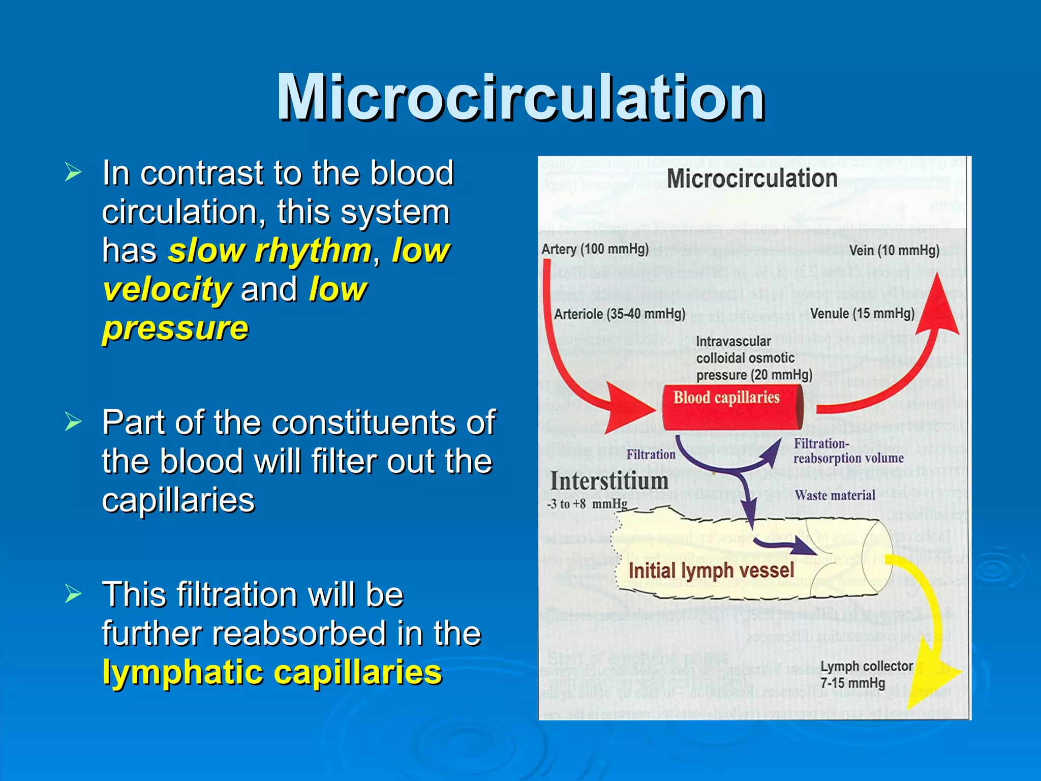
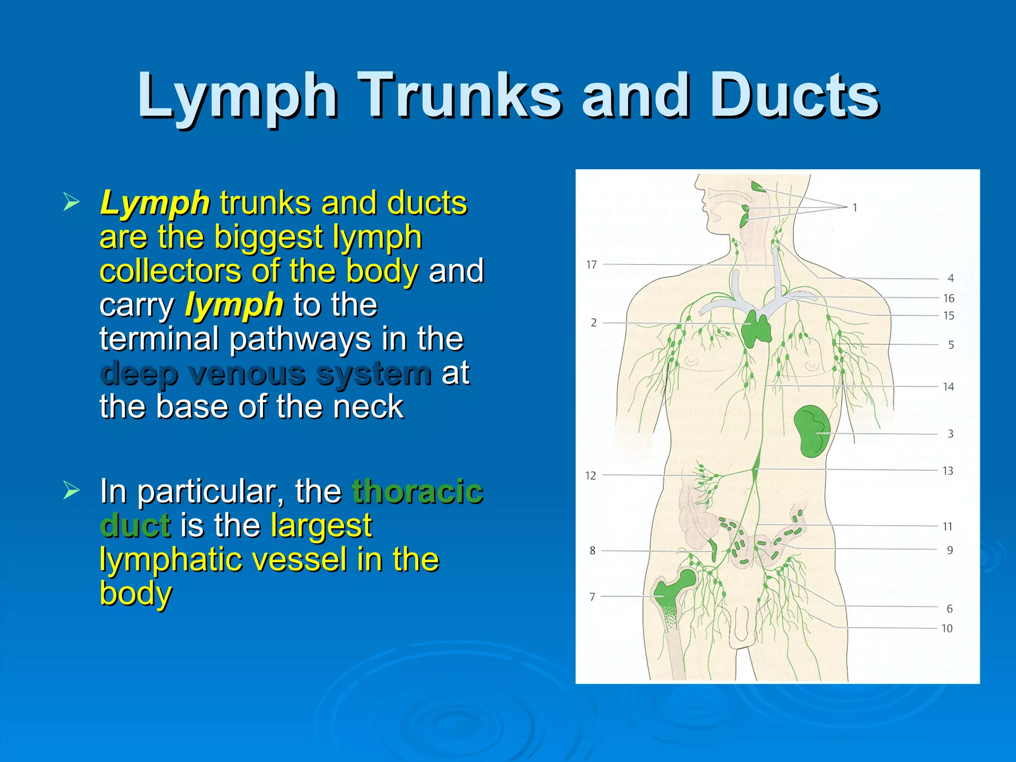
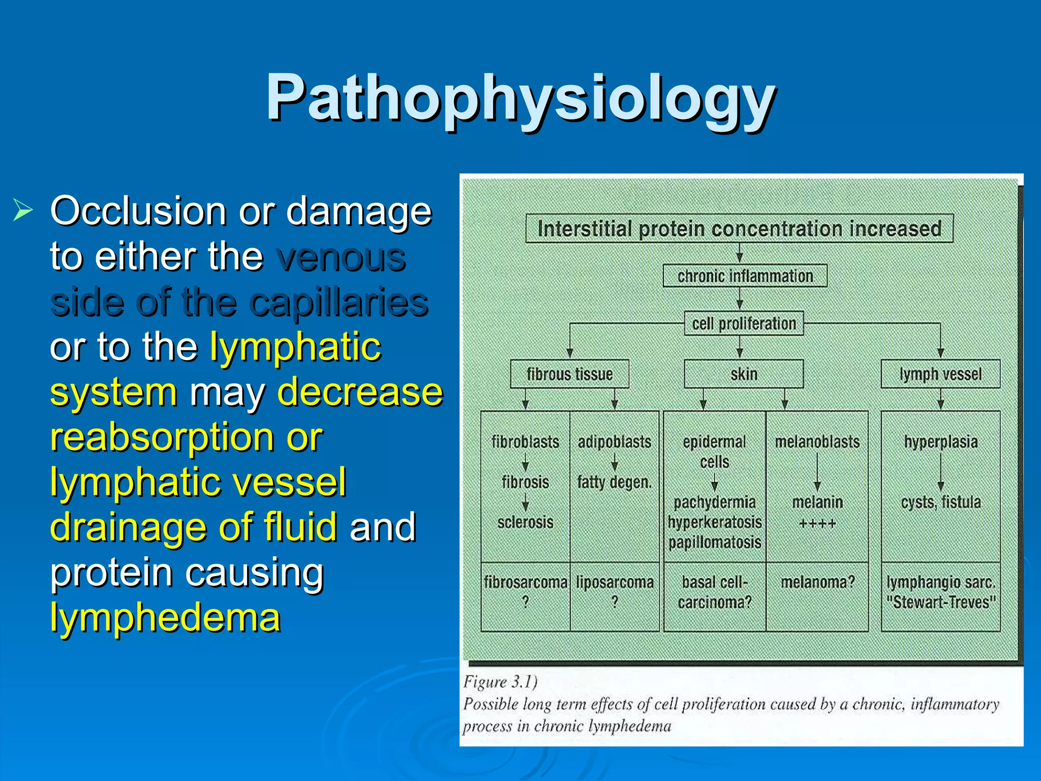
Lymphedema: an overview Margarita Correa MD, FAAPMR Physical Medicine and Rehabilitation Physical Medicine Institute 2020 Oakley Seaver Dr, Ste 1 Clermont, FL
Blood and Lymphatic Circulation It belongs to the circulatory system: one way for the blood to leave the  heart , the  arterial  system, and 2 ways to return (the  venous  and  lymphatic  pathways) Lymph   is a fluid which originates in the connective tissue spaces of the body  Once it has entered the first  lymph capillaries  (initial lymph capillaries) this fluid is called  lymph
Microcirculation In contrast to the blood circulation, this system has  slow rhythm ,  low velocity  and  low pressure Part of the constituents of the blood will filter out the capillaries  This filtration will be further reabsorbed in the  lymphatic capillaries
Lymphatic System In fact  if  the  lymphatic system  did  not recover the protein rich fluid  the body will probably develop  major systemic edema, protein loss & autointoxication and die in 24 - 48 hr
Lymphatic System The lymphatics gradually increase in size:  lymph capillaries      precollectors      lymphatic collectors      ducts and trunks      major venous circulation  just before reaching the heart behind the clavicles  ( angulus venosus )
Composition of the Lymph   Lymph   is usually  clear ,  transparent  or  yellowish alkaline  fluid, slightly less viscuos than blood  When the  lymph  is filled with the absorbed fat of the digestion in the intestines, becomes whitish and viscous and is called “ chyle ” – thoracic duct
Composition of the Lymph Water Proteins Lipids Carbohydrates Enzymes Urea Minerals Hormones Some dissolved gases (i.e. CO2 ) Cells (lymphocytes, macrophages, erythrocytes) Toxins Bacteria Body waste, bits of cell debris, etc.
Composition of Lymph 75-100 gm proteins/day  escape from the blood circulation; this is about  50%  of the protein circulating in the blood plasma per day These  proteins are transported  in  the  lymphatic vessels Lymph  production  2.1 – 3.0 lt/day
Lymphatic Capillaries The  lymphatic circulation  is a  one way structure  beginning with the  lymph capillaries Lymph capillaries  have no valves in their central  endothelial channels   ->   aspirating force ,  pinocytosis   &   micropinocytosis
Lymphatic Capillaries Anchoring filaments  help the  lymph capillaries  to  open widely if there’s significant fluid pressure  in the connective tissue This helps when the tissue is moved manually by  Lymph Drainage Therapy  or similar lymphatic therapies
Lymphatic Capillaries These edges overlap allowing the entry of protein
Pre-collectors Have  one-way valves  spaced about every 2-3 mm These valves help to prevent backflow and to move   lymph  in one direction to the big collectors  These  valves  are essentially invaginations of the  lymph  vessel walls; usually consist of two leaflets  (“bicuspid” valves)
Lymph Collectors The  lymph collectors  are the  main transporting vessels of the lymphatic system They are  large vessels  with  valves  and  muscular units , which carry the lymph to the surrounding  lymph   nodes Consist of  three (3) layers  corresponding more or less to the  intima ,  media , and  adventitia  layers of arteries and veins
Lymph Collectors The  bicuspid valve  is found  at each end  of the  lymphangions  or at specific inter-valvular units of the  lymph   collectors Lymphangions  give the lymphatic vessels the characteristic appearance of a “ pearl necklace” Lymphangions   are considered the  functional unit of the lymphatic system
Lymph Trunks and Ducts Lymph   trunks and ducts are the biggest lymph collectors of the body  and carry  lymph   to the terminal pathways in the  deep venous system  at the base of the neck In particular, the  thoracic duct  is the  largest lymphatic vessel in the body
Lymph Nodes Functions: Biological filtration  -  specific immune cells destroys foreign or unwanted substances which can be then handled by the liver and eliminated by the urinary tract, digestive system, skin and lungs  Immunological  - provide an environment for capturing and destroying microbes through phagocytosis and other process Lymphocyte production and maturation
Lymph Circulation   Superficial circulation , just under the  dermo-epidermal junction . It accounts for about  70%  of upper limb  lymph   flow.  Not directly stimulated by exercise. Deep circulation  of the muscles, below the fascia Very deep circulation  of  the viscera, which is stimulated by muscle contraction
Physiology 80-90%  of the fluid filtered into the interstitium from  capillaries  is reabsorbed into the  venous side The remaining  10-20% of the fluid and protein  is removed from the interstitium by the  lymphatic vessels
Physiology Any condition that increases the capillary pressure, interstitial fluid proteins or capillary permeability or decreases plasma colloid osmotic pressure alters the equilibrium ( Starling equilibrium ) causes fluid movement out of arterial capillaries into the interstitium.
Pathophysiology Occlusion or damage to either the  venous side of the capillaries  or to the  lymphatic system  may  decrease reabsorption or lymphatic vessel drainage   of fluid  and protein causing  lymphedema
Primary Lymphedema
Primary Lymphedema   Lymphatic vessels or nodes are either  absent  ( aplastic ),  underdeveloped  ( hypoplastic ) or too  large and incompetent  ( hyperplastic ) 70-90% woman The earlier it appears the worse the prognosis  Non-pitting edema
Kinmonth’s Age Classification Lymphedema Congenitum and Hereditary Lymphedema Type 1: Milroy’s Syndrome Hereditary lymphedema represent only 1-2 %  of  the primary lymphedema Lymphedema present at birth (congenital)  accounts for 15 % of primary lymphedema
Lymphedema Type 1: Milroy’s Syndrome Autosomal dominant is more common that the recessive form Usually bilateral in the lower limbs
Lymphedema Praecox and Hereditary Lymphedema Type 2: Meige’s Syndrome 75-80% of primary lymphedema  Onset at puberty Usually result of lymphatic hypoplasia  Also affects the distal lymphatics of one or  both legs
Primary Lymphedema Lymphedema Tardum Similar to lymphedema praecox, the only difference is that it appears after age 35
Secondary Lymphedema   Surgery, biopsy, dissection, radiation Numerous surgical interventions, especially for cancer involve the removal of lymph nodes or their destruction by radiation therapy causing fibrosis Lymph vessels can regenerate after being cut but it can be inhibited by the formation of scar tissue
Secondary Lymphedema Metastatic carcinomas   can cause obstruction  of the lymphatic system Trauma and burns physically destroys lymph vessels or nodes and provoke local or regional lymphedema, at least temporarily
Secondary Lymphedema Filariasis   Endemic lymphedema, elephantiasis,   involves the infection by parasitic nematodes (worms) of the order  Filarioidea
Secondary Lymphedema   Lipolymphedema   Lipedema   begins as a pathological accumulation  of fat in which lymphatic circulation is normal, later lipedema can develop into a lymphostatic edema called  lipo-lymphedema The mechanism is compression of the lymph vessels by the adipose layer
Lipolymphedema - Lipedema
Conditions Related to Lymphedema Cardiofaciocutaneous syndrome Cholestasis-oedema syndrome, Norwegian type Cumming syndrome Hennekam syndrome Klippel-Trenaunay syndrome Lymphoedema-distichiasis syndrome Noonan’s syndrome Rheumatoid disease Turner’s syndrome
Breast CA  Lymphedema Incidence Lumpectomy with  sentinel node technique   ->   2%   risk for developing lymphedema Sentinel node  (inject the surrounding tumor tissue with radioisotope and visualize the lymph nodes uptake at the axillary region – during surgery inject blue dye and remove these nodes)
Clinical Diagnosis of Lymphedema Lymphedema  is considered  chronic after 3 months If the patient persists with a  difference of  2 to 3 cm  in circumference  or  10%  volume difference  between the affected and unaffected limb for  more than 6 weeks after surgery
Lymphedema First Signs Puffiness Stiffness Fatigue, malaise, discomfort Tightness Skin tension Heaviness Heat Pain, affecting the skin or the articulation Numbness and paresthesias
Lymphedema Other Signs Associated with Swelling Fibrosis: thickening, and dryness of the skin (hyperkeratosis), papillomas Decreased ROM, stiffness in the affected limb or contractures of the joints
Stemmer’s sign It reflects the  degree of distal fibrosis , usually making it an especially accurate indicator for  primary lymphedema  of the lower extremity The sign is  positive  in  82% to 92% of primary  lower limb lymphedemas and in  56% of cases of secondary  lower limb lymphedema
Lymphedema Diagnosis Special Diagnostic Tools:  Imaging These are generally used for difficult diagnoses to help assess whether the patient is suffering from a  pure lymphedema  or a  mixed form :  veno or phlebo-lymphedema lipolymphedema lymphedema secondary to an arterial condition angiodysplasia, etc
Lymphedema Diagnosis Lymphoscintigraphy Is the gold standard for diagnosis and follow up of lymphedema .  Tc99 colloid  is injected between digits and the radiotracer distributes along the lymphatic system Yields more functional than anatomical information
Lymphoscintigraphy
Diagnosis Computed tomography (CT) Provides more anatomic than functional information Specific images in lymphedema: Enlargement of the skin Subcutaneous tissue is heterogenous Lymphatic lakes Muscle compartment
 
Diagnosis Magnetic Resonance Imaging (MRI) Diffuse dermal and subcutaneous edema  Thickness between fat lobules is  increased The fat cells is isolated by fibrosis  (intralobular fibrosis) Specific “ honeycomb ” aspect of the  subcutaneous compartment Increased size of the subcutaneous  fat compartment
Diagnosis Consider  Arterial  and  Venous  Duplex if a deep venous thrombosis (DVT) or arterial insufficiency is suspected
Differential Diagnosis of  Chronic Leg Edema elevated pulmonary artery pressure (often due to obstructive sleep apnea) congestive heart failure (CHF) venous insufficiency use of non-steroidal anti-inflammatory drugs (NSAID's) proteinuria (>1 g daily)  idiopathic causes
Lymphedema Stages Stage 0 – Latent Mild lymphedema – Stage 1 – still pitting < 3 cm larger than the opposite extremity Moderate lymphedema – Stage 2 – non pitting 3 - 5 cm difference Severe lymphedema – Stage 3 - lymphostatic elephantiasis  > 5 cm difference
Stage 0 Stage 0:  Subclinical or Latent Stage This stage cannot be detected clinically Microscopic examination usually reveals abnormally tortuous and dilated lymphatic vessels and widening of subcutaneous spaces caused by increased pressure in lymphatic capillaries
Stage 1 Lymphedema Stage 1 (Reversible) The swelling usually resolves spontaneously Pitting, soft edema High protein edema Focal fibrosclerotic tissue alterations
Stage 1 Lymphedema
Stage 2 Lymphedema Stage 2 (Spontaneously irreversible) The skin does not move much and is chronically inflamed Fibroblasts predominate in the stagnant, protein rich liquid Skin becomes thickened developing hyperkeratosis This condition will not reverse on its own, but can usually be reduced through CDT
Stage 2 Lymphedema
Stage 3 Lymphedema Stage 3 (Elephantiasis) Non pitting Very hard edema, with no skin movement Significant fibrosis and hardening of the skin; proliferation of adipose tissue Lymphostatic verucosis (warts), papilloma formation, skin fissuring Significant loss of joint mobility and movement
Elephantiasis
Complications Lymphangitis patient with erythema, increased swelling, calor, fever, chills; treat as an  EMERGENCY !!  Rx:  oral vs. I.V. antibiotic, if skin laceration add topical antibiotic Lymphangiosarcoma – Stewart-Treves Syndrome
Complications  Infection and inflammation  are believed to damage the lymphatic system and trigger lymphedema Infection  is often difficult to treat because of lymphostasis and may cause fibrosis, which further compromises the lymphatic draining system
Absolute Contraindications for Decongestive Therapy Hemorrhage (bleeding) Acute enuresis – renal failure * Carcinomatous invasion to the  extremity   * No large scientific studies have shown any increase in metastasis in cancer patients treated with lymphatic drainage  (palliative) Sensory impairment i.e. Brachial or  lumbosacral plexus injury
Absolute Contraindications Acute infection, fever, early inflammatory disease -   caution with chronic tuberculosis or toxoplasmosis which can be reactivated and become acute Serious circulatory problems  -   venous thrombosis (risk of embolism)  and  peripheral arterial insufficiency Major cardiac conditions  -  unstable angina pectoris, acute myocardial infarction and uncompensated congestive heart failure
Contraindications Major Cardiac conditions It’s not recommended to work on cardiac edemas if the cardiac problem is not under control due to additional liquid coming from the tissue drainage increasing the cardiac pre-load This arises a degree of uncertainty in respect to patients with compensated congestive heart failure and a severe left ventricular dysfunction  (LVEF <30%)   Work close with physician !
Thanks! http://www.lymphedemaphysmedi.com Join Lymphedema Support

Lymphedema an overview

  • 1.
    Lymphedema: an overviewMargarita Correa MD, FAAPMR Physical Medicine and Rehabilitation Physical Medicine Institute 2020 Oakley Seaver Dr, Ste 1 Clermont, FL
  • 2.
    Blood and LymphaticCirculation It belongs to the circulatory system: one way for the blood to leave the heart , the arterial system, and 2 ways to return (the venous and lymphatic pathways) Lymph is a fluid which originates in the connective tissue spaces of the body Once it has entered the first lymph capillaries (initial lymph capillaries) this fluid is called lymph
  • 3.
    Microcirculation In contrastto the blood circulation, this system has slow rhythm , low velocity and low pressure Part of the constituents of the blood will filter out the capillaries This filtration will be further reabsorbed in the lymphatic capillaries
  • 4.
    Lymphatic System Infact if the lymphatic system did not recover the protein rich fluid the body will probably develop major systemic edema, protein loss & autointoxication and die in 24 - 48 hr
  • 5.
    Lymphatic System Thelymphatics gradually increase in size: lymph capillaries  precollectors  lymphatic collectors  ducts and trunks  major venous circulation just before reaching the heart behind the clavicles ( angulus venosus )
  • 6.
    Composition of theLymph Lymph is usually clear , transparent or yellowish alkaline fluid, slightly less viscuos than blood When the lymph is filled with the absorbed fat of the digestion in the intestines, becomes whitish and viscous and is called “ chyle ” – thoracic duct
  • 7.
    Composition of theLymph Water Proteins Lipids Carbohydrates Enzymes Urea Minerals Hormones Some dissolved gases (i.e. CO2 ) Cells (lymphocytes, macrophages, erythrocytes) Toxins Bacteria Body waste, bits of cell debris, etc.
  • 8.
    Composition of Lymph75-100 gm proteins/day escape from the blood circulation; this is about 50% of the protein circulating in the blood plasma per day These proteins are transported in the lymphatic vessels Lymph production 2.1 – 3.0 lt/day
  • 9.
    Lymphatic Capillaries The lymphatic circulation is a one way structure beginning with the lymph capillaries Lymph capillaries have no valves in their central endothelial channels -> aspirating force , pinocytosis & micropinocytosis
  • 10.
    Lymphatic Capillaries Anchoringfilaments help the lymph capillaries to open widely if there’s significant fluid pressure in the connective tissue This helps when the tissue is moved manually by Lymph Drainage Therapy or similar lymphatic therapies
  • 11.
    Lymphatic Capillaries Theseedges overlap allowing the entry of protein
  • 12.
    Pre-collectors Have one-way valves spaced about every 2-3 mm These valves help to prevent backflow and to move lymph in one direction to the big collectors These valves are essentially invaginations of the lymph vessel walls; usually consist of two leaflets (“bicuspid” valves)
  • 13.
    Lymph Collectors The lymph collectors are the main transporting vessels of the lymphatic system They are large vessels with valves and muscular units , which carry the lymph to the surrounding lymph nodes Consist of three (3) layers corresponding more or less to the intima , media , and adventitia layers of arteries and veins
  • 14.
    Lymph Collectors The bicuspid valve is found at each end of the lymphangions or at specific inter-valvular units of the lymph collectors Lymphangions give the lymphatic vessels the characteristic appearance of a “ pearl necklace” Lymphangions are considered the functional unit of the lymphatic system
  • 15.
    Lymph Trunks andDucts Lymph trunks and ducts are the biggest lymph collectors of the body and carry lymph to the terminal pathways in the deep venous system at the base of the neck In particular, the thoracic duct is the largest lymphatic vessel in the body
  • 16.
    Lymph Nodes Functions:Biological filtration - specific immune cells destroys foreign or unwanted substances which can be then handled by the liver and eliminated by the urinary tract, digestive system, skin and lungs Immunological - provide an environment for capturing and destroying microbes through phagocytosis and other process Lymphocyte production and maturation
  • 17.
    Lymph Circulation Superficial circulation , just under the dermo-epidermal junction . It accounts for about 70% of upper limb lymph flow. Not directly stimulated by exercise. Deep circulation of the muscles, below the fascia Very deep circulation of the viscera, which is stimulated by muscle contraction
  • 18.
    Physiology 80-90% of the fluid filtered into the interstitium from capillaries is reabsorbed into the venous side The remaining 10-20% of the fluid and protein is removed from the interstitium by the lymphatic vessels
  • 19.
    Physiology Any conditionthat increases the capillary pressure, interstitial fluid proteins or capillary permeability or decreases plasma colloid osmotic pressure alters the equilibrium ( Starling equilibrium ) causes fluid movement out of arterial capillaries into the interstitium.
  • 20.
    Pathophysiology Occlusion ordamage to either the venous side of the capillaries or to the lymphatic system may decrease reabsorption or lymphatic vessel drainage of fluid and protein causing lymphedema
  • 21.
  • 22.
    Primary Lymphedema Lymphatic vessels or nodes are either absent ( aplastic ), underdeveloped ( hypoplastic ) or too large and incompetent ( hyperplastic ) 70-90% woman The earlier it appears the worse the prognosis Non-pitting edema
  • 23.
    Kinmonth’s Age ClassificationLymphedema Congenitum and Hereditary Lymphedema Type 1: Milroy’s Syndrome Hereditary lymphedema represent only 1-2 % of the primary lymphedema Lymphedema present at birth (congenital) accounts for 15 % of primary lymphedema
  • 24.
    Lymphedema Type 1:Milroy’s Syndrome Autosomal dominant is more common that the recessive form Usually bilateral in the lower limbs
  • 25.
    Lymphedema Praecox andHereditary Lymphedema Type 2: Meige’s Syndrome 75-80% of primary lymphedema Onset at puberty Usually result of lymphatic hypoplasia Also affects the distal lymphatics of one or both legs
  • 26.
    Primary Lymphedema LymphedemaTardum Similar to lymphedema praecox, the only difference is that it appears after age 35
  • 27.
    Secondary Lymphedema Surgery, biopsy, dissection, radiation Numerous surgical interventions, especially for cancer involve the removal of lymph nodes or their destruction by radiation therapy causing fibrosis Lymph vessels can regenerate after being cut but it can be inhibited by the formation of scar tissue
  • 28.
    Secondary Lymphedema Metastaticcarcinomas can cause obstruction of the lymphatic system Trauma and burns physically destroys lymph vessels or nodes and provoke local or regional lymphedema, at least temporarily
  • 29.
    Secondary Lymphedema Filariasis Endemic lymphedema, elephantiasis, involves the infection by parasitic nematodes (worms) of the order Filarioidea
  • 30.
    Secondary Lymphedema Lipolymphedema Lipedema begins as a pathological accumulation of fat in which lymphatic circulation is normal, later lipedema can develop into a lymphostatic edema called lipo-lymphedema The mechanism is compression of the lymph vessels by the adipose layer
  • 31.
  • 32.
  • 33.
    Conditions Related toLymphedema Cardiofaciocutaneous syndrome Cholestasis-oedema syndrome, Norwegian type Cumming syndrome Hennekam syndrome Klippel-Trenaunay syndrome Lymphoedema-distichiasis syndrome Noonan’s syndrome Rheumatoid disease Turner’s syndrome
  • 34.
    Breast CA Lymphedema Incidence Lumpectomy with sentinel node technique -> 2% risk for developing lymphedema Sentinel node (inject the surrounding tumor tissue with radioisotope and visualize the lymph nodes uptake at the axillary region – during surgery inject blue dye and remove these nodes)
  • 35.
    Clinical Diagnosis ofLymphedema Lymphedema is considered chronic after 3 months If the patient persists with a difference of 2 to 3 cm in circumference or 10% volume difference between the affected and unaffected limb for more than 6 weeks after surgery
  • 36.
    Lymphedema First SignsPuffiness Stiffness Fatigue, malaise, discomfort Tightness Skin tension Heaviness Heat Pain, affecting the skin or the articulation Numbness and paresthesias
  • 37.
    Lymphedema Other SignsAssociated with Swelling Fibrosis: thickening, and dryness of the skin (hyperkeratosis), papillomas Decreased ROM, stiffness in the affected limb or contractures of the joints
  • 38.
    Stemmer’s sign Itreflects the degree of distal fibrosis , usually making it an especially accurate indicator for primary lymphedema of the lower extremity The sign is positive in 82% to 92% of primary lower limb lymphedemas and in 56% of cases of secondary lower limb lymphedema
  • 39.
    Lymphedema Diagnosis SpecialDiagnostic Tools: Imaging These are generally used for difficult diagnoses to help assess whether the patient is suffering from a pure lymphedema or a mixed form : veno or phlebo-lymphedema lipolymphedema lymphedema secondary to an arterial condition angiodysplasia, etc
  • 40.
    Lymphedema Diagnosis LymphoscintigraphyIs the gold standard for diagnosis and follow up of lymphedema . Tc99 colloid is injected between digits and the radiotracer distributes along the lymphatic system Yields more functional than anatomical information
  • 41.
  • 42.
    Diagnosis Computed tomography(CT) Provides more anatomic than functional information Specific images in lymphedema: Enlargement of the skin Subcutaneous tissue is heterogenous Lymphatic lakes Muscle compartment
  • 43.
  • 44.
    Diagnosis Magnetic ResonanceImaging (MRI) Diffuse dermal and subcutaneous edema Thickness between fat lobules is increased The fat cells is isolated by fibrosis (intralobular fibrosis) Specific “ honeycomb ” aspect of the subcutaneous compartment Increased size of the subcutaneous fat compartment
  • 45.
    Diagnosis Consider Arterial and Venous Duplex if a deep venous thrombosis (DVT) or arterial insufficiency is suspected
  • 46.
    Differential Diagnosis of Chronic Leg Edema elevated pulmonary artery pressure (often due to obstructive sleep apnea) congestive heart failure (CHF) venous insufficiency use of non-steroidal anti-inflammatory drugs (NSAID's) proteinuria (>1 g daily) idiopathic causes
  • 47.
    Lymphedema Stages Stage0 – Latent Mild lymphedema – Stage 1 – still pitting < 3 cm larger than the opposite extremity Moderate lymphedema – Stage 2 – non pitting 3 - 5 cm difference Severe lymphedema – Stage 3 - lymphostatic elephantiasis > 5 cm difference
  • 48.
    Stage 0 Stage0: Subclinical or Latent Stage This stage cannot be detected clinically Microscopic examination usually reveals abnormally tortuous and dilated lymphatic vessels and widening of subcutaneous spaces caused by increased pressure in lymphatic capillaries
  • 49.
    Stage 1 LymphedemaStage 1 (Reversible) The swelling usually resolves spontaneously Pitting, soft edema High protein edema Focal fibrosclerotic tissue alterations
  • 50.
  • 51.
    Stage 2 LymphedemaStage 2 (Spontaneously irreversible) The skin does not move much and is chronically inflamed Fibroblasts predominate in the stagnant, protein rich liquid Skin becomes thickened developing hyperkeratosis This condition will not reverse on its own, but can usually be reduced through CDT
  • 52.
  • 53.
    Stage 3 LymphedemaStage 3 (Elephantiasis) Non pitting Very hard edema, with no skin movement Significant fibrosis and hardening of the skin; proliferation of adipose tissue Lymphostatic verucosis (warts), papilloma formation, skin fissuring Significant loss of joint mobility and movement
  • 54.
  • 55.
    Complications Lymphangitis patientwith erythema, increased swelling, calor, fever, chills; treat as an EMERGENCY !! Rx: oral vs. I.V. antibiotic, if skin laceration add topical antibiotic Lymphangiosarcoma – Stewart-Treves Syndrome
  • 56.
    Complications Infectionand inflammation are believed to damage the lymphatic system and trigger lymphedema Infection is often difficult to treat because of lymphostasis and may cause fibrosis, which further compromises the lymphatic draining system
  • 57.
    Absolute Contraindications forDecongestive Therapy Hemorrhage (bleeding) Acute enuresis – renal failure * Carcinomatous invasion to the extremity * No large scientific studies have shown any increase in metastasis in cancer patients treated with lymphatic drainage (palliative) Sensory impairment i.e. Brachial or lumbosacral plexus injury
  • 58.
    Absolute Contraindications Acuteinfection, fever, early inflammatory disease - caution with chronic tuberculosis or toxoplasmosis which can be reactivated and become acute Serious circulatory problems - venous thrombosis (risk of embolism) and peripheral arterial insufficiency Major cardiac conditions - unstable angina pectoris, acute myocardial infarction and uncompensated congestive heart failure
  • 59.
    Contraindications Major Cardiacconditions It’s not recommended to work on cardiac edemas if the cardiac problem is not under control due to additional liquid coming from the tissue drainage increasing the cardiac pre-load This arises a degree of uncertainty in respect to patients with compensated congestive heart failure and a severe left ventricular dysfunction (LVEF <30%) Work close with physician !
  • 60.