Central Nervous System(CNS)
Central Nervous System (CNS)
• Adult brain has 100 billion neurons.
• Weight of brain---1300 g
• Lies in cranial cavity
• That provide it protection from damage and
injury.
3.
Protection of theCentral Nervous
Protection of the Central Nervous
System
System
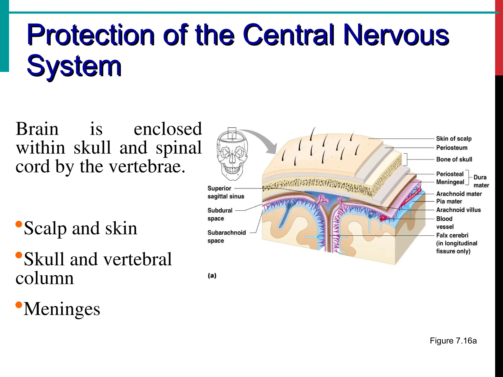
Brain is enclosed
within skull and spinal
cord by the vertebrae.
Scalp and skin
Skull and vertebral
column
Meninges
Figure 7.16a
4.
Protection of theCentral Nervous
Protection of the Central Nervous
System
System
Cerebrospinal fluid
Blood brain barrier
Figure 7.16a
5.
Meninges
Meninges
The brain andspinal cord are completely surrounded by
3 layers of tissue, The Meninges;
Dura mater
Double-layered external covering:
Periosteum – attached to surface of the skull
Meningeal layer – outer covering of the brain
6.
Meninges
Meninges
Arachnoid mater
Middle layer
CSF is present in the space below arachnoid
mater (Sub arachnoid space)
Pia mater
Internal layer
Adheres to the surface of the brain
Pia mater & Arachnoid mater are collectively
called Lepto meninges.(thin, or delicate.)
7.
Cerebrospinal fluid (CSF)
•Fluid found in and around the central nervous
system (CNS), the brain and spinal cord.
• 80- 90 % CSF is produced by ependymal cells
within the lateral ventricles; remainder is
produced by similar cells in third and fourth
ventricles.
• These ependymal cells, their supporting tissue ,
and the associated blood vessels are called
choroid plexuses.
8.
Ventricles and Locationof the
Ventricles and Location of the
Cerebrospinal Fluid
Cerebrospinal Fluid
Figure 7.17a
9.
Ventricles and Locationof the
Ventricles and Location of the
Cerebrospinal Fluid
Cerebrospinal Fluid
Figure 7.17b
10.
CSF Composition
CSF Composition
Secretedcontinuously at a rate of about 0.5ml per
minute; 720ml per day. The volume remains fairly
constant at about 150ml, as absorption keeps pace with
the secretion.
CSF pressure Normal 8 -10cm H2O
Appearance; clear, colorless
Water
Mineral salts
Glucose: 40–85 mg/dL.
Protein (total): 15–45 mg/dL.
Leukocytes (WBC): 0–5/µL (adults / children)
Specific gravity: 1.005–1.009
11.
CSF circulation:
• Choroidplexus produces ~720ml/day of CSF
• body produces, absorbs and replenishes the total volume
of CSF about 3-4 times daily.)
• CSF produced in the lateral ventricles flows into the 3rd
ventricle through an opening called interventricular
foramen
• Flows into through a canal called cerebral aqueduct into
the 4th
ventricle
• Passes through paired lateral apertures and a median
aperture into the subarachnoid space
• Flows through the subarachnoid space surrounding the
brain (small amount enters the central canal of the SC)
14.
Functions of CSF
Functionsof CSF
Supports and protects the brain & spinal cord.
Keeps the brain and spinal cord moist
Supplies nutrients to the nervous system tissue.
Removes waste products from cerebral metabolism.
15.
Brain Anatomy
Brain Anatomy
TheBrain constitutes about one fiftieth of the
The Brain constitutes about one fiftieth of the
body weight & lies within the cranial cavity.
body weight & lies within the cranial cavity.
16.
Regions of theBrain
Regions of the Brain
Cerebrum(Cerebral
hemispheres)
Diencephalon
Thalamus
Hypothalamus
Epithalamus
Brain stem
Midbrain
Pons
Medulla Oblangata
Cerebellum
Figure 7.12
17.
Cerebrum -The largestpart of the brain. It is
divided into two hemispheres, each of which is
divided into four lobes.
Cerebrum
Cerebrum
Cerebellum
It is comprised of right and left hemispheres connected by the
a band of white mater called as corpus callosum.
The surface of the brain is convoluted to create more surface
area.
CEREBRAL FEATURES
Gyri orConvulsions– Elevated ridges “winding” around of
the brain.
Sulci – Small grooves dividing the gyri
e.g. 1) Central Sulcus – Divides the Frontal Lobe from
the Parietal Lobe
2) Parietal-occipital sulcus
Fissures – Deep grooves, generally dividing large
regions/lobes of the brain
1) Longitudinal Fissure – Divides the two Cerebral
2) Transverse Fissure – Separates the Cerebrum from
the Cerebellum
3) Sylvian/Lateral Fissure – Divides the Temporal Lobe
from the Frontal and Parietal Lobes
21.
Lobes of theCerebrum
Lobes of the Cerebrum
Fissures (deep grooves)
divide the cerebrum into
4 lobes
Lobes of the cerebrum
Frontal lobe
Parietal lobe
Occipital lobe
Temporal lobe
22.
Cerebral Lobes andtheir General Functions
Frontal lobe. The top, front regions of each of the cerebral
hemispheres used for reasoning, emotions, judgment, and
voluntary motor movement
Parietal lobe. The middle lobe of each cerebral
hemisphere between the frontal and occipital lobes; it
contains important sensory centers.
Occipital lobe. The region at the back of each cerebral
hemisphere that contains the centers of vision and reading
ability (located at the back of the head).
Temporal lobe. The region at the lower side of each
cerebral hemisphere; contains centers of hearing
and smell (located at the sides of the head).
23.
Functional Areas ofthe Cerebrum
Functional Areas of the Cerebrum
Sensory areas – receives impulses from the
body’s sensory receptors
Motor areas – sends impulses to skeletal
muscles
Association areas – concerned with
integration and processing of complex
mental functions such as intelligence,
memory, reasoning, judgement & emotions.
24.
Functional Areas ofCerebral Cortex
Functional Areas of Cerebral Cortex
Motor areas of Cerebral Cortex
Primary motor area – lies in the frontal lobe ant. To central sulcus
Broca’s area – motor speech area, lies in frontal lobe above lateral sulcus
Sensory areas of Cerebral Cortex
The somatosensory area – lies immediately behind the central sulcus in the
parietal lobe.
The auditory area - Hearing area, lies in temporal lobe
The Olfactory area – Smell area, lies deep within the temporal lobe
The Gustatory area – Taste area, lies just above the lateral sulcus
The visual area – lies in occipital lobe
Association Areas
The Premotor area – lies in frontal lobe anterior to motor area
The prefrontal area - extend anteriorly from premotor area.
Wernicke’s area – Sensory speech, area lies in temporal lobe.
26.
Sensory and MotorAreas of the
Sensory and Motor Areas of the
Cerebral Cortex
Cerebral Cortex
Figure 7.14
27.
Layers of theCerebrum
Layers of the Cerebrum
Gray matter
Outer layer
Composed of
neuron cell bodies
Figure 7.13a
28.
Layers of theCerebrum
Layers of the Cerebrum
White matter
Middle part of the
cerebrum
Composed of
nerve cell
processes
Figure 7.13a
29.
Layers of theCerebrum
Layers of the Cerebrum
Basal Ganglia
Groups of cell bodies (nuclei) deep within the
cerebral hemispheres.
Act as a unified functional unit.
Their functions include initiation and fine
control of complex movement & learned
coordinated activities such as posture and
walking
Figure 7.13a
30.
Functions of CerebralCortex
Functions of Cerebral Cortex
Mental activities involved in memory,
intelligence, sense of responsibility, thinking,
reasoning, moral sense and learning.
Sensory perception, including the perception of
pain, touch, sight, hearing, taste & smell.
Initiation and control of skeletal muscle
contraction (voluntary movement)
Figure 7.13a
Editor's Notes
#24 1. Premotor Area: Responsible for planning, sequencing, and coordinating movements.
Helps in learning skilled and patterned motor activities (e.g., writing, playing piano).
2. Prefrontal Area: Involved in higher cognitive functions: reasoning, decision-making, planning, judgment.
Wernicke’s Area
:Responsible for language comprehension (understanding spoken and written language).