OPPORTUNISTIC INFECTIONS
• Opportunistic infections (Ois): An infection by a
microorganism when the body's immune system is
impaired and unable to fight off infection, as in AIDS,
Infants, neutropenia, and cancer
• Immunocompromised patients are those whose host
defense mechanisms are impaired by an inherited
deficit, disease, or treatment.
• The immunocompromised state increases the risk of
infection with many of the common pathogens as well as
with low-virulence organisms present in the normal flora
or environment.
Definitions
 Many opportunistic infections are acquired in
hospital. Opportunistic infections are a major
cause of illness and death in oncology patients and
the leading cause of death in recipients of renal
transplants.
 Severely immunocompromised patients may
develop simultaneous infections with several
different types of ‘opportunist’ organisms.
 Opportunistic infections are the usual cause of
death in patients with the acquired
immunodeliciency syn­
drome (AIDS(
The organisms involved are those most able to take
advantage of situations such as disruption of the skin or
mucosal barriers and the more specific immune defects,
including:
(1) Defects in the phagocytic response,
(2) Defects in the complement system,
(3) Defects in antibody-mediated immunity,
(4) Defects in cell-mediated immunity,
(5) loss of reticuloendothelial function.
Each of these defects tends to be associated with infections caused
by specific groups of organisms (Table). For example, neutropenia
and disorders of phagocytosis are associated with infections by
Gram-positive cocci, Enterobacteriaceae, Pseudomonas, and fungi.
In contrast, patients with defects in cell-mediated immunity tend to
have severe viral, parasitic, and fungal infections or disease caused
by bacteria that can multiply intracellular (e.g, mycobacteria).
IMMUNE DEFICITS ASSOCIATED WITH INFECTION
Defects in Number or Function of Phagocytes
Neutropenia : the usual value of leucocytes is 2500 to
7500 cells/mm3 of blood in adults.
Neutropenia may result from inherited or acquired
diseases, malignancies, use of cytotoxic drugs, or adverse
reactions to therapeutic agents such as chloramphenicol.
If the absolute neutrophil count decreases to fewer than
500 cells/mm3, the incidence of infections increases
markedly, and counts below 100 cells/mm3 are
associated with bacteremia.

IMMUNE DEFICITS ASSOCIATED WITH INFECTION
Antibody Deficiency
Several congenital and acquired disorders can lead to
inadequate synthesis of immunoglobulins as a result of
deficiency or dysfunction of B lymphocytes
(hypogammaglobulinemia or agammaglobulinemia).
These Individuals are prone to recurrent infections
especially with encapsulated organisms and invasive
pathogen .
SO repeated injections of immunoglobulins or giving
protected vaccines may decrease the incidence and
morbidity of infections in these patients.
IMMUNE DEFICITS ASSOCIATED WITH INFECTION
Disorders in Cell-Mediated Immunity
Both congenital and acquired abnormalities of the cell-
mediated immune system occur.
AIDS the most important cause of acquired cellular
immunodeficiency and transplant recipients due to
treatment with immunosuppressive or cytotoxic agents
that damage both macrophage precursors and T
lymphocytes.
Cytotoxic chemotherapy for cancer has these effects and
also inhibits humoral immune responses.
Infections in the Compromised Host
TYPE OF
COMPROMISE
EXAMPLE PATHOGEN
Leukocyte
number or
function
 Myelocytic leukemias
 Chronic granulomatous
disease
 Granulocytopenia
 Acidosis
 Burns
 Extracellular
bacteria
 Opportunistic fungi
Infections in the Compromised Host
TYPE OF
COMPROMISE
EXAMPLE PATHOGEN
Humoral
immune response
 Lymphocytic leukemias
 Multiple myeloma
 Nephrotic syndrome
 Antimetabolites
 Hypogammaglobulinema
 AIDS
 Encapsulated
bacteria
 Enteroviruses
Infections in the Compromised Host
TYPE OF
COMPROMISE
EXAMPLE PATHOGEN
Cellular
immune response
 AIDS
 Hodgkin’s disease
 Transplantation
 Steroids
 Uremia
 Antimetabolites
 Malnutrition
 Pneumocystis
 Intracellular
bacteriae
 Nocardia
 Candida and fungi
of systemic mycoses
 Viruses, especially
herpes viruses
OPPORTUNISTIC CONDITIONS
 When the immune system isn’t working properly,
normal flora can overpopulate or move into areas
of the body where they do not normally occur.
 When the balance of normal microbes is
disrupted, for example when a person takes broad
spectrum antibiotics.
 Disease can result when normal flora are
traumatically introduced to an area of the body
that is not originally present.
Risk Factors
1-Elders
2- Malnutrition
3-HIV
4-Inflammatory bowel disease
5- Leukopenia
6-Diabetes mellitus
7-Cancer
8- Immunosuppressant drugs
‘OPPORTUNIST’ Organisms
The term ‘opportunist’ is not an exact one. ‘Opportunist’ organisms have
three main characteristics:
1.They are usually organisms of low pathogenicity, e.g.
 Pseudomonas aeruginosa
 Staph. Epidermidis
 Candida albicans
 Cytomegalovirus
 Pneumocysus carinii
2 .They cause serious infections mainly when the host’s defense mechanisms
against infection are impaired, e.g.
 In patients receiving treatment for acute leukaemia or lymphoma, recipi­
ents of
renal or other transplants who are immunosuppressed, and patients with AIDS
3.They can behave as conventional pathogens’ but under opportunistic condi­
tions may cause atypical clinical presenta­
tions or disseminated lesions, e.g.
 Mycobacterium tuberculosis causing a PUO illness in immunosuppressed
patients due to miliary TB
1- Bacterial causes:
Gram negative: pseudomonas aeruginosa,
Stenotrophomonas and acinetobacter
baumannii
Acid–Fast bacilli: Mycobacterium Avium
Complex .
Gram positive: Enterococci, Enterobacter,
Citrobacter and Nocardia
Opportunistic pathogens
2- Viral causes:
Cytomegalovirus (CMV)
Varicella zoster virus (VZV)
Herpes simplex virus (HSV)
Human papilloma virus(HPV)
Hepatitis B virus (HBV)
Molluscum contagiosum
Opportunistic pathogens
2- Fungal causes:
Candida albicans and other Candida species
Pneumocystis jiroveci
Aspergillus fumigatus and other Aspergillus
species
Cryptococcus neoformans
Mucor
Histoplasma capsulatum
Coccidioides immitis.
Opportunistic pathogens
‘Opportunist’ bacteria
Gram­negative bacilli
 Gram­negative bacilli are the most common ‘opportunist’
pathogens and their sources are either
A. Endogenous: from the patient’s alimentary tract flora,
the most frequent source, causing ‘autogenous’ infections
(‘self­infections(
OR
B. Exogenous from infected or colonized lesions from other
patients or from moist contaminated sites in the hospital
environment
 Cross­ infection may be asso­
ciated with the spread of
Gram­negative bacilli by the hands of hospital staff
Pseudomonas aeruginosa as an opportunistic pathogen
- Gram-negative, aerobic rod, oxidase-positive
Infections by Pseudomonas aeroginosa
 urinary tract infections,
 respiratory system infections,
 dermatitis,
 bacteremia,
 bone and joint infections,
 gastrointestinal infections
- Natural habitat in soil, water and on surfaces in contact with
soil or water
- Resistance to antibiotics ( due to the impermeabiliity of its Gram-
negative outer membrane) and phagocytes
Virulence factors & Pathogenesis of
Pseudomonas aeruginosa
• Attachment and colonization (by Flagella, Pilli
Exopolysaccharide (alginate or slime)
• Invasion ( by Elastase , Alkaline protease,
hemolysins and cytotoxins), that produce break
down physical barriers, damage host cells and
immune defense.
• Dissemination Involves spread of infection to
other parts mediated by same extracellular
products that produce localized infection
Lab. diagnosis of pseudomonas aeruginosa
A-Specimens:
• Sputum
• urine
• blood .
B- Direct detection :
Gram-negative bacilli, Non – spore – forming, Motile
C- Cultivation on culture media:
• P. aeruginosa grows on simple media >>>>> Nutrient agar
Yellow pyoverdin + blue pyocyanin pigments → Diffusible exopigments
(bright green colour of colonies).
• Blood agar (alpha haemolysis)
• Maconckey agar (Lactose non fermenters)
Biochemical Activities
• Oxidase-positive
• Carbohydrate non-
fermenter
Nocardia
 Nocardia is a gram positive actinomycete.
 Human infection is rare and contracted through inhalation.
 Infection is more common among immunocompromised
patients, especially those with impaired cell mediated
immunity.
 The patient may have other infections e.g tuberculosis
Members of the Genus Mycobacterium
1. Mycobacterium tuberculosis Complex: M.
tuberculosis, M. bovis, M. africanum & M. microti.
2. Non-tuberculous mycobacteria (NTM): e.g. M.
avium complex, M. ulcerans & M. fortuitum.
3. Mycobacterium leprae: causes leprosy.
4. Saprophytic mycobacteria species.
Mycobacterium tuberculosis
• Major world wide co-infection.
• Clinical features:-cough, -hemoptysis, - weight loss, -
evening rise of temp.
• Diagnosis: sputum for AFB, chest X-ray, culture of
specimen from the site (in case of extra PTB), Skin
test (PPD)
Mycobacterium Avium Complex(MAC)
• MAC is a common opportunistic infection in patients
with AIDS.
• Mycobacterium avium complex is a group of
mycobacteria comprising Mycobacterium intracellulare
and Mycobacterium avium
• Clinical features:-Flu like fever, -chills, sweats, -anemia,
fatigue.
Laboratory Diagnosis
A) Specimens: include sputum,
BAL, gastric lavage…etc.
Early morning sputum specimens
collected on 3 consecutive days,
from a deep productive cough, give
the best results.
B) Direct Detection:
Smears:
a. Ziehl-Neelsen (Z-N) stain
Under the ordinary light microscope,
AFB appear pink in a blue background.
c) Cultivation:
1. Conventional culture
media: (incubated in 5 –
10% CO2 at 37o
C):
L-J tube.
- Results may be relatively
delayed (2-8 weeks).
Cultural Characters
• Solid media include:
- Lowenstein-Jensen (L-J) medium
(egg-based)
- Middlebrook's medium (agar-based)
Results may be relatively delayed (2-8 weeks).
• Fluid medium (Middlebrook broth).
1. Bactec AFB system
2. Mycobacteria Growth Indicator Tube (MGIT)
(4-14 days identification)
E) Identification:
1. Cultures examined twice weekly.
2. Colonies appear after about 2-4 weeks of incubation
up to 8 weeks.
Then tested for:
- acid-fast stain (M. tuberculosis are AFB),
- biochemical reactions (M. tuberculosis are
positive for nitrate reduction & niacin production).
Viral opportunistic infections
• Cytomegalovirus
• Human papilloma virus
• Varicella Zoster virus
Cytomegalovirus
• Cytomegalovirus is a family of opportunistic viruses, most
frequently associated with respiratory infection
• CMV can infect multiple parts of the body and cause
pneumonia, gastroenteritis (especially abdominal pain
caused by infection of the colon), encephalitis (infection)
of the brain, and sight-threatening retinitis (infection of the
retina at the back of eye).
• People with CMV retinitis have difficulty with vision that
worsens over time. CMV retinitis is a medical emergency
because it can cause blindness if not treated promptly
Lab diagnosis of CMV
1. Isolation of the virus from throat washing or
urine on tissue culture
2. Detection of viral DNA by PCR
3. Detection of viral Ags in urine or saliva
4. Serodiagnosis: Detection of CMV IgM or rising
titre of IgG
5. Detection of intranuclear inclusion bodies
which is oval shape called owl’s eye in tisues or
in desquamated cells in urine
Owl’s eye in CMV
Human papilloma virus
• Human papilloma viruses are tropic for epithelial
cells of the skin and mucous membranes.
– Produce hyperplastic epithelial lesions
1. induce benign lesions of the skin (warts) and
mucous membranes (condylomas).
2. Some HPVs have also been implicated in the
development of epithelial malignancies, especially
cancer of the uterine cervix.
Laboratory Diagnosis
• HPV cannot be cultivated to diagnose infections.
• The following methods are used for cervical
cancer screening.
• Cervical swabs or biopsy samples are subjected
to:
– Cytology (Pap smear)
– Detection of cervical biomarkers: include E6 and E7
– PCR molecular assays to detect HPV DNA sequences,
E6 or E7 mRNA
Cytology
• HPV infects squamous epithelial cells and
induces within those cells a characteristic
cytoplasmic vacuole.
• These vacuolated cells, called koilocytes, are
the hallmark of infection by these viruses.
• These koilocytes are squamous epithelial cells
that contain an acentric, hyperchromatic
nucleus that is displaced by a large perinuclear
vacuole in the differentiated layers of the
squamous epithelium.
Cytology
Varicella zoster virus
• Varicella-zoster virus (VZV) usually causes
localized zoster in adults.
• However, in immunocompromised patients, it
can cause systemic infection accompanied by
complications such as pneumonia,
encephalitis, and hepatitis.
Lab diagnosis of VZV
• The signs and symptoms of herpes zoster are usually
distinctive enough to make an accurate clinical
diagnosis once the rash has appeared
• The most sensitive method for confirming a diagnosis
of varicella is the use of polymerase chain reaction
(PCR) to detect VZV in skin lesions
Lab diagnosis of VZV
• Other viral isolation techniques for confirming
varicella are direct fluorescent antibody assay (DFA)
and viral culture. they are less sensitive than PCR and,
in the case of viral culture, will take longer to generate
results.
• IgM serologic testing provide evidence for a recent
active VZV infection or reactivation
Multinucleated Giant cells
Herpes simplex virus (HSV) and varicella zoster
virus (VZV) infections induce the formation of
intraepidermal vesicles containing
multinucleated giant cells in the skin.
Candidiasis
They can produce :
Cutaneous, subcutaneous or systemic mycosis.
The most pathogenic spp. is candida albicans.
CUTANEOUS INFECTION:
Oral thrush.
Onychomycosis
Paronychia
MUCOUS INFECTIONS: oesophagitis,vaginitis
SYSTEMIC INFECTION: endophthalmitis, liver eye
bone,lung
Diagnosis: C/F, KOH preparation of the scrapings
Diagnosis
• 1- specimens:
Blood, urine, CSF, sputum and skin.
2- Direct microscopy:
KOH, Gram stain
3- Culture :
SDA ,corn meal agar or CHROM agar colonies are
pasty and creamy.
Diagnosis
Identification:
-sugar fermentation
-germ tube test
4- Molecular : PCR
5- Serology
Cryptococcosis is an
infection of the lungs
(Pneumonia) & brain
(Meningitis)
- Mortality rate is 40%.
Cryptococcosis
- It is caused by Cryptococcus neoformans.
Morphology: it is an oval, budding yeast with a narrow-based bud surrounded
by a wide polysaccharide capsule.
- The organism is present in the soil contaminated with excreta of
birds particularly pigeons.
Mode of transmission: inhalation
of fungal spores (common in bird breeders)
)
Dr Azza Abdulazim
Cryptococcus (capsule on India Ink)
Cryptococcosis
• Clinical features:
- Mild sublinical lung infection is the most
common presentation.
• CNS : meningitis, meningoencephalitis .
Cutaneous lesions .
• Bone infection.
• In AIDS patients, cryptococcosis is the second
most common fungal infection (after
candidiasis
India ink
.
• Laboratory Diagnosis:
A- Specimens: CSF by lumber puncture under complete a septic
condition
B-Direct detection:
1- Microscopic: spinal fluid mixed with India ink, the yeast cell is
seen microscopically surrounded by a wide, unstained capsule.
2-Cryptococcal antigen test: capsular antigen can be detected by
the latex particle agglutination test.
C- Culture on SDA media: The colonies are highly mucoid due
to the large amount of capsular polysaccharide produced by the
organism.
Cryptococcus neoformans
MUCOR & RHIZOPUS
• Mucormycosis (zygomycosis, phycomycosis) is a disease
caused by saprophytic moulds (e.g., Mucor, Rhizopus,
and Absidia) found widely in the environment.
Especially in diabetic ketoacidosis, burns, bone
marrow transplants, or leukemic patients.
• Mode of transmission: airborne inhalation of spores
and invade tissues of patients with reduced host defenses.
Mucor on Lactophenol Cotton Blue staining
• Pathogenesis and clinical picture:
Rhinocerebral mucormycosis, in which mold spores in
the sinuses germinate to form hyphae that invade
blood vessels that supply the brain.
The disease can progress rapidly with invasion of
eyes, cranial bones, and brain. Blood vessels and
nerves are damaged, and patients develop edema of
the involved facial area, a bloody nasal exudate, and
orbital cellulitis.
Laboratory Diagnosis:
A- Specimens: nasal discharge and tissue
B-Direct detection:
1- Microscopic: reveal irregular, broad and non septate hyphae.
C- Culture on SDA media: These fungi grow rapidly (3–5
days) and incubated at 25–30°C, producing abundant cottony colonies.
Irregular, broad and non septate hyphae. Cottony colonies on SDA
Mucormycosis
• The most common form of the disease which can
be fatal within 1 week, is rhinocerebral
mucormycosis.
• The infection begins in the nasal mucosa or
sinuses and progresses to the orbits, palate, and
brain.
• The disease is so aggressive, many cases are not
diagnosed until after death..
Aspergillosis
• Aspergillus spp.:
- fumigatus – niger - flavus.
- Clinical picture:
• The most severe, and often fatal is acute invasive
infection of the lung.
• Disseminated: to the brain, GI tract, and other
organs.
• Aspergilloma: A less severe, a mass of hyphal
tissue that can form in lung cavities.
Diagnosis
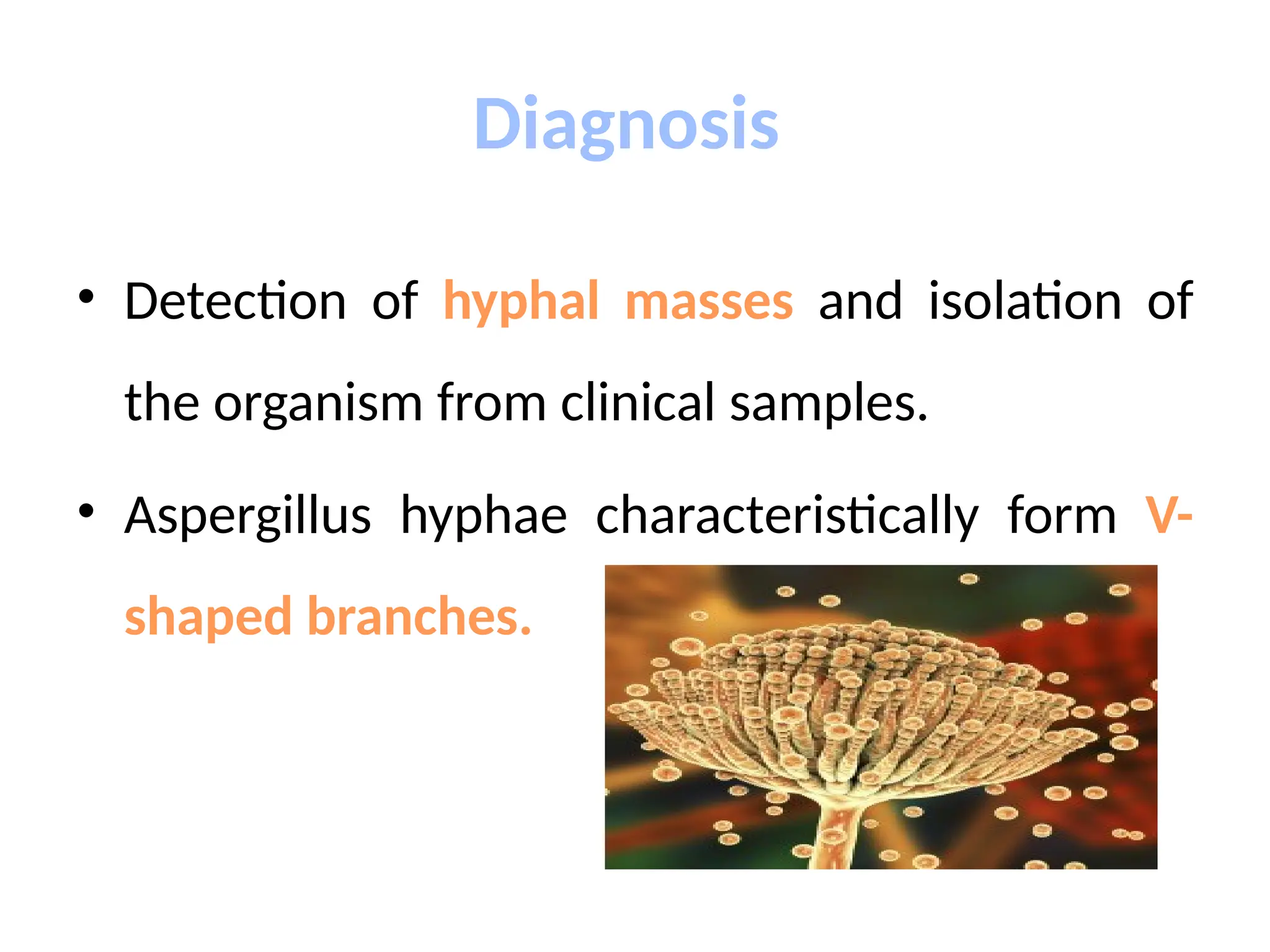
• Detection of hyphal masses and isolation of
the organism from clinical samples.
• Aspergillus hyphae characteristically form V-
shaped branches.
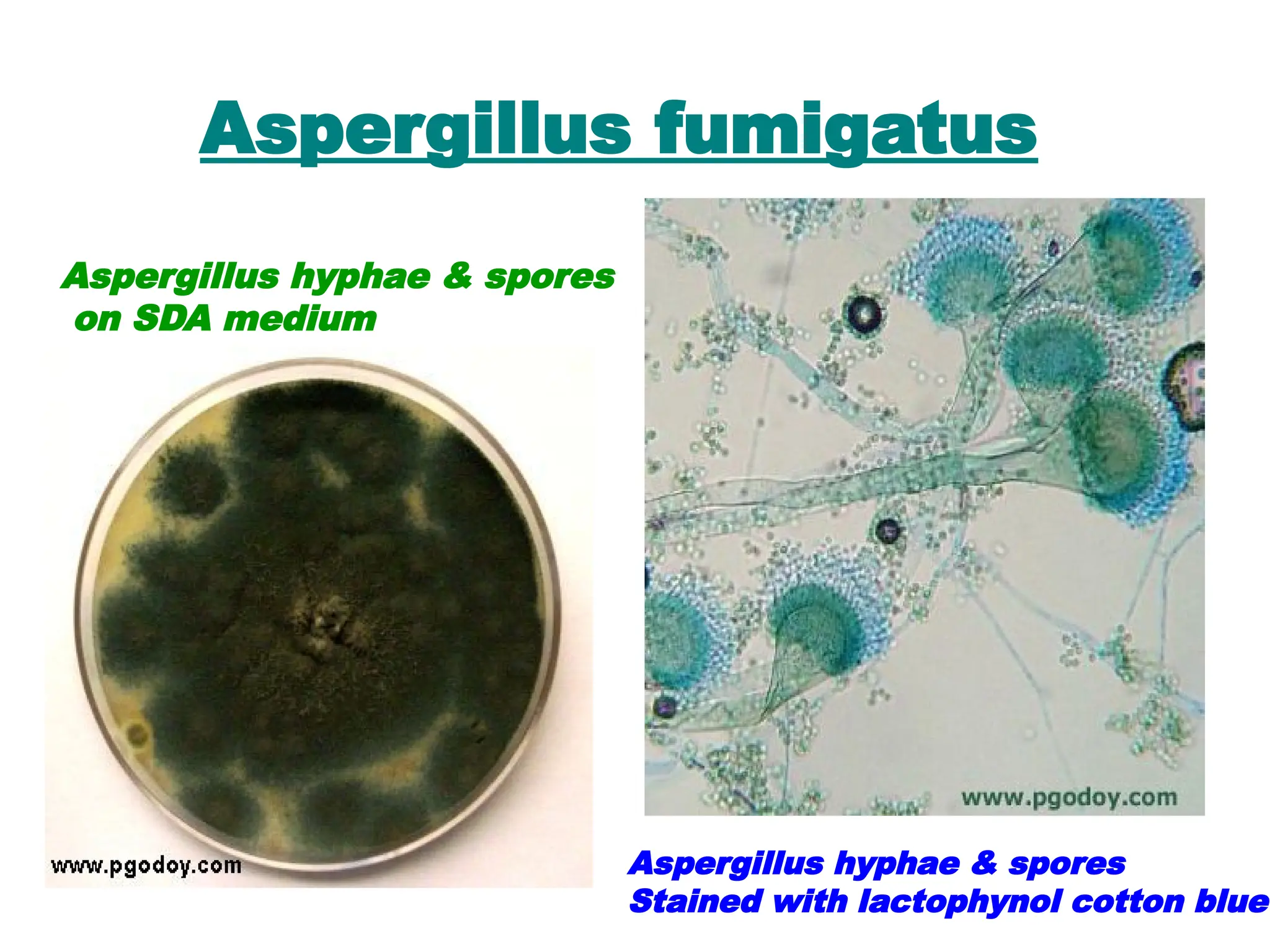
Aspergillus fumigatus
Aspergillus hyphae & spores
on SDA medium
Aspergillus hyphae & spores
Stained with lactophynol cotton blue
Pneumocystis Jiroveci
• Mainly interstitial pneumonia
• Major cause of death in AIDS patients.
• The disease is not transmitted from person to person. It
is developed of by activation of in the lungs.
• The encysted forms induce inflammation of alveoli,
resulting in production of an exudate that blocks gas
exchange.
Diagnosis
• P. jirovecii cannot be cultivated
• Diagnosis is based on microscopic examination
of biopsied lung tissue or washings using special
stains(Giemsa, silver).
• Immunofluorescence
• It shows the characteristic cysts. The cysts
resemble crushed ping-pong balls and are
present in aggregates of two to eight
• PCR
Cysts of Pneumocystis jiroveci in smear from
bronchoalveolar lavage by silver stain.
DIAGNOSIS OF OPPORTUNISTIC
INFECTIONS
 General considerations;
 A clear bacteriological diagnosis, achieved in only 20- ­
40%
of patients. This is due to:
1- Clinical features are often lacking in immuno­
compromised
patients. The infection may be relatively silent due to
impairment of the immune response.
2- Signs of infection may be lacking due to deficiency in
‘pus’ cells and impaired inflammatory response
 Fever maybe the only obvious feature. However,
fever may also be due to non-­
infective causes
including malignancy or drug reactions.
3- Diagnosis of the exact site of infection is difficult in
severely neutropenic patients.
 Common sites of infection should be considered;
Lungs, Pharynx and Oesophagus, and Peri­
anal
Region.
4- Physical examination: chest X-­
ray may also show
only minimal abnormalities if sputum is not be produced.
5-Laboratory diagnosis can also be difficult, because
many of the organisms involved require special culture
media and grow slowly such as Pneumocystis jiroveci
cannot be grown at all.
 The increased involvement of low-virulence organisms
commonly found in the normal flora may make it difficult
to distinguish colonization from infection.
 Thus, isolation of C. albicans from the urine or the
pharynx does not prove that it is the cause of a
concurrent renal abscess or pneumonitis.
 So diagnostic procedures such as biopsy of involved
organs are often needed to identify the causative agent.
The following lab. Investigation done to
patients
1- Blood cultures:
preferably two or three sets, should be collected
before the start of prompt ‘blind chemotherapy.
2- urine sample: if possible, should also be
collected for culture before treatment starts.
3- appropriate swabs should be collected
in addition to blood and urine cultures
 Diagnosis of Pneumocystis and other opportunistic lung infections by
open lung biopsy is the method of choice since adequate samples of
tissue can be stained by silver stains However, open lung biopsy is not
practical.
 less invasive procedures such as bronchoscopy with the collection of
bronchial washings.
 Histology may also reveal infection by Mycobacrerium tuberculosis
or cytomegalovirus.
 ‘Induced sputum’ samples can be examined by microscopy and
culture for fungi and acid­
fast bacilli.
 .
 Serological investigations :
Candida albicans, Aspergillus fumigatus , Crvptococcus neoformans
CMV show a rising ‘specific’ antibody titer of four­
fold or greater, or a
single high titer or a positive antigen test.
 A POSITIVE SPECIFIC IGM ANTIBODY result is particularly
helpful if cytomegalovirus or legionella infection is suspected.
 These antibody tests are unhelpful.
 ‘False negative’ or only low serum antibody titers result in some
patients with established infections, because the immune response is
too poor to generate significant antibody titers.
 Molecular biology :
These tests are likely to have an important role in
diagnosis of certain opportunistic infections such as
pneumocystis and cytomegalovirus disease
Treatment
Successful treatment of infections in the compromised host
depends on recognition of the deficit, early diagnosis, and prompt
intervention.
 Usually it is necessary to initiate antimicrobial treatment before
results of culture and antibiotic susceptibility tests are available.
 Broad-spectrum antibiotics is used initially and replaced with
narrower spectrum, when the etiologic agent and its susceptibility
are known, to reduce the risk of superinfection.
 Bactericidal antibiotics are needed to control infections when host
defenses are inadequate, and with severe infections a
combination of synergistic agents may be necessary to provide
increased bactericidal action.
Treatment
 Patients with neutropenia have high rates of infection,
and mortality 20 to 30% if bacteremia develops.
 Therefore, short-term prophylactic antibiotic treatment
has been advocated and can be effective in preventing
infection until the neutrophil count improves.
 Neutropenia can be ameliorated by the use of cytokines
(eg, granulocyte-colony stimulating factor).
PREVENTION
Prevention of opportunistic infections in patients disposed to them.
For example, patients undergoing bone marrow transplantation may
receive prophylactic acyclovir or ganciclovir to prevent herpes virus
and cytomegalovirus infection.
AIDS patients receive prophylactic trimethoprim – sulfamethoxazole to
prevent P. jirovici pneumonia as well as toxoplasmosis.
General preventive measures for infected patients:
• Prevent exposure to ill patients.
• Personal hygiene (washing hands etc.)
• Avoid contact with raw food, soil, cats, bird excreta, litter boxes etc.
• Wash vegetables before cooking, avoid raw meat intake, drink boiled
water.
• Use condoms during sexual contact.
•
General preventive measures for
infected patients
 The patient should be placed in isolation room
 Cough etiquettes measured should be followed.
 For prevention of air born infections the patients should
wear N95 and surgical mask for prevention of droplet
infections
 All surrounding and surfaces should be cleaned with
proper disinfectants
 All instruments used for patients should be sterilized
 Decreased the invasive procedures as can as possible
Opportunistic infections including bacteria, viruses and fungi

Opportunistic infections including bacteria, viruses and fungi

  • 1.
  • 2.
    • Opportunistic infections(Ois): An infection by a microorganism when the body's immune system is impaired and unable to fight off infection, as in AIDS, Infants, neutropenia, and cancer • Immunocompromised patients are those whose host defense mechanisms are impaired by an inherited deficit, disease, or treatment. • The immunocompromised state increases the risk of infection with many of the common pathogens as well as with low-virulence organisms present in the normal flora or environment. Definitions
  • 3.
     Many opportunisticinfections are acquired in hospital. Opportunistic infections are a major cause of illness and death in oncology patients and the leading cause of death in recipients of renal transplants.  Severely immunocompromised patients may develop simultaneous infections with several different types of ‘opportunist’ organisms.  Opportunistic infections are the usual cause of death in patients with the acquired immunodeliciency syn­ drome (AIDS(
  • 4.
    The organisms involvedare those most able to take advantage of situations such as disruption of the skin or mucosal barriers and the more specific immune defects, including: (1) Defects in the phagocytic response, (2) Defects in the complement system, (3) Defects in antibody-mediated immunity, (4) Defects in cell-mediated immunity, (5) loss of reticuloendothelial function. Each of these defects tends to be associated with infections caused by specific groups of organisms (Table). For example, neutropenia and disorders of phagocytosis are associated with infections by Gram-positive cocci, Enterobacteriaceae, Pseudomonas, and fungi. In contrast, patients with defects in cell-mediated immunity tend to have severe viral, parasitic, and fungal infections or disease caused by bacteria that can multiply intracellular (e.g, mycobacteria).
  • 5.
    IMMUNE DEFICITS ASSOCIATEDWITH INFECTION Defects in Number or Function of Phagocytes Neutropenia : the usual value of leucocytes is 2500 to 7500 cells/mm3 of blood in adults. Neutropenia may result from inherited or acquired diseases, malignancies, use of cytotoxic drugs, or adverse reactions to therapeutic agents such as chloramphenicol. If the absolute neutrophil count decreases to fewer than 500 cells/mm3, the incidence of infections increases markedly, and counts below 100 cells/mm3 are associated with bacteremia. 
  • 6.
    IMMUNE DEFICITS ASSOCIATEDWITH INFECTION Antibody Deficiency Several congenital and acquired disorders can lead to inadequate synthesis of immunoglobulins as a result of deficiency or dysfunction of B lymphocytes (hypogammaglobulinemia or agammaglobulinemia). These Individuals are prone to recurrent infections especially with encapsulated organisms and invasive pathogen . SO repeated injections of immunoglobulins or giving protected vaccines may decrease the incidence and morbidity of infections in these patients.
  • 7.
    IMMUNE DEFICITS ASSOCIATEDWITH INFECTION Disorders in Cell-Mediated Immunity Both congenital and acquired abnormalities of the cell- mediated immune system occur. AIDS the most important cause of acquired cellular immunodeficiency and transplant recipients due to treatment with immunosuppressive or cytotoxic agents that damage both macrophage precursors and T lymphocytes. Cytotoxic chemotherapy for cancer has these effects and also inhibits humoral immune responses.
  • 8.
    Infections in theCompromised Host TYPE OF COMPROMISE EXAMPLE PATHOGEN Leukocyte number or function  Myelocytic leukemias  Chronic granulomatous disease  Granulocytopenia  Acidosis  Burns  Extracellular bacteria  Opportunistic fungi
  • 9.
    Infections in theCompromised Host TYPE OF COMPROMISE EXAMPLE PATHOGEN Humoral immune response  Lymphocytic leukemias  Multiple myeloma  Nephrotic syndrome  Antimetabolites  Hypogammaglobulinema  AIDS  Encapsulated bacteria  Enteroviruses
  • 10.
    Infections in theCompromised Host TYPE OF COMPROMISE EXAMPLE PATHOGEN Cellular immune response  AIDS  Hodgkin’s disease  Transplantation  Steroids  Uremia  Antimetabolites  Malnutrition  Pneumocystis  Intracellular bacteriae  Nocardia  Candida and fungi of systemic mycoses  Viruses, especially herpes viruses
  • 11.
    OPPORTUNISTIC CONDITIONS  Whenthe immune system isn’t working properly, normal flora can overpopulate or move into areas of the body where they do not normally occur.  When the balance of normal microbes is disrupted, for example when a person takes broad spectrum antibiotics.  Disease can result when normal flora are traumatically introduced to an area of the body that is not originally present.
  • 12.
    Risk Factors 1-Elders 2- Malnutrition 3-HIV 4-Inflammatorybowel disease 5- Leukopenia 6-Diabetes mellitus 7-Cancer 8- Immunosuppressant drugs
  • 13.
    ‘OPPORTUNIST’ Organisms The term‘opportunist’ is not an exact one. ‘Opportunist’ organisms have three main characteristics: 1.They are usually organisms of low pathogenicity, e.g.  Pseudomonas aeruginosa  Staph. Epidermidis  Candida albicans  Cytomegalovirus  Pneumocysus carinii 2 .They cause serious infections mainly when the host’s defense mechanisms against infection are impaired, e.g.  In patients receiving treatment for acute leukaemia or lymphoma, recipi­ ents of renal or other transplants who are immunosuppressed, and patients with AIDS 3.They can behave as conventional pathogens’ but under opportunistic condi­ tions may cause atypical clinical presenta­ tions or disseminated lesions, e.g.  Mycobacterium tuberculosis causing a PUO illness in immunosuppressed patients due to miliary TB
  • 14.
    1- Bacterial causes: Gramnegative: pseudomonas aeruginosa, Stenotrophomonas and acinetobacter baumannii Acid–Fast bacilli: Mycobacterium Avium Complex . Gram positive: Enterococci, Enterobacter, Citrobacter and Nocardia Opportunistic pathogens
  • 15.
    2- Viral causes: Cytomegalovirus(CMV) Varicella zoster virus (VZV) Herpes simplex virus (HSV) Human papilloma virus(HPV) Hepatitis B virus (HBV) Molluscum contagiosum Opportunistic pathogens
  • 16.
    2- Fungal causes: Candidaalbicans and other Candida species Pneumocystis jiroveci Aspergillus fumigatus and other Aspergillus species Cryptococcus neoformans Mucor Histoplasma capsulatum Coccidioides immitis. Opportunistic pathogens
  • 17.
    ‘Opportunist’ bacteria Gram­negative bacilli Gram­negative bacilli are the most common ‘opportunist’ pathogens and their sources are either A. Endogenous: from the patient’s alimentary tract flora, the most frequent source, causing ‘autogenous’ infections (‘self­infections( OR B. Exogenous from infected or colonized lesions from other patients or from moist contaminated sites in the hospital environment  Cross­ infection may be asso­ ciated with the spread of Gram­negative bacilli by the hands of hospital staff
  • 18.
    Pseudomonas aeruginosa asan opportunistic pathogen - Gram-negative, aerobic rod, oxidase-positive Infections by Pseudomonas aeroginosa  urinary tract infections,  respiratory system infections,  dermatitis,  bacteremia,  bone and joint infections,  gastrointestinal infections - Natural habitat in soil, water and on surfaces in contact with soil or water - Resistance to antibiotics ( due to the impermeabiliity of its Gram- negative outer membrane) and phagocytes
  • 19.
    Virulence factors &Pathogenesis of Pseudomonas aeruginosa • Attachment and colonization (by Flagella, Pilli Exopolysaccharide (alginate or slime) • Invasion ( by Elastase , Alkaline protease, hemolysins and cytotoxins), that produce break down physical barriers, damage host cells and immune defense. • Dissemination Involves spread of infection to other parts mediated by same extracellular products that produce localized infection
  • 20.
    Lab. diagnosis ofpseudomonas aeruginosa A-Specimens: • Sputum • urine • blood . B- Direct detection : Gram-negative bacilli, Non – spore – forming, Motile C- Cultivation on culture media: • P. aeruginosa grows on simple media >>>>> Nutrient agar Yellow pyoverdin + blue pyocyanin pigments → Diffusible exopigments (bright green colour of colonies). • Blood agar (alpha haemolysis) • Maconckey agar (Lactose non fermenters)
  • 22.
  • 23.
    Nocardia  Nocardia isa gram positive actinomycete.  Human infection is rare and contracted through inhalation.  Infection is more common among immunocompromised patients, especially those with impaired cell mediated immunity.  The patient may have other infections e.g tuberculosis
  • 25.
    Members of theGenus Mycobacterium 1. Mycobacterium tuberculosis Complex: M. tuberculosis, M. bovis, M. africanum & M. microti. 2. Non-tuberculous mycobacteria (NTM): e.g. M. avium complex, M. ulcerans & M. fortuitum. 3. Mycobacterium leprae: causes leprosy. 4. Saprophytic mycobacteria species.
  • 26.
    Mycobacterium tuberculosis • Majorworld wide co-infection. • Clinical features:-cough, -hemoptysis, - weight loss, - evening rise of temp. • Diagnosis: sputum for AFB, chest X-ray, culture of specimen from the site (in case of extra PTB), Skin test (PPD)
  • 27.
    Mycobacterium Avium Complex(MAC) •MAC is a common opportunistic infection in patients with AIDS. • Mycobacterium avium complex is a group of mycobacteria comprising Mycobacterium intracellulare and Mycobacterium avium • Clinical features:-Flu like fever, -chills, sweats, -anemia, fatigue.
  • 28.
    Laboratory Diagnosis A) Specimens:include sputum, BAL, gastric lavage…etc. Early morning sputum specimens collected on 3 consecutive days, from a deep productive cough, give the best results.
  • 29.
    B) Direct Detection: Smears: a.Ziehl-Neelsen (Z-N) stain Under the ordinary light microscope, AFB appear pink in a blue background.
  • 30.
    c) Cultivation: 1. Conventionalculture media: (incubated in 5 – 10% CO2 at 37o C): L-J tube. - Results may be relatively delayed (2-8 weeks).
  • 31.
    Cultural Characters • Solidmedia include: - Lowenstein-Jensen (L-J) medium (egg-based) - Middlebrook's medium (agar-based) Results may be relatively delayed (2-8 weeks). • Fluid medium (Middlebrook broth). 1. Bactec AFB system 2. Mycobacteria Growth Indicator Tube (MGIT) (4-14 days identification)
  • 32.
    E) Identification: 1. Culturesexamined twice weekly. 2. Colonies appear after about 2-4 weeks of incubation up to 8 weeks. Then tested for: - acid-fast stain (M. tuberculosis are AFB), - biochemical reactions (M. tuberculosis are positive for nitrate reduction & niacin production).
  • 33.
    Viral opportunistic infections •Cytomegalovirus • Human papilloma virus • Varicella Zoster virus
  • 34.
    Cytomegalovirus • Cytomegalovirus isa family of opportunistic viruses, most frequently associated with respiratory infection • CMV can infect multiple parts of the body and cause pneumonia, gastroenteritis (especially abdominal pain caused by infection of the colon), encephalitis (infection) of the brain, and sight-threatening retinitis (infection of the retina at the back of eye). • People with CMV retinitis have difficulty with vision that worsens over time. CMV retinitis is a medical emergency because it can cause blindness if not treated promptly
  • 35.
    Lab diagnosis ofCMV 1. Isolation of the virus from throat washing or urine on tissue culture 2. Detection of viral DNA by PCR 3. Detection of viral Ags in urine or saliva 4. Serodiagnosis: Detection of CMV IgM or rising titre of IgG 5. Detection of intranuclear inclusion bodies which is oval shape called owl’s eye in tisues or in desquamated cells in urine
  • 36.
  • 37.
    Human papilloma virus •Human papilloma viruses are tropic for epithelial cells of the skin and mucous membranes. – Produce hyperplastic epithelial lesions 1. induce benign lesions of the skin (warts) and mucous membranes (condylomas). 2. Some HPVs have also been implicated in the development of epithelial malignancies, especially cancer of the uterine cervix.
  • 39.
    Laboratory Diagnosis • HPVcannot be cultivated to diagnose infections. • The following methods are used for cervical cancer screening. • Cervical swabs or biopsy samples are subjected to: – Cytology (Pap smear) – Detection of cervical biomarkers: include E6 and E7 – PCR molecular assays to detect HPV DNA sequences, E6 or E7 mRNA
  • 40.
    Cytology • HPV infectssquamous epithelial cells and induces within those cells a characteristic cytoplasmic vacuole. • These vacuolated cells, called koilocytes, are the hallmark of infection by these viruses.
  • 41.
    • These koilocytesare squamous epithelial cells that contain an acentric, hyperchromatic nucleus that is displaced by a large perinuclear vacuole in the differentiated layers of the squamous epithelium. Cytology
  • 42.
    Varicella zoster virus •Varicella-zoster virus (VZV) usually causes localized zoster in adults. • However, in immunocompromised patients, it can cause systemic infection accompanied by complications such as pneumonia, encephalitis, and hepatitis.
  • 43.
    Lab diagnosis ofVZV • The signs and symptoms of herpes zoster are usually distinctive enough to make an accurate clinical diagnosis once the rash has appeared • The most sensitive method for confirming a diagnosis of varicella is the use of polymerase chain reaction (PCR) to detect VZV in skin lesions
  • 44.
    Lab diagnosis ofVZV • Other viral isolation techniques for confirming varicella are direct fluorescent antibody assay (DFA) and viral culture. they are less sensitive than PCR and, in the case of viral culture, will take longer to generate results. • IgM serologic testing provide evidence for a recent active VZV infection or reactivation
  • 45.
    Multinucleated Giant cells Herpessimplex virus (HSV) and varicella zoster virus (VZV) infections induce the formation of intraepidermal vesicles containing multinucleated giant cells in the skin.
  • 46.
    Candidiasis They can produce: Cutaneous, subcutaneous or systemic mycosis. The most pathogenic spp. is candida albicans. CUTANEOUS INFECTION: Oral thrush. Onychomycosis Paronychia MUCOUS INFECTIONS: oesophagitis,vaginitis SYSTEMIC INFECTION: endophthalmitis, liver eye bone,lung Diagnosis: C/F, KOH preparation of the scrapings
  • 48.
    Diagnosis • 1- specimens: Blood,urine, CSF, sputum and skin. 2- Direct microscopy: KOH, Gram stain 3- Culture : SDA ,corn meal agar or CHROM agar colonies are pasty and creamy.
  • 49.
  • 50.
    Cryptococcosis is an infectionof the lungs (Pneumonia) & brain (Meningitis) - Mortality rate is 40%. Cryptococcosis - It is caused by Cryptococcus neoformans. Morphology: it is an oval, budding yeast with a narrow-based bud surrounded by a wide polysaccharide capsule. - The organism is present in the soil contaminated with excreta of birds particularly pigeons. Mode of transmission: inhalation of fungal spores (common in bird breeders) ) Dr Azza Abdulazim Cryptococcus (capsule on India Ink)
  • 51.
    Cryptococcosis • Clinical features: -Mild sublinical lung infection is the most common presentation. • CNS : meningitis, meningoencephalitis . Cutaneous lesions . • Bone infection. • In AIDS patients, cryptococcosis is the second most common fungal infection (after candidiasis
  • 52.
  • 53.
    . • Laboratory Diagnosis: A-Specimens: CSF by lumber puncture under complete a septic condition B-Direct detection: 1- Microscopic: spinal fluid mixed with India ink, the yeast cell is seen microscopically surrounded by a wide, unstained capsule. 2-Cryptococcal antigen test: capsular antigen can be detected by the latex particle agglutination test. C- Culture on SDA media: The colonies are highly mucoid due to the large amount of capsular polysaccharide produced by the organism. Cryptococcus neoformans
  • 54.
    MUCOR & RHIZOPUS •Mucormycosis (zygomycosis, phycomycosis) is a disease caused by saprophytic moulds (e.g., Mucor, Rhizopus, and Absidia) found widely in the environment. Especially in diabetic ketoacidosis, burns, bone marrow transplants, or leukemic patients. • Mode of transmission: airborne inhalation of spores and invade tissues of patients with reduced host defenses. Mucor on Lactophenol Cotton Blue staining
  • 55.
    • Pathogenesis andclinical picture: Rhinocerebral mucormycosis, in which mold spores in the sinuses germinate to form hyphae that invade blood vessels that supply the brain. The disease can progress rapidly with invasion of eyes, cranial bones, and brain. Blood vessels and nerves are damaged, and patients develop edema of the involved facial area, a bloody nasal exudate, and orbital cellulitis.
  • 56.
    Laboratory Diagnosis: A- Specimens:nasal discharge and tissue B-Direct detection: 1- Microscopic: reveal irregular, broad and non septate hyphae. C- Culture on SDA media: These fungi grow rapidly (3–5 days) and incubated at 25–30°C, producing abundant cottony colonies. Irregular, broad and non septate hyphae. Cottony colonies on SDA
  • 57.
    Mucormycosis • The mostcommon form of the disease which can be fatal within 1 week, is rhinocerebral mucormycosis. • The infection begins in the nasal mucosa or sinuses and progresses to the orbits, palate, and brain. • The disease is so aggressive, many cases are not diagnosed until after death..
  • 58.
    Aspergillosis • Aspergillus spp.: -fumigatus – niger - flavus. - Clinical picture: • The most severe, and often fatal is acute invasive infection of the lung. • Disseminated: to the brain, GI tract, and other organs. • Aspergilloma: A less severe, a mass of hyphal tissue that can form in lung cavities.
  • 60.
    Diagnosis • Detection ofhyphal masses and isolation of the organism from clinical samples. • Aspergillus hyphae characteristically form V- shaped branches.
  • 61.
    Aspergillus fumigatus Aspergillus hyphae& spores on SDA medium Aspergillus hyphae & spores Stained with lactophynol cotton blue
  • 62.
    Pneumocystis Jiroveci • Mainlyinterstitial pneumonia • Major cause of death in AIDS patients. • The disease is not transmitted from person to person. It is developed of by activation of in the lungs. • The encysted forms induce inflammation of alveoli, resulting in production of an exudate that blocks gas exchange.
  • 63.
    Diagnosis • P. jiroveciicannot be cultivated • Diagnosis is based on microscopic examination of biopsied lung tissue or washings using special stains(Giemsa, silver). • Immunofluorescence • It shows the characteristic cysts. The cysts resemble crushed ping-pong balls and are present in aggregates of two to eight • PCR
  • 64.
    Cysts of Pneumocystisjiroveci in smear from bronchoalveolar lavage by silver stain.
  • 65.
    DIAGNOSIS OF OPPORTUNISTIC INFECTIONS General considerations;  A clear bacteriological diagnosis, achieved in only 20- ­ 40% of patients. This is due to: 1- Clinical features are often lacking in immuno­ compromised patients. The infection may be relatively silent due to impairment of the immune response. 2- Signs of infection may be lacking due to deficiency in ‘pus’ cells and impaired inflammatory response
  • 66.
     Fever maybethe only obvious feature. However, fever may also be due to non-­ infective causes including malignancy or drug reactions. 3- Diagnosis of the exact site of infection is difficult in severely neutropenic patients.  Common sites of infection should be considered; Lungs, Pharynx and Oesophagus, and Peri­ anal Region. 4- Physical examination: chest X-­ ray may also show only minimal abnormalities if sputum is not be produced.
  • 67.
    5-Laboratory diagnosis canalso be difficult, because many of the organisms involved require special culture media and grow slowly such as Pneumocystis jiroveci cannot be grown at all.  The increased involvement of low-virulence organisms commonly found in the normal flora may make it difficult to distinguish colonization from infection.  Thus, isolation of C. albicans from the urine or the pharynx does not prove that it is the cause of a concurrent renal abscess or pneumonitis.  So diagnostic procedures such as biopsy of involved organs are often needed to identify the causative agent.
  • 68.
    The following lab.Investigation done to patients 1- Blood cultures: preferably two or three sets, should be collected before the start of prompt ‘blind chemotherapy. 2- urine sample: if possible, should also be collected for culture before treatment starts. 3- appropriate swabs should be collected in addition to blood and urine cultures
  • 69.
     Diagnosis ofPneumocystis and other opportunistic lung infections by open lung biopsy is the method of choice since adequate samples of tissue can be stained by silver stains However, open lung biopsy is not practical.  less invasive procedures such as bronchoscopy with the collection of bronchial washings.  Histology may also reveal infection by Mycobacrerium tuberculosis or cytomegalovirus.  ‘Induced sputum’ samples can be examined by microscopy and culture for fungi and acid­ fast bacilli.  .
  • 70.
     Serological investigations: Candida albicans, Aspergillus fumigatus , Crvptococcus neoformans CMV show a rising ‘specific’ antibody titer of four­ fold or greater, or a single high titer or a positive antigen test.  A POSITIVE SPECIFIC IGM ANTIBODY result is particularly helpful if cytomegalovirus or legionella infection is suspected.  These antibody tests are unhelpful.  ‘False negative’ or only low serum antibody titers result in some patients with established infections, because the immune response is too poor to generate significant antibody titers.
  • 71.
     Molecular biology: These tests are likely to have an important role in diagnosis of certain opportunistic infections such as pneumocystis and cytomegalovirus disease
  • 72.
    Treatment Successful treatment ofinfections in the compromised host depends on recognition of the deficit, early diagnosis, and prompt intervention.  Usually it is necessary to initiate antimicrobial treatment before results of culture and antibiotic susceptibility tests are available.  Broad-spectrum antibiotics is used initially and replaced with narrower spectrum, when the etiologic agent and its susceptibility are known, to reduce the risk of superinfection.  Bactericidal antibiotics are needed to control infections when host defenses are inadequate, and with severe infections a combination of synergistic agents may be necessary to provide increased bactericidal action.
  • 73.
    Treatment  Patients withneutropenia have high rates of infection, and mortality 20 to 30% if bacteremia develops.  Therefore, short-term prophylactic antibiotic treatment has been advocated and can be effective in preventing infection until the neutrophil count improves.  Neutropenia can be ameliorated by the use of cytokines (eg, granulocyte-colony stimulating factor).
  • 74.
    PREVENTION Prevention of opportunisticinfections in patients disposed to them. For example, patients undergoing bone marrow transplantation may receive prophylactic acyclovir or ganciclovir to prevent herpes virus and cytomegalovirus infection. AIDS patients receive prophylactic trimethoprim – sulfamethoxazole to prevent P. jirovici pneumonia as well as toxoplasmosis. General preventive measures for infected patients: • Prevent exposure to ill patients. • Personal hygiene (washing hands etc.) • Avoid contact with raw food, soil, cats, bird excreta, litter boxes etc. • Wash vegetables before cooking, avoid raw meat intake, drink boiled water. • Use condoms during sexual contact. •
  • 75.
    General preventive measuresfor infected patients  The patient should be placed in isolation room  Cough etiquettes measured should be followed.  For prevention of air born infections the patients should wear N95 and surgical mask for prevention of droplet infections  All surrounding and surfaces should be cleaned with proper disinfectants  All instruments used for patients should be sterilized  Decreased the invasive procedures as can as possible

Editor's Notes

  • #37 Epithelium lines both the outside (skin) and the inside cavities and lumina of bodies. The outermost layer of human skin is composed of dead stratified squamous, keratinized epithelial cells.[13] Tissues that line the inside of the mouth, the esophagus and part of the rectum are composed of nonkeratinized stratified squamous epithelium. Other surfaces that separate body cavities from the outside environment are lined by simple squamous, columnar, or pseudostratified epithelial cells. Other epithelial cells line the insides of the lungs, the gastrointestinal tract, the reproductive and urinary tracts, and make up the exocrine and endocrine glands. The outer surface of the cornea is covered with fast-growing, easily regenerated epithelial cells. A specialised form of epithelium – endothelium forms the inner lining of blood vessels and the heart, and is known as vascular endothelium, and lining lymphatic vessels as lymphatic endothelium. Another type, mesothelium, forms the walls of the pericardium, pleurae, and peritoneum