Downloaded 35 times


This document discusses various optical and non-optical methods of measuring axial length of the eye. It begins by defining axial length and noting its importance in intraocular lens power calculations. It then describes ultrasonic (A-scan) biometry, the historical standard, and optical biometry techniques like partial coherence interferometry used in devices like the IOLMaster 500. Key advantages of optical techniques are discussed as well as limitations of ultrasound. Details are provided on performing both immersion and non-immersion ultrasound techniques and interpreting the results.
Introduction to optical and non-optical methods for measuring axial length presented by Rabindra Adhikary.
Definition of axial length with rapid growth statistics in infants. Key data: Birth - 16.8 mm, 12 months - 20 mm. Significant errors impact post-operative refraction.
Normal adult mean axial length: 23.67 mm; cataractous eyes: 23.65 mm. Noted diurnal fluctuations of 15-40μ.
Comparison of axial lengths for emmetropia and ametropia in different age groups with specific values.
Description of ultrasonic biometry, methodology using ultrasonic waves to determine axial length with accuracy ±0.1 mm.
Details on echogram peaks, their significance in measuring ocular structures, and factors affecting amplitude.
Applanation technique for accurate axial length measurement, discussing its method and potential measurement errors.
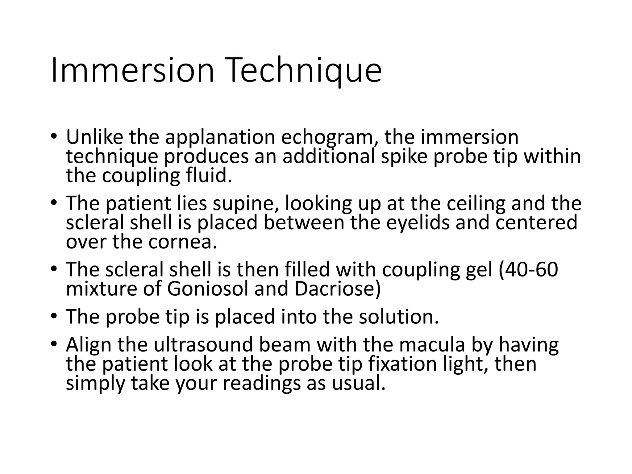
Description and methodology of immersion technique for biometry, highlighting its accuracy advantages.
Comparison results of axial length measurements by immersion and applanation techniques, significant findings on accuracy.
Identifies common errors in ultrasound measurement of axial length and limitations of immersion technique.
Introduction of optical methods like Partial Coherence Interferometry as a gold standard for axial length measurement.
Different types of interferometers and their relevance in optical biometers for accurate measurements.
The IOLMaster 500 biometer's features, including key measurements made for comprehensive ocular analysis.
Critical requirements for measurements with IOLMaster devices, emphasizing fixation and measurement variability.
Discusses limitations of IOLMaster 500, particularly in dense cataract cases and alternative ultrasound use.
Research findings comparing PCI optical biometry with ultrasound, emphasizing predictive accuracy.
Introduction and advantages of the IOLMaster 700, exploring its advanced technology in biometric measurements.
Comparison study results on IOLMaster 700 and IOLMaster 500 efficacy in various eye conditions.
Discusses the practical applications and future topics related to IOL calculations and best practices for biometry.


![Types of pediatric contact lens [autosaved]](https://cdn.slidesharecdn.com/ss_thumbnails/typesofpediatriccontactlensautosaved-200210123904-thumbnail.jpg?width=640&height=640&fit=bounds)


