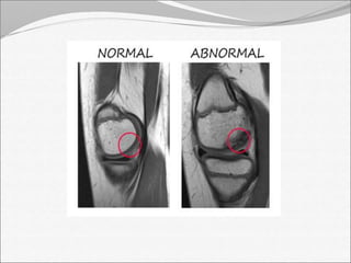
Causes : 
• Bacterial (staphylococcus aureus, E.coli, proteus and 
streptococcus) 
• viral 
• mycobacterial 
• fungal
Predisposing factors: 
 Rheumatoid arthritis 
 Intravenous drug abuse 
 Immunosuppressive drug therapy 
 AIDS 
 DM 
 Old age >80 years 
 Chronic disorder
Hematogenous spread 
 Most common form of spread 
 Usually affect people with underlying medical 
problem 
Direct inoculation 
• May result from penetrating trauma 
• Introduction of organism during diagnostic and 
surgical procedure e.g. arthroscopy and intra-articular 
injection 
Direct spread from adjacent focal infection
Clinical manifestations 
 Fever 
 Acute severe pain 
 Swelling of the joint 
 Tenderness 
 Warmth 
 Limited joint mobility
Imaging: 
X-RAY: 
 Early stage –soft tissue swelling, loss of tissue planes, 
widening of joint space and slight sublaxation due to 
fluid in the joint. 
 Gas may be seen with E.coli infection 
 Late stage – narrowing and irregularity of joint space 
 Periosteal reaction, bone destruction and 
sequestrum formation
Lateral view of ankle joint, exaggerated soft tissue swelling, 
decreased joint space with sclerotic ends of bone
Underlying bony erosions and reduced joint space in 
talus AP view
Joint space 
decreased 
osteophytes AP view
Head of femur deformed and 
necrosed,loss of joint space LEFT SIDE
Ultrasonography 
 More reliable in revealing a joint effusion in early 
cases. 
 Widening of space between capsule and bone of 
>2mm indicates effusion. 
 Echo free  transient synovitis 
 Positively echogenic  septic arthritis 
 Ultrasound can detect joint-swelling.
Being a non- ionizing, easily available, non-invasive 
and relatively cheap modality, Ultrasound should be 
first line of investigation in a suspected case of 
infective arthritis.
computed tomography: 
 Early findings: 
 Soft tissue swelling 
 Joint space widening
 Late findings(2-3 w): 
 Joint space narrowing 
 Blurring of fat planes 
 Increased density of fatty marrow 
 Periosteal reaction 
 Cortical erosion or destruction 
 Intraosseous gas
CT Scan demonstrating soft tissue oedema localised to the 
right sternoclavicular joint (A) and appearances of an 
associated joint effusion, erosion and destruction of sternal end
CT of left hip joint The dd included septic 
arthritis/osteomyelitis joint
Sagittal CT scan of C-spine shows a bone 
erosion of the anterior portion of odontoid 
process of C2 (arrow)
MRI findings: 
• Synovial enhancement 
• Perisynovial edema and joint effusion. 
• Single or multiple radiolucent abscesses 
• Assessment of the extent of tissue affected
Significance of CT and MRI: 
• Cross-sectional imaging modalities such as CT and 
MRI are now considered standard in the diagnosis of 
septic arthritis because they have, 
• Excellent spatial resolution 
• Early detection 
• Assessment of the extent of tissue affected 
 Although expensive, they are sensitive and specific.
Nuclear medicine imaging: 
 Nuclear medicine imaging can detect septic arthritis 
10 to 14 days before changes are visible on plain 
radiographs. 
 Highly Sensitive but Nonspecific 
 Inexpensive 
 Focal hyper perfusion 
 Focal hyperemia 
 Focal bone uptake
Complications: 
 bone destruction and dislocation of the joint 
(especially hip) 
 Cartilage destruction 
 May lead to either fibrosis or bony ankylosis 
 In adult partial destruction of the joint will result in 
secondary osteoarthritis 
 Growth disturbance 
 Presenting as either localized deformity or shortening 
of the bone
Thank you

Septic arthritis

  • 4.
    Causes : •Bacterial (staphylococcus aureus, E.coli, proteus and streptococcus) • viral • mycobacterial • fungal
  • 5.
    Predisposing factors: Rheumatoid arthritis  Intravenous drug abuse  Immunosuppressive drug therapy  AIDS  DM  Old age >80 years  Chronic disorder
  • 7.
    Hematogenous spread Most common form of spread  Usually affect people with underlying medical problem Direct inoculation • May result from penetrating trauma • Introduction of organism during diagnostic and surgical procedure e.g. arthroscopy and intra-articular injection Direct spread from adjacent focal infection
  • 10.
    Clinical manifestations Fever  Acute severe pain  Swelling of the joint  Tenderness  Warmth  Limited joint mobility
  • 11.
    Imaging: X-RAY: Early stage –soft tissue swelling, loss of tissue planes, widening of joint space and slight sublaxation due to fluid in the joint.  Gas may be seen with E.coli infection  Late stage – narrowing and irregularity of joint space  Periosteal reaction, bone destruction and sequestrum formation
  • 13.
    Lateral view ofankle joint, exaggerated soft tissue swelling, decreased joint space with sclerotic ends of bone
  • 14.
    Underlying bony erosionsand reduced joint space in talus AP view
  • 15.
    Joint space decreased osteophytes AP view
  • 16.
    Head of femurdeformed and necrosed,loss of joint space LEFT SIDE
  • 17.
    Ultrasonography  Morereliable in revealing a joint effusion in early cases.  Widening of space between capsule and bone of >2mm indicates effusion.  Echo free  transient synovitis  Positively echogenic  septic arthritis  Ultrasound can detect joint-swelling.
  • 18.
    Being a non-ionizing, easily available, non-invasive and relatively cheap modality, Ultrasound should be first line of investigation in a suspected case of infective arthritis.
  • 21.
    computed tomography: Early findings:  Soft tissue swelling  Joint space widening
  • 22.
     Late findings(2-3w):  Joint space narrowing  Blurring of fat planes  Increased density of fatty marrow  Periosteal reaction  Cortical erosion or destruction  Intraosseous gas
  • 23.
    CT Scan demonstratingsoft tissue oedema localised to the right sternoclavicular joint (A) and appearances of an associated joint effusion, erosion and destruction of sternal end
  • 24.
    CT of lefthip joint The dd included septic arthritis/osteomyelitis joint
  • 25.
    Sagittal CT scanof C-spine shows a bone erosion of the anterior portion of odontoid process of C2 (arrow)
  • 26.
    MRI findings: •Synovial enhancement • Perisynovial edema and joint effusion. • Single or multiple radiolucent abscesses • Assessment of the extent of tissue affected
  • 27.
    Significance of CTand MRI: • Cross-sectional imaging modalities such as CT and MRI are now considered standard in the diagnosis of septic arthritis because they have, • Excellent spatial resolution • Early detection • Assessment of the extent of tissue affected  Although expensive, they are sensitive and specific.
  • 32.
    Nuclear medicine imaging:  Nuclear medicine imaging can detect septic arthritis 10 to 14 days before changes are visible on plain radiographs.  Highly Sensitive but Nonspecific  Inexpensive  Focal hyper perfusion  Focal hyperemia  Focal bone uptake
  • 33.
    Complications:  bonedestruction and dislocation of the joint (especially hip)  Cartilage destruction  May lead to either fibrosis or bony ankylosis  In adult partial destruction of the joint will result in secondary osteoarthritis  Growth disturbance  Presenting as either localized deformity or shortening of the bone
  • 34.

Editor's Notes