Agenda
Background
StentThrombosis (ST)
Classification
Management – Interventional and Pharmaceutical
In-Stent Restenosis (ISR)
Mechanisms, Classification and Imaging
Management
Prevention
Take Home Message
3.
Introduction
• Stent failureremains the major drawback to the use of coronary stents as a
revascularisation strategy.
• Stent failure constitutes stent thrombosis(ST) and In-stent restenosis (ISR).
• Recent advances in imaging have substantially improved our understanding of the
mechanisms underlying the stent failure.
• Both have in common numerous clinical risk factors and mechanical elements at the
time of stent implantation.
Background
• ST isan acute or subacute thrombotic occlusion that usually presents
as an acute MI or acute coronary syndrome and is associated with high
rates of morbidity and mortality.
Incidence: 0.5-1% in the first year
0.2-0.6% in every subsequent year
Higher incidence with STEMI
Lower incidence with elective stent placement
6.
Risk factors forstent thrombosis
Clinical
ACS
(STEMI/NSTEMI)
Left ventricular
dysfunction
Chronic kidney
disease
Diabetes
mellitus
COVID -19
Procedural
Stent length
Stent
underexpansion
No reflow
Residual
stenosis
Dissection
Multiple
stents
Bifurcation
stenting
Lesion
related
Necrotic core
Bifurcation
lesions
Prior
brachytherapy
Multivessel
disease
Inflow and
outflow
obstruction
Stent
related
Biocompatible
polymers
Polymer/stent
thickness
Drug dosage
Antiplatelet
related
adherence
CYP2C19
polymorphisms
High- on
treatment
platelet activity
Antiplatelet
type
DAPT
duration
7.
Early ST (<30 days) Late ST (1-12 months) Very Late ST (>12 months)
Platelet rich thrombi formation
Mainly due to Inadequate procedural
result
Impaired neointimal healing
Acute < 24 hours Predisposing factors:
Stenting across major branches
Bifurcations
Overlapped areas
Exaggerated response to healing in DES
Stents are placed in stenosed segments
with high lipid core
Predisposing factors
Malapposition
Uncovered struts
Neoatherosclerosis
Stent underexpansion
Sub acute – 24 hours to 30 days
Emergency PCI – when presentation is
acute
Optimal reperfusion only in
2/3rds
Types of ST
8.
Image guidance :a must to explore cause and
decide on management
SCAI Expert Consensus Statement on Management of In-Stent Restenosis and Stent Thrombosis, JSCAI. 2023
Early restenosis due to protrusion
of a calcified nodule (white
arrows). After Stent thrombosis, the
repeat OCT showed protruding
calcified
nodule within the stent.
9.
Management Algorithm
• Mostare dealt with balloon dilatation (NC, Scoring, Cutting)
• Thrombus aspiration if the clot burden is large
• Address stent-related mechanical issues
• Additional stent implantation should ordinarily be limited to significant residual
dissections, especially if recent DAPT has been discontinued.
• Optimise pharmacotherapy
• Assess the aetiology of stent closure after the establishment of flow
10.
SCAI Expert ConsensusStatement on Management of In-Stent Restenosis and Stent Thrombosis, JSCAI. 2023
11.
Pharmacologic Management
• Evaluatethe compliance with DAPT
• Restart or intensify dual antiplatelet therapy (DAPT)
• More potent drugs: prasugrel/ticagrelor
• Sustained administration of 150mg Clopidogrel if platelet aggregation studies reveal insufficient
(<50%) inhibition of platelet aggregation
• Consider prolonged therapy in high-risk patients.
• Glycoprotein IIb/IIIa antagonists – prolonged infusions up to 72 hours: to prevent
distal embolisation
• Long-term anticoagulation – rare; for recurrent ST
12.
Case
• 52 F
•Diabetes, hypertension
• IWMI
• CHB
• Cardiogenic shock
• CAG – Distal RCA total occlusion, 90 % stenosis in LAD and LCX
• Primary PCI to the distal RCA
• Had chest pain with in 24 hours
• ST elevation in inferior leads
13.
Final Result
Negotiated run-throughwire
1.25*6 mm balloon dilation
Heavy thrombus burden After 3.0*15 NC balloon dilation
Acute Stent Thrombosis
Background
• Recurrent diameterstenosis at the stent segment >50% of the vessel diameter.
• The rate of ISR is higher in patients with Diabetes (5.7% vs 8.7%).
• Recurrent ISR is seen in approximately 20% of all cases.
• Recurrence is independently predicted by the number of stents placed at the location. (43.1%
increase in TLR ).
• A third layer of metal should be avoided as associated with underexpansion.
Incidence of ISR – 10%
25% cases present with Acute MI
30-day mortality (AMI)- 10%-25%.
16.
Mechanisms
of ISR
Biological
Neointimal tissue
proliferationor
hyperplasia
Neoatherosclero
sis
Mechanical
Stent
underexpansion
– primary cause
MSA
IVUS > 5.0mm2
OCT -> 4.5mm2
Target MSA > 90% of the reference
segment
Stent fracture
Edge
dissection
>60 °, > 3 mm in
length,
penetrating the
media
Late acquired
malapposition
17.
Classification
• Temporally
• EarlyISR (<30 days)
• Late ISR (30 days – 1 year)
• Very Late ISR (>1 year)
• Coronary angiography remains the standard diagnostic method.
• IVUS and OCT provide a detailed assessment of the native artery and stented
segment
18.
Morphological classification
MEHRAN CLASSIFICATION-
•based on coronary angiography
1. Class I-focal involvement
2. Class II-diffuse intrastent
3. Class III-diffuse proliferative
4. Class IV-total occlusion.
This was highly relevant to bare metal stenting (BMS), but its applicability to DES ISR
is uncertain.
19.
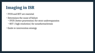
Imaging in ISR
•IVUS and OCT are essential
• Determines the cause of failure
• IVUS (better penetration) for stent underexpansion
• OCT ( high resolution) for neoatherosclerosis
• Guide re-intervention strategy
21.
THE WAKSMAN ISRCLASSIFICATION
• Based on intracoronary imaging.
• Type I -Mechanical
TYPE 1 A-
UNDEREXPANTION
TYPE 1 B- STENT FRACTURE
OCT OCT
IVUS IVUS
22.
• Type II-
•Type III-mixed pattern
• Type IV-chronic total occlussions
• Type V-lesions previously treated with > 2 stents
TYPE 2 A-NEOINTIMAL
HYPERPLASIA
TYPE 2B-NEOATHEROSCLEROIS NONCALCIFED AND
CALCIFIED (TYPE 2C)
OCT IVUS OCT IVUS
23.
SCAI Algorithm ofISR Mx
Critical principle
Obtain the largest acute
lumen gain as possible by
maximising the
immediate postprocedural
MLA
24.
Treatment Modalities
Balloon Angioplasty
Forunderdeployed stent, focal ISR,
short DAPT
Underexpansion, focal stent gap,
stent fracture – high NC pressure
balloons
Hyperplasia – scoring/ cutting
balloon
Drug coated balloons
Class I indication (ESC)
Inflation time > 60 sec
Balloon: artery ratio > 0.91
New DES
After appropriate sizing and expansion
of original DES
Minimise stent coverage
Atherectomy
Orbital/ rotational
Arc of calcium > 270°, > 0.67 mm in
thickness
Risk of entrapment
ELCA
Brachytherapy
IVL
For highly calcific
neoatherosclerosis
25.
Considerations for CABGin refractory or
recurrent ISR
• Multivessel CAD especially LM or proximal LAD involvement
• Prior CABG
• Suitability of distal vessel for grafting (including diffuseness of CAD, extent of “metal
jacket,” and size of vessel)
• Global and regional LV function including viability (especially the segment
subtended by the involved vessel)
• Comorbid conditions (including age, frailty, life expectancy, and activity level)
• Anticipated completeness of revascularization
• Response to optimal medical therapy
26.
Prevention of Stentfailure
• Adequate bed preparation – plaque modification
• Optimisation of the stent by imaging modality
• Addressing the post-stent complications
• Compliance with the drug therapy (DAPT)
27.
Case
• 55-year-old male
•Hypertensive, Diabetic
• H/o PTCA in 2010
• Noncompliant with medications after a year
of PCI
• Presented with unstable angina
• CAG – DVD – RCA ISR, mid LAD 90% stenosis
28.
OCT images ofISR
Neointimal hyperplasia of BMS in mid RCA
MLA 3.06mm²
After NC balloon dilation
Increase in MLA to 4.9 mm²
Take Home Message
Stentfailure remains the major drawback to the use of coronary stents as a
revascularisation strategy.
Incidence of Stent Thrombosis is 0.5 to 1.0%; higher incidence in STEMI.
Incidence of ISR is 10%. It is higher in diabetics, patients on hemodialysis and those
with multiple stents.
Recurrence of ISR is seen in almost 20% cases.
Imaging-guided PCI is needed to establish the cause and management strategy.
Optimise the vessel preparation, especially in complex lesions.
Drug Compliance must be revisited in all cases of stent thrombosis.
Editor's Notes
#5 The rate is lower for elective stent placement (0.3%-0.5%) but higher in acute coronary syndrome (3.4%) and MI
In contemporary practice, the observed mortality rate (~30%) is high, although recent clinical trials and studies requiring au topsy confirmation suggest a better survival, with an average rate of <10%.
The ST rate is higher in ST-elevation myocardial infarction presentations treated with primary stenting.
Approximately 20% of patients with a first ST expe rience a recurrent ST episode within 2 years.
#7 Stent thrombosis is classified by the Academic Research Consortium criteria based on the presenting clinical scenario and timing after initial stent placement.
#8 Early restenosis due to re-protrusion of a calcified nodule. This patient underwent percutaneous coronary intervention to treat lesions in the distal and mid right coronary ar tery. Optical coherence tomography (OCT) showed an eruptive calcified nodule (white ar rows)inbothlesions. A calcified noduleischaracterized byanaccumulationofsmallcalcium fragmentstypically with strong signalattenuation duetoaccompanyingandoverlyingfibrin. The patient came back for staged procedure of LAD (left anterior descending artery) 6 weeks later. OCTshowed reprotruding calcified nodules within the stent.
#11 Prevention of ST is dependent on optimal stent implantation and the duration and compliance with DAPT.
Congenital or acquired hyporesponder DAPT status seen with clopidogrel is uncommon with prasugrel or ticagrelor. Prior generation stents were susceptible to ST with discontinuation of DAPT out to 5 years and longer in anecdotal cases. With the newest generation of stents, the duration of treatment can be decreased safely to 3 months or 1 month.
#15 Additional criteria for clinically relevant ISR include: recurrent angina, objective signs of ischemia, or abnormal fractional flow reserve
Second-generation drug-eluting stents (DES) have a 5.7% ISR rate in patients without diabetes, and 8.7% rate in those with diabetes.
Beyond 1 year, there is a gradual increase in major adverse cardiovascular events (MACE); the 5-year ISR rate is 9% to 12% in noncomplex lesions.
Recurrent ISR occurs in approximately 20% of all ISR cases.
Recurresnt ISR is independently predicted by the number of stents placed at the location.
The 1-year MACE (43.1%) and target lesion revascularization (41.2%) rates were significantly higher in the 3 stent layer group than in the 1-stent-layer and 2-stent-layer groups. The number of metallic layers and hemodialysis requirement were identified as independent predictors of MACE. A third layer of metal is almost always associated with underexpansion and should be avoided.
#16 Waksman ISR Classification – type II
Neointima – SMCs and ECM
Neoatherosclerosis – Inhibition of endothelialization by DES allows LDL into vessel wall. So at later stages, the healed neointima is prone to atherosclerosis.
#17 Early – undersizing, underexpansion, stent fracture
Late – delayed healing, uncovered stent struts, intimal hyperplasia
Very late – neoatherosclerosis, , intimal hyperplasia, stent fracture
#19 The most common treatment approach for the first episode of ISR is to implant a second DES, based on the rationale that DES therapy has superior efficacy over balloon angioplasty alone. However, this is not always necessary and may not be the bestsolution,particularly when the reference vessel and the resultant minimal lumen area are small.
If the underlying etiology is not directly addressed and corrected, there is a high likelihood of recurrent ISR, and the rate of ISR in second layer DES is high: 12% to 16% at 12 months and 33% at 3 to 5 years.56–58
#24 ELCA – Debulks neointimal hyperplasia; for breaking peri-stent calcium
IVL – for breaking calcium
Focal ISR – Balloon angioplasty; ELCA or atherectomy may be beneficial in selected cases
For diffuse ISR, atherectomy or scoring/cutting balloon angioplasty followed by repeat DES implantation is typically advised
For focal calcific nodule – double layer NC super high pressure balloons sustaining pressures of 30-35 mm Hg