Jordan University of Science and Technology
Faculty of Applied Medical Sciences
Department of Rehabilitation science
Physical Therapy Program
Elbow Joint Assessment
First Semester 2023-2024
Elbow Joint Anatomy
• The elbow is a complex hinged joint whose function is to facilitate
the placement of the hand in space.
• It allows flexion–extension and pronation–supination of the
forearm.
• It is composed of three bones: the humerus, radius, and ulna, and
three articulations: humeroulnar, humeroradial, and the less
important proximal radioulnar.
• The humeroulnar joint is the largest and most stable of the elbow
articulations.
• The humeroradial joint lies lateral to the humeroulnar articulation.
• It is composed of a shallow disc (radial head) articulating on the
spherical humeral capitellum.
• Pronation and supination are accomplished by rotation of the
radius along its long axis about the ulna
• Rotation toward the palm down is pronation, whereas rotation
toward the palm up is supination.
Elbow Joint Anatomy
3
Patient History
• After taking proper history from the patient a number of additional questions may be asked:
1. Any repeated activity previous to the injury?
2. Does he/she play any sport? (tennis elbow/ golfer’s elbow)
3. Any neurological signs? (numbness, tingling, … etc.)
4. Any trauma at that area?
4
Observation
• Notice how the patient is posturing the upper extremity.
• Is the arm relaxed at the side or is the patient cradling it for protection?
• If the elbow is swollen, the patient may posture it at 70 degrees of flexion (the resting position), which allows for
the most space for the fluid.
• Swelling may be easily noticed at the triangular space bordered by the lateral epicondyle, radial head, and the
olecranon.
• How willing is the patient to use the upper extremity?
• Pain may be altered by changes in position?
• Note whether there is any atrophy present in the biceps.
5
Observation
• The therapist should observe for any generalized swelling (indicating a trauma or an inflammatory process) or
localized swelling (indicating olecranon bursitis “student’s elbow”)
• The therapist should:
1. Look at the bony contours of the joint
2. Look at the skin covering the joint
3.Look for any abnormalities in the bone above and below the elbow joint
6
Palpation - Anterior Aspect
1. Cubital Fossa
• The base of the triangle is formed by a line between the
medial and lateral epicondyles of the humerus.
• The medial side is formed by the pronator teres and the
lateral side by the brachioradialis.
• The floor is composed of the brachialis and the supinator.
• The fossa contains the following structures: biceps tendon,
distal part of the brachial artery and veins, the origins of
the radial and ulnar arteries, and parts of the median and
radial nerves.
2. Biceps Muscle and Tendon
3. Brachial Artery
• The brachial artery is located in the cubital fossa medial to
the biceps tendon.
• The brachial pulse can be readily assessed at this point.
4. Median Nerve
7
Palpation-Medial Aspect
8
1. Medial epicondyle
• Tenderness in this area can be due to inflammation of the
common aponeurosis of the flexor and pronator tendons of the
forearm and wrist and is commonly referred to as golfer’s elbow
(medial epicondylitis).
2. Medial (Ulnar) Collateral Ligament (is not distinctly palpable)
3. Wrist Flexor–Pronator
Palpation- Lateral Aspect
• Lateral Epicondyle and Supracondylar Ridge
• Tenderness in this area can be due to inflammation of the
common aponeurosis of the extensor tendons of the wrist and
is commonly referred to as tennis elbow (lateral
epicondylitis).
• Radial Head
• Lateral (Radial) Collateral Ligament
• Annular Ligament
• Wrist Extensor–Supinator
9
Palpation- Posterior Aspect
• Olecranon flexes the arm, bringing the olecranon out of the
olecranon fossa.
• Olecranon Fossa
• Triceps Muscles
• Olecranon Bursa
• Ulna Border
10
Examination
• The therapist should examine the main movements actively and passively which include:
1. Flexion (0-150)
2. Extension
3. Supination (0-80)
4. Pronation (0-80)
• The therapist should feel the end-range of all movements where it is soft tissue approximation in
flexion, bony in extension, and tissue stretch for pronation and supination.
• Note: sometimes extension can go to hyperextension (up to 10 degrees, commonly in females).
• Additionally, myotomes, dermatomes and reflexes should be examined in the elbow area.
144
Elbow Joint ROM assessment
Elbow Flexion and Extension
• Start Position: The patient is supine or sitting.
• The arm is in the anatomical position with the elbow in extension (0°).
• A towel is placed under the distal end of the humerus to accommodate the ROM.
• Owing to biceps muscle tension, unusually muscular men may not be able to achieve 0°.
• Stabilization: The therapist stabilizes the humerus.
• Goniometer Axis: The axis is placed over the lateral epicondyle of the humerus.
• Stationary Arm: Parallel to the longitudinal axis of the humerus, pointing toward the tip of the acromion process.
• Movable Arm: Parallel to the longitudinal axis of the radius, pointing toward the styloid process of the radius.
• End Position: From the start position of elbow extension, the forearm is moved in an anterior direction so that the hand
approximates the shoulder to the limit of elbow flexion (150°).
• Extension/Hyperextension: The forearm is moved in a posterior direction to the limit of elbow extension (0°)/
hyperextension (up to 15°)
Elbow Flexion and Extension
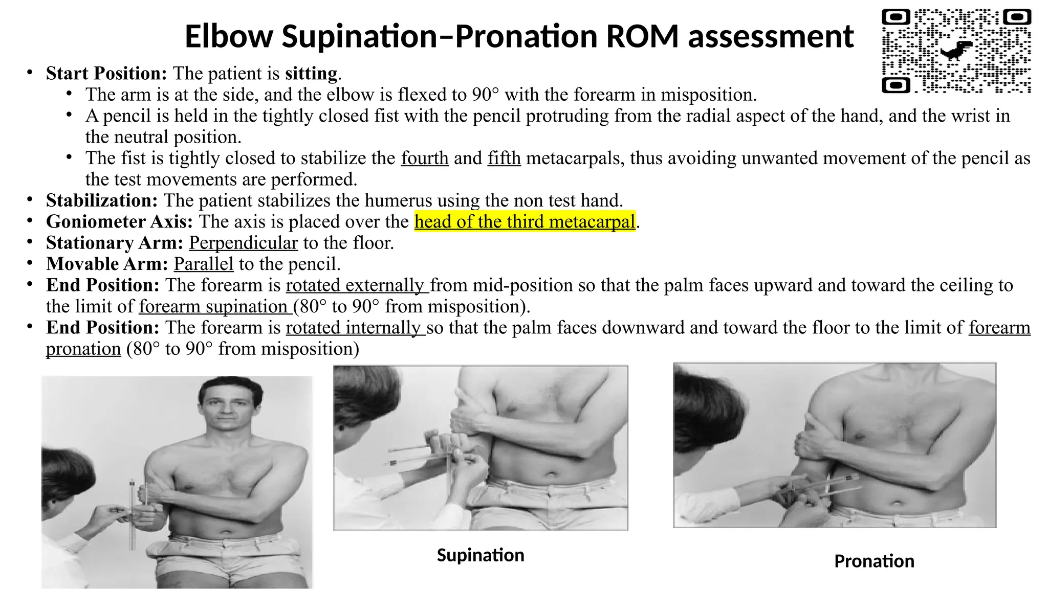
Elbow Supination–Pronation ROM assessment
• Start Position: The patient is sitting.
• The arm is at the side, and the elbow is flexed to 90° with the forearm in misposition.
• A pencil is held in the tightly closed fist with the pencil protruding from the radial aspect of the hand, and the wrist in
the neutral position.
• The fist is tightly closed to stabilize the fourth and fifth metacarpals, thus avoiding unwanted movement of the pencil as
the test movements are performed.
• Stabilization: The patient stabilizes the humerus using the non test hand.
• Goniometer Axis: The axis is placed over the head of the third metacarpal.
• Stationary Arm: Perpendicular to the floor.
• Movable Arm: Parallel to the pencil.
• End Position: The forearm is rotated externally from mid-position so that the palm faces upward and toward the ceiling to
the limit of forearm supination (80° to 90° from misposition).
• End Position: The forearm is rotated internally so that the palm faces downward and toward the floor to the limit of forearm
pronation (80° to 90° from misposition)
Supination Pronation
Elbow Joint MMT Assessment
ELBOW FLEXION
(Biceps, Brachialis, and Brachioradialis)
Grade 5 (Normal), Grade 4 (Good), and Grade 3 (Fair)
• Position of Patient: Short sitting with arms at sides.
• The following are the positions of choice, but it is doubtful whether the individual muscles can be
separated when strong effort is used.
Biceps brachii: forearm in
supination
Brachialis: forearm in
pronation
Brachioradialis: forearm in
misposition between
pronation and supination
Grade 5 (Normal), Grade 4 (Good), and Grade 3 (Fair)
• Position of Therapist: Stand in front of patient toward the test side.
• Hand giving resistance is contoured over the flexor surface of the forearm proximal to the wrist.
• The other hand applies counterforce by cupping the palm over the anterior superior surface of the shoulder.
• Test (All Three Forearm Positions): Patient flexes elbow through range of motion.
• Instructions to Patient (All Three Tests)
Grades 5 and 4: "Bend your elbow. Hold it. Don't let me pull it down."
• Grade 3: "Bend your elbow."
• Grade 5 (Normal): Completes available range and holds firmly against maximal resistance.
• Grade 4 (Good): Completes available range against strong to moderate resistance.
• Grade 3 (Fair): Completes available range with each forearm position with no manual resistance.
Grade 2 (Poor)
• Position of Patient
• All Elbow Flexors: Short sitting with arm abducted to 90° and supported by
examiner. Forearm is supinated (biceps), pronated (brachialis), and in misposition
(brachioradialis).
• Alternate Position for Patients Unable to Sit:
• Supine. Elbow is flexed to about 45° with forearm supinated (for biceps), pronated
(for brachialis), and in misposition (for brachioradialis).
• Test: Patient attempts to flex the elbow.
• Instructions to Patient: "Try to bend your elbow."
• Grade 2 (Poor): Completes range of motion (in each of the muscles tested).
Grade 1 (Trace) and Grade 0 (Zero)
• Positions of Patient and Therapist: Supine for all three muscles with therapist standing at test side.
• All other aspects are the same as for the Grade 2 test.
• Test: Patient attempts to bend elbow with hand supinated, pronated, and in misposition.
• Grade 1 (Trace): Examiner can palpate a contractile response in each of the three muscles for which a Trace grade is given.
• Grade 0 (Zero): No palpable contractile activity.
ELBOW EXTENSION
Grade 5 (Normal), Grade 4 (Good), and Grade 3 (Fair)
• Position of Patient: Prone on table.
• The patient starts the test with the arm in 90° of abduction and the forearm flexed and hanging vertically over the side of
the table.
• Position of Therapist: For the prone patient, the therapist provides support just above the elbow. The other hand is used to
apply downward resistance on the dorsal surface of the forearm.
• Test: Patient extends elbow to end of available range or until the forearm is horizontal to the floor.
• Instructions to Patient: "Straighten your elbow. Hold it. Don't let me bend it." Do not allow hyper- extension.
• Grade 5 (Normal): Completes available range and holds firmly against maximal resistance.
• Grade 4 (Good): Completes available range against strong resistance, but there is a "give" to the resistance at the end range.
• Grade 3 (Fair): Completes available range with no manual resistance.
Grade 2 (Poor), Grade 1 (Trace), and Grade 0 (Zero)
• Position of Patient: Short sitting.
• The arm is abducted to 90° with the shoulder in neutral rotation and the elbow flexed to about 45°.
• The entire limb is horizontal to the floor.
• Position of Therapist: Stand at test side of patient.
• For the Grade 2 test, support the limb at the elbow. For a Grade 1 or 0 test, support the limb under the forearm and palpate the triceps on
the posterior surface of the arm just proximal to the olecranon process.
• Test: Patient attempts to extend the elbow.
• Instructions to Patient: "Try to straighten your elbow."
• Grade 2 (Poor): Completes available range in the absence of gravity.
• Grade 1 (Trace): Examiner can feel tension in the triceps tendon just proximal to the olecranon or contractile activity in the muscle fibers on
the posterior surface of the arm.
• Grade 0 (Zero): No evidence of any muscle activity.
FOREARM SUPINATION (Supinator and Biceps brachii)
Grade 5 (Normal), Grade 4 (Good), and Grade 3 (Fair)
• Position of Patient: Short sitting; arm at side and elbow flexed 90: forarm in pronation.
• Position of Therapist: Stand at side or in front of patient.
• One hand supports the elbow.
• For resistance, grasp the forearm on the volar surface at the wrist.
• Test: Patient begins in pronation and supinates the forearm until the palm faces the ceiling.
• Therapist resists motion in the direction of pronation. (No resistance is given for Grade 3.)
• Alternate Test: Grasp patient's hand as if shaking hands; cradle the elbow and resist via the hand grip. This test is used if the
patient has Grade 5 or 4 wrist and hand strength.
• If wrist flexion is painful, give resistance at the wrist a more difficult level, but less painful.
• Instructions to Patient: "Turn your palm up. Hold it. Don't let me turn it down. Keep your wrist and fingers relaxed." For
Grade 3: "Turn your palm up."
• Grade 5 (Normal): Completes full available range of motion and holds against maximal resistance.
• Grade 4 (Good): Completes full range of motion against strong to moderate resistance.
• Grade 3 (Fair): Completes available range of motion without resistance.
Forearm Supination- Grade 5 (Normal), Grade 4 (Good), and Grade 3 (Fair)
Grade 2 (Poor)
• Position of Patient: Short sitting with shoulder flexed between 45° and
90° and elbow flexed to 90°. Forearm in neutral.
• Position of Therapist: Support the test arm by cup- ping the hand under
the elbow.
• Test: Patient supinates forearm through partial range of motion.
• Instructions to Patient: "Turn your palm toward your face."
• Grade 2 (Poor): Completes a full range of motion.
Grade 1 (Trace) and Grade 0 (Zero)
• Position of Patient: Short sitting. Arm and elbow are flexed as
for the Grade 3 test.
• Position of Therapist: Support the forearm just distal to the
elbow. Palpate the supinator distal to the head of the radius on the
dorsal aspect of the forearm.
• Test: Patient attempts to supinate the forearm.
• Instructions to Patient: "Try to turn your palm so it faces the
ceiling."
• Grade 1 (Trace): Slight contractile activity but no limb
movement.
• Grade 0 (Zero): No contractile activity.
FOREARM PRONATION
(Pronator teres and Pronator quadratus)
FOREARM PRONATION
Grade 5 (Normal), Grade 4 (Good), and Grade 3 (Fair)
• Position of Patient: Short sitting or may sit at a table.
• Arm at side with elbow flexed to 90° and forearm in supination.
• Position of Therapist: Standing at side or in front of patient. Support the elbow.
• Hand used for resistance grasps the forearm over the dorsal surface at the wrist.
• Test: Patient pronates the forearm until the palm faces downward.
• Therapist resists motion at the wrist in the direction of supination for Grades 4 and 5. (No resistance is given
for Grade 3.)
• Alternate Test: Grasp patient's hand as if to shake hands, cradling the elbow with the other hand and resisting
pronation via the hand grip.
• Instructions to Patient: "Turn your palm down. Hold it. Don't let me turn it up. Keep your wrist and fingers
relaxed."
• Grade 5 (Normal): Completes available range of motion and holds against maximal resistance.
• Grade 4 (Good): Completes all available range against strong to moderate resistance.
• Grade 3 (Fair): Completes available range without resistance.
Grade 2 (Poor)
• Position of Patient: Short sitting with shoulder flexed between
45° and 90° and elbow flexed to 90°. Forearm in neutral.
• Position of Therapist: Support the test arm by cup- ping the hand
under the elbow.
• Test: Patient pronates forearm.
Instructions to Patient: "Turn your palm facing outward away
from your face."
• Grade 2 (Poor): Complete range of motion.
Grade 1 (Trace) and Grade 0 (Zero)
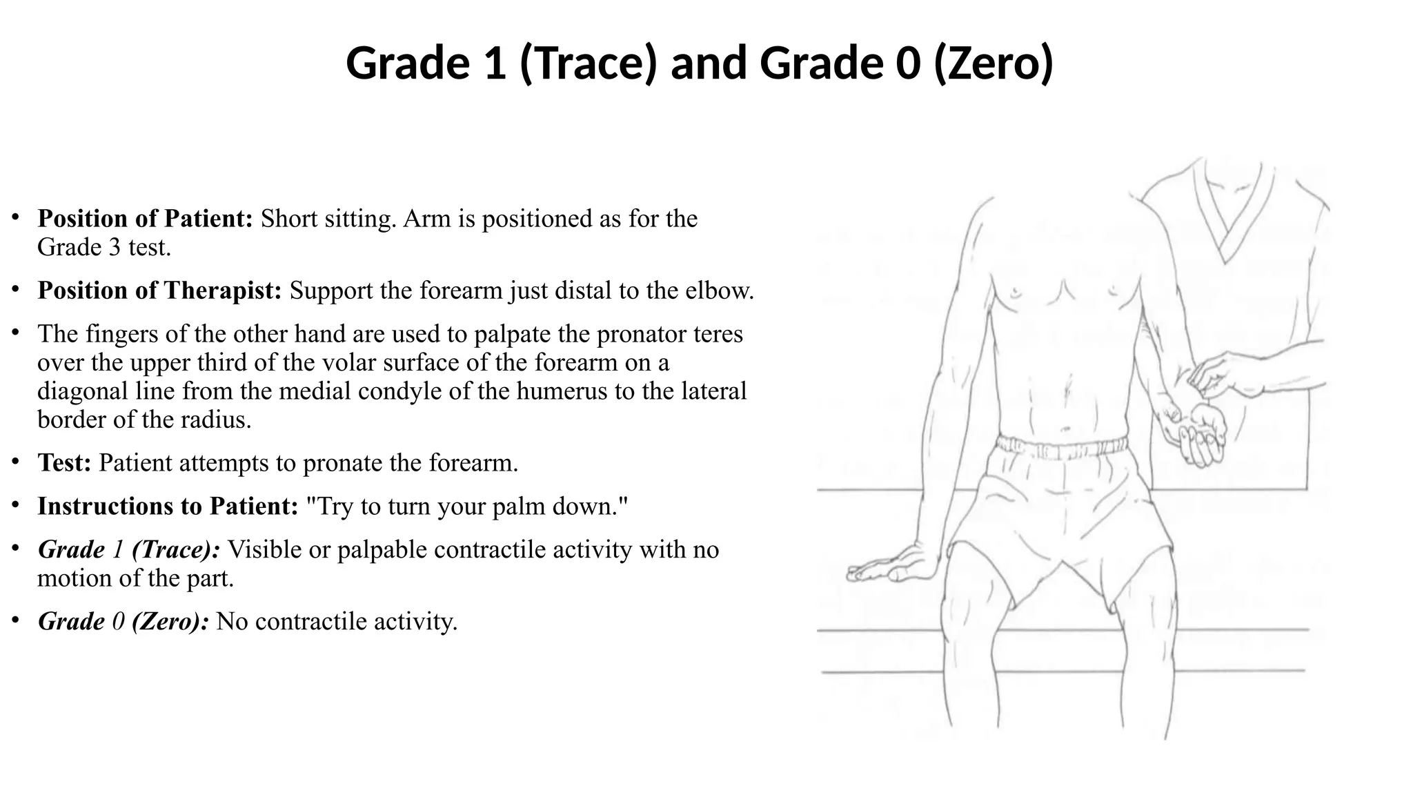
• Position of Patient: Short sitting. Arm is positioned as for the
Grade 3 test.
• Position of Therapist: Support the forearm just distal to the elbow.
• The fingers of the other hand are used to palpate the pronator teres
over the upper third of the volar surface of the forearm on a
diagonal line from the medial condyle of the humerus to the lateral
border of the radius.
• Test: Patient attempts to pronate the forearm.
• Instructions to Patient: "Try to turn your palm down."
• Grade 1 (Trace): Visible or palpable contractile activity with no
motion of the part.
• Grade 0 (Zero): No contractile activity.

The Elbow Joint 1_a93e492f7cfcdba2d5a0fde7c8fecec5(2).pptx

  • 1.
    Jordan University ofScience and Technology Faculty of Applied Medical Sciences Department of Rehabilitation science Physical Therapy Program Elbow Joint Assessment First Semester 2023-2024
  • 2.
    Elbow Joint Anatomy •The elbow is a complex hinged joint whose function is to facilitate the placement of the hand in space. • It allows flexion–extension and pronation–supination of the forearm. • It is composed of three bones: the humerus, radius, and ulna, and three articulations: humeroulnar, humeroradial, and the less important proximal radioulnar. • The humeroulnar joint is the largest and most stable of the elbow articulations. • The humeroradial joint lies lateral to the humeroulnar articulation. • It is composed of a shallow disc (radial head) articulating on the spherical humeral capitellum. • Pronation and supination are accomplished by rotation of the radius along its long axis about the ulna • Rotation toward the palm down is pronation, whereas rotation toward the palm up is supination.
  • 3.
  • 4.
    Patient History • Aftertaking proper history from the patient a number of additional questions may be asked: 1. Any repeated activity previous to the injury? 2. Does he/she play any sport? (tennis elbow/ golfer’s elbow) 3. Any neurological signs? (numbness, tingling, … etc.) 4. Any trauma at that area? 4
  • 5.
    Observation • Notice howthe patient is posturing the upper extremity. • Is the arm relaxed at the side or is the patient cradling it for protection? • If the elbow is swollen, the patient may posture it at 70 degrees of flexion (the resting position), which allows for the most space for the fluid. • Swelling may be easily noticed at the triangular space bordered by the lateral epicondyle, radial head, and the olecranon. • How willing is the patient to use the upper extremity? • Pain may be altered by changes in position? • Note whether there is any atrophy present in the biceps. 5
  • 6.
    Observation • The therapistshould observe for any generalized swelling (indicating a trauma or an inflammatory process) or localized swelling (indicating olecranon bursitis “student’s elbow”) • The therapist should: 1. Look at the bony contours of the joint 2. Look at the skin covering the joint 3.Look for any abnormalities in the bone above and below the elbow joint 6
  • 7.
    Palpation - AnteriorAspect 1. Cubital Fossa • The base of the triangle is formed by a line between the medial and lateral epicondyles of the humerus. • The medial side is formed by the pronator teres and the lateral side by the brachioradialis. • The floor is composed of the brachialis and the supinator. • The fossa contains the following structures: biceps tendon, distal part of the brachial artery and veins, the origins of the radial and ulnar arteries, and parts of the median and radial nerves. 2. Biceps Muscle and Tendon 3. Brachial Artery • The brachial artery is located in the cubital fossa medial to the biceps tendon. • The brachial pulse can be readily assessed at this point. 4. Median Nerve 7
  • 8.
    Palpation-Medial Aspect 8 1. Medialepicondyle • Tenderness in this area can be due to inflammation of the common aponeurosis of the flexor and pronator tendons of the forearm and wrist and is commonly referred to as golfer’s elbow (medial epicondylitis). 2. Medial (Ulnar) Collateral Ligament (is not distinctly palpable) 3. Wrist Flexor–Pronator
  • 9.
    Palpation- Lateral Aspect •Lateral Epicondyle and Supracondylar Ridge • Tenderness in this area can be due to inflammation of the common aponeurosis of the extensor tendons of the wrist and is commonly referred to as tennis elbow (lateral epicondylitis). • Radial Head • Lateral (Radial) Collateral Ligament • Annular Ligament • Wrist Extensor–Supinator 9
  • 10.
    Palpation- Posterior Aspect •Olecranon flexes the arm, bringing the olecranon out of the olecranon fossa. • Olecranon Fossa • Triceps Muscles • Olecranon Bursa • Ulna Border 10
  • 11.
    Examination • The therapistshould examine the main movements actively and passively which include: 1. Flexion (0-150) 2. Extension 3. Supination (0-80) 4. Pronation (0-80) • The therapist should feel the end-range of all movements where it is soft tissue approximation in flexion, bony in extension, and tissue stretch for pronation and supination. • Note: sometimes extension can go to hyperextension (up to 10 degrees, commonly in females). • Additionally, myotomes, dermatomes and reflexes should be examined in the elbow area. 144
  • 12.
    Elbow Joint ROMassessment
  • 13.
    Elbow Flexion andExtension • Start Position: The patient is supine or sitting. • The arm is in the anatomical position with the elbow in extension (0°). • A towel is placed under the distal end of the humerus to accommodate the ROM. • Owing to biceps muscle tension, unusually muscular men may not be able to achieve 0°. • Stabilization: The therapist stabilizes the humerus. • Goniometer Axis: The axis is placed over the lateral epicondyle of the humerus. • Stationary Arm: Parallel to the longitudinal axis of the humerus, pointing toward the tip of the acromion process. • Movable Arm: Parallel to the longitudinal axis of the radius, pointing toward the styloid process of the radius. • End Position: From the start position of elbow extension, the forearm is moved in an anterior direction so that the hand approximates the shoulder to the limit of elbow flexion (150°). • Extension/Hyperextension: The forearm is moved in a posterior direction to the limit of elbow extension (0°)/ hyperextension (up to 15°)
  • 14.
  • 15.
    Elbow Supination–Pronation ROMassessment • Start Position: The patient is sitting. • The arm is at the side, and the elbow is flexed to 90° with the forearm in misposition. • A pencil is held in the tightly closed fist with the pencil protruding from the radial aspect of the hand, and the wrist in the neutral position. • The fist is tightly closed to stabilize the fourth and fifth metacarpals, thus avoiding unwanted movement of the pencil as the test movements are performed. • Stabilization: The patient stabilizes the humerus using the non test hand. • Goniometer Axis: The axis is placed over the head of the third metacarpal. • Stationary Arm: Perpendicular to the floor. • Movable Arm: Parallel to the pencil. • End Position: The forearm is rotated externally from mid-position so that the palm faces upward and toward the ceiling to the limit of forearm supination (80° to 90° from misposition). • End Position: The forearm is rotated internally so that the palm faces downward and toward the floor to the limit of forearm pronation (80° to 90° from misposition) Supination Pronation
  • 16.
    Elbow Joint MMTAssessment
  • 17.
  • 18.
    Grade 5 (Normal),Grade 4 (Good), and Grade 3 (Fair) • Position of Patient: Short sitting with arms at sides. • The following are the positions of choice, but it is doubtful whether the individual muscles can be separated when strong effort is used. Biceps brachii: forearm in supination Brachialis: forearm in pronation Brachioradialis: forearm in misposition between pronation and supination
  • 19.
    Grade 5 (Normal),Grade 4 (Good), and Grade 3 (Fair) • Position of Therapist: Stand in front of patient toward the test side. • Hand giving resistance is contoured over the flexor surface of the forearm proximal to the wrist. • The other hand applies counterforce by cupping the palm over the anterior superior surface of the shoulder. • Test (All Three Forearm Positions): Patient flexes elbow through range of motion. • Instructions to Patient (All Three Tests) Grades 5 and 4: "Bend your elbow. Hold it. Don't let me pull it down." • Grade 3: "Bend your elbow." • Grade 5 (Normal): Completes available range and holds firmly against maximal resistance. • Grade 4 (Good): Completes available range against strong to moderate resistance. • Grade 3 (Fair): Completes available range with each forearm position with no manual resistance.
  • 20.
    Grade 2 (Poor) •Position of Patient • All Elbow Flexors: Short sitting with arm abducted to 90° and supported by examiner. Forearm is supinated (biceps), pronated (brachialis), and in misposition (brachioradialis). • Alternate Position for Patients Unable to Sit: • Supine. Elbow is flexed to about 45° with forearm supinated (for biceps), pronated (for brachialis), and in misposition (for brachioradialis). • Test: Patient attempts to flex the elbow. • Instructions to Patient: "Try to bend your elbow." • Grade 2 (Poor): Completes range of motion (in each of the muscles tested).
  • 21.
    Grade 1 (Trace)and Grade 0 (Zero) • Positions of Patient and Therapist: Supine for all three muscles with therapist standing at test side. • All other aspects are the same as for the Grade 2 test. • Test: Patient attempts to bend elbow with hand supinated, pronated, and in misposition. • Grade 1 (Trace): Examiner can palpate a contractile response in each of the three muscles for which a Trace grade is given. • Grade 0 (Zero): No palpable contractile activity.
  • 22.
  • 23.
    Grade 5 (Normal),Grade 4 (Good), and Grade 3 (Fair) • Position of Patient: Prone on table. • The patient starts the test with the arm in 90° of abduction and the forearm flexed and hanging vertically over the side of the table. • Position of Therapist: For the prone patient, the therapist provides support just above the elbow. The other hand is used to apply downward resistance on the dorsal surface of the forearm. • Test: Patient extends elbow to end of available range or until the forearm is horizontal to the floor. • Instructions to Patient: "Straighten your elbow. Hold it. Don't let me bend it." Do not allow hyper- extension. • Grade 5 (Normal): Completes available range and holds firmly against maximal resistance. • Grade 4 (Good): Completes available range against strong resistance, but there is a "give" to the resistance at the end range. • Grade 3 (Fair): Completes available range with no manual resistance.
  • 24.
    Grade 2 (Poor),Grade 1 (Trace), and Grade 0 (Zero) • Position of Patient: Short sitting. • The arm is abducted to 90° with the shoulder in neutral rotation and the elbow flexed to about 45°. • The entire limb is horizontal to the floor. • Position of Therapist: Stand at test side of patient. • For the Grade 2 test, support the limb at the elbow. For a Grade 1 or 0 test, support the limb under the forearm and palpate the triceps on the posterior surface of the arm just proximal to the olecranon process. • Test: Patient attempts to extend the elbow. • Instructions to Patient: "Try to straighten your elbow." • Grade 2 (Poor): Completes available range in the absence of gravity. • Grade 1 (Trace): Examiner can feel tension in the triceps tendon just proximal to the olecranon or contractile activity in the muscle fibers on the posterior surface of the arm. • Grade 0 (Zero): No evidence of any muscle activity.
  • 25.
    FOREARM SUPINATION (Supinatorand Biceps brachii)
  • 26.
    Grade 5 (Normal),Grade 4 (Good), and Grade 3 (Fair) • Position of Patient: Short sitting; arm at side and elbow flexed 90: forarm in pronation. • Position of Therapist: Stand at side or in front of patient. • One hand supports the elbow. • For resistance, grasp the forearm on the volar surface at the wrist. • Test: Patient begins in pronation and supinates the forearm until the palm faces the ceiling. • Therapist resists motion in the direction of pronation. (No resistance is given for Grade 3.) • Alternate Test: Grasp patient's hand as if shaking hands; cradle the elbow and resist via the hand grip. This test is used if the patient has Grade 5 or 4 wrist and hand strength. • If wrist flexion is painful, give resistance at the wrist a more difficult level, but less painful. • Instructions to Patient: "Turn your palm up. Hold it. Don't let me turn it down. Keep your wrist and fingers relaxed." For Grade 3: "Turn your palm up." • Grade 5 (Normal): Completes full available range of motion and holds against maximal resistance. • Grade 4 (Good): Completes full range of motion against strong to moderate resistance. • Grade 3 (Fair): Completes available range of motion without resistance.
  • 27.
    Forearm Supination- Grade5 (Normal), Grade 4 (Good), and Grade 3 (Fair)
  • 28.
    Grade 2 (Poor) •Position of Patient: Short sitting with shoulder flexed between 45° and 90° and elbow flexed to 90°. Forearm in neutral. • Position of Therapist: Support the test arm by cup- ping the hand under the elbow. • Test: Patient supinates forearm through partial range of motion. • Instructions to Patient: "Turn your palm toward your face." • Grade 2 (Poor): Completes a full range of motion.
  • 29.
    Grade 1 (Trace)and Grade 0 (Zero) • Position of Patient: Short sitting. Arm and elbow are flexed as for the Grade 3 test. • Position of Therapist: Support the forearm just distal to the elbow. Palpate the supinator distal to the head of the radius on the dorsal aspect of the forearm. • Test: Patient attempts to supinate the forearm. • Instructions to Patient: "Try to turn your palm so it faces the ceiling." • Grade 1 (Trace): Slight contractile activity but no limb movement. • Grade 0 (Zero): No contractile activity.
  • 30.
    FOREARM PRONATION (Pronator teresand Pronator quadratus)
  • 31.
    FOREARM PRONATION Grade 5(Normal), Grade 4 (Good), and Grade 3 (Fair) • Position of Patient: Short sitting or may sit at a table. • Arm at side with elbow flexed to 90° and forearm in supination. • Position of Therapist: Standing at side or in front of patient. Support the elbow. • Hand used for resistance grasps the forearm over the dorsal surface at the wrist. • Test: Patient pronates the forearm until the palm faces downward. • Therapist resists motion at the wrist in the direction of supination for Grades 4 and 5. (No resistance is given for Grade 3.) • Alternate Test: Grasp patient's hand as if to shake hands, cradling the elbow with the other hand and resisting pronation via the hand grip. • Instructions to Patient: "Turn your palm down. Hold it. Don't let me turn it up. Keep your wrist and fingers relaxed." • Grade 5 (Normal): Completes available range of motion and holds against maximal resistance. • Grade 4 (Good): Completes all available range against strong to moderate resistance. • Grade 3 (Fair): Completes available range without resistance.
  • 33.
    Grade 2 (Poor) •Position of Patient: Short sitting with shoulder flexed between 45° and 90° and elbow flexed to 90°. Forearm in neutral. • Position of Therapist: Support the test arm by cup- ping the hand under the elbow. • Test: Patient pronates forearm. Instructions to Patient: "Turn your palm facing outward away from your face." • Grade 2 (Poor): Complete range of motion.
  • 34.
    Grade 1 (Trace)and Grade 0 (Zero) • Position of Patient: Short sitting. Arm is positioned as for the Grade 3 test. • Position of Therapist: Support the forearm just distal to the elbow. • The fingers of the other hand are used to palpate the pronator teres over the upper third of the volar surface of the forearm on a diagonal line from the medial condyle of the humerus to the lateral border of the radius. • Test: Patient attempts to pronate the forearm. • Instructions to Patient: "Try to turn your palm down." • Grade 1 (Trace): Visible or palpable contractile activity with no motion of the part. • Grade 0 (Zero): No contractile activity.

Editor's Notes

  • #9 Radial Head Ask the patient to flex the elbow to 90 degrees. Place your fingers on the lateral epicondyle and move them distally. You will first palpate a small indentation and then come to the rounded surface of the radial head (Figure 9.10). If you place your fingers more laterally, the radial head is more difficult to locate because it is covered by the thick bulk of the extensor mass. To confirm your hand placement, ask the patient to supinate and pronate the forearm and you will feel the radial head turning under your fingers.