Introduction
◦ Why tostudy Histology?
◦ For Example, the diagnosis of OSCC is based
on clinical examination, biopsy and imaging
techniques to determine the extent of the
disease.
◦ Malignancy is confirmed histologically.
3.
Introduction
◦ Histology isthe scientific and microscopic study of cells and tissues of plants and
animals using staining techniques.
◦ Histological techniques encompasses the processes involved in preparation of
tissue samples for their study using microscopes.
4.
Histological techniques
steps
Tissue Processing
Describesthe steps required to take animal or
human tissue from fixation to the state where it is
completely infiltrated with a suitable histological
wax and can be embedded ready for section
cutting on microtome.
5.
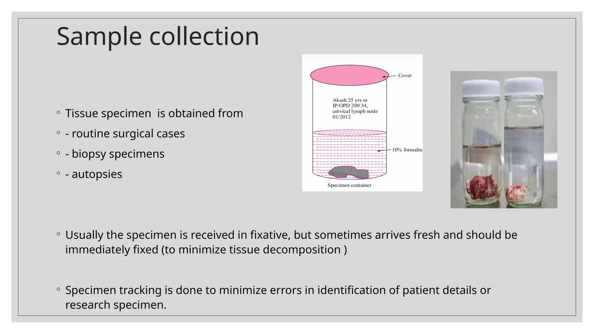
Sample collection
◦ Tissuespecimen is obtained from
◦ - routine surgical cases
◦ - biopsy specimens
◦ - autopsies
◦ Usually the specimen is received in fixative, but sometimes arrives fresh and should be
immediately fixed (to minimize tissue decomposition )
◦ Specimen tracking is done to minimize errors in identification of patient details or
research specimen.
6.
Grossing the specimen
◦Tissue sample received is examined, described and trimmed to proper size.
◦ Steps for better grossing:
◦ - fixation status is checked
◦ - uniform thin slices prepared
◦ - specimen trauma avoided
◦ - cross contamination avoided
◦ - osmotic injury avoided
◦ - tissue kept moistened
◦ - excessive blood mucus washed off
◦ - appropriate cassette size used
◦ - overloading of cassettes not done
◦ - cassettes clearly labelled
7.
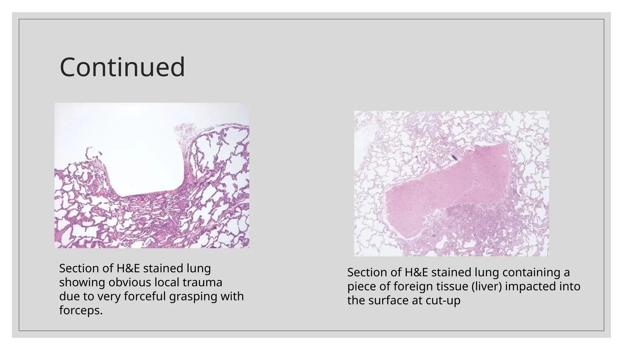
Continued
Section of H&Estained lung
showing obvious local trauma
due to very forceful grasping with
forceps.
Section of H&E stained lung containing a
piece of foreign tissue (liver) impacted into
the surface at cut-up
8.
Fixation of thetissue section
◦ Is done so that the tissue section can withstand subsequent steps for histological studies
◦ Aim of fixation-
◦ Prevention of tissue autolysis
◦ Prevention of bacterial attack
◦ Stabilization of protein components
◦ Maintainnece of shape and volume of molecular structures of tissue during
subsequent procedures
◦ To keep tissues as close to their natural state
◦ To prevent loss of tissue substances or rearrangement of tissue ingredients
◦ To allow cell parts to be selectively and clearly visible when stained
9.
Criteria for a“good” fixative
◦ Produces immediate death of cells in such a way that they retain closest possible resemblance of their life
like appearance
◦ Prevents autolysis
◦ Prevents putrefaction
◦ Reacts rapidly and completely with the tissue to stabilize it
◦ Fixes all constituents of the tissue
◦ Neither shrinks nor swells any tissue components
◦ Makes specimen hard enough to handle
◦ Raises RI of some of the cell contents for better visualization
◦ Has no rigid upper limit fixing time
◦ Cheap, non toxic, non inflammable, non irritant, non- deterioting, easy to prepare
10.
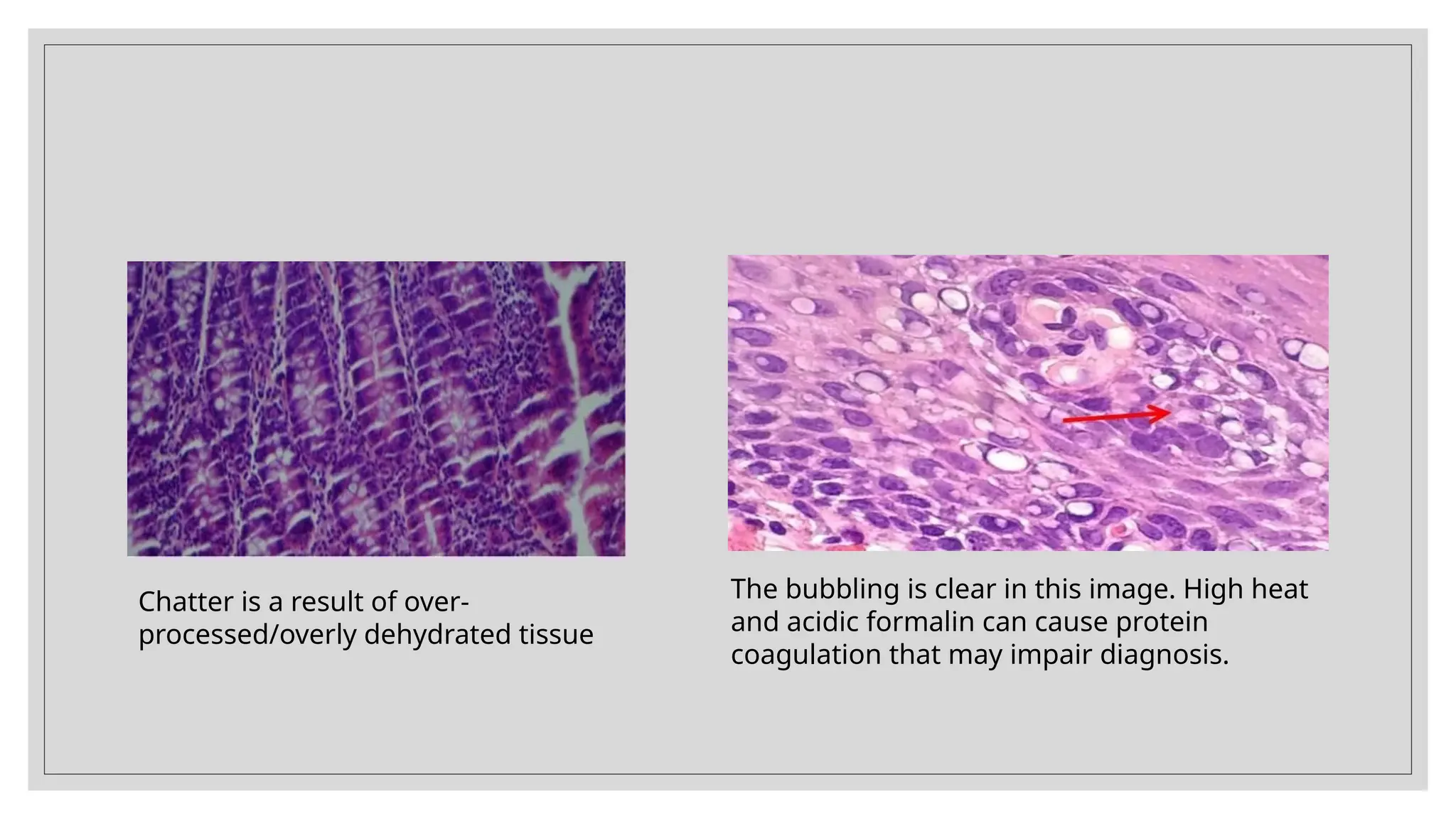
Chatter is aresult of over-
processed/overly dehydrated tissue
The bubbling is clear in this image. High heat
and acidic formalin can cause protein
coagulation that may impair diagnosis.
11.
Methods of Fixation
◦Fixation by immersion
◦ -- 10- 20 times the volume of specimen
◦ -- usually for 8 hours at Room temperature
◦ Fixation by perfusion (method of choice for EM)
◦ the fixative solution is introduced through the vascular system and reaches all the cells of the tissue via the
capillary net
12.
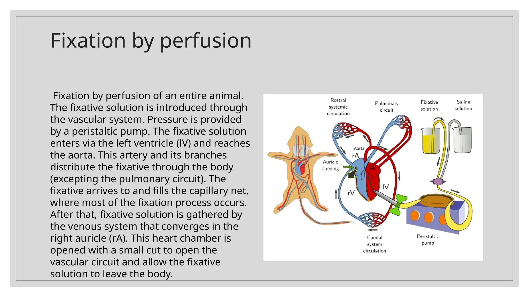
Fixation by perfusion
Fixationby perfusion of an entire animal.
The fixative solution is introduced through
the vascular system. Pressure is provided
by a peristaltic pump. The fixative solution
enters via the left ventricle (lV) and reaches
the aorta. This artery and its branches
distribute the fixative through the body
(excepting the pulmonary circuit). The
fixative arrives to and fills the capillary net,
where most of the fixation process occurs.
After that, fixative solution is gathered by
the venous system that converges in the
right auricle (rA). This heart chamber is
opened with a small cut to open the
vascular circuit and allow the fixative
solution to leave the body.
13.
Most common fixative
◦The most common fixative for light microscopy is 10% neutral buffered formalin.
◦ It preserves tissues by irreversibly cross linking proteins.
◦ Ideal time 2-8 hours (less than 24 hours) at room temperature.
14.
Factors involved intissue fixation
◦ Temperature: Usually done at room temperature. For electron microscopy and some
histochemistry low temperature (0–4°C) is preferred to slow down the autolysis. For urgent
biopsy, formalin may be heated up to 60°C.
◦ ™™pH: (hydrogen ion concentration): Good fixation is achieved at a pH of 6–8..
◦ ™™ Duration: Primary fixation in buffered formalin for 2–8 hours
◦ Tissue penetration
™™ : The depth of penetration is proportional to the square root of time(t) and
can be expressed as d = K t; where K (in tissue)is the constant and it is the coefficient of
√
diffusibility of the fixative in tissues or gel. K value (tissue) is high (1.33) in potassium dichromate
fixative whereas it is low (0.25) in chromium and glutaraldehyde fixative.
15.
Factors involved intissue fixation
◦ Osmolality: The preferred osmolality is slightly hypertonic solution or isotonic
solution.
◦ Volume changes
™™ : Volume of tissue may be changed during fixation. Nucleuses in
frozen sections are usually bigger whereas prolonged fixation in formalin causes
shrinkage. Some intercellular material like collagen swells when they are fixed.
◦ ™™ Concentration of fixative: Ideal concentration should be used for good fixation,
e.g. 10% buffered formalin, 3% glutaraldehyde or saturated solution of picric acid
and mercuric chloride.